logo

Pathophysiology and Medical Sonography of Hydrocele


Added on  2022-11-24

7 Pages1192 Words85 Views
 | 
 | 
 | 
Running Head: PATHOPHYSIOLOGY AND MEDICAL SONOGRAPHY OF HYDROCELE
Pathophysiology and Medical Sonography of Testes Hydrocele
Student’s Name
University Name
Author Note
Pathophysiology and Medical Sonography of Hydrocele_1

PATHOPHYSIOLOGY AND MEDICAL SONOGRAPHY OF HYDROCELE1
PART A:
Pathophysiology of the disease:
Normally, the testis is bounded by a smooth protective tissue sac. It generates a small amount of
lubricating fluid which allows the testis to move freely. The excess fluid generally drains away
into the veins present in the scrotum. Thus, Hydrocele arises due to the accumulation of excess
fluid inside the scrotum. This results in the enlargement one of the testicles. This is mostly
common in premature new-born babies. This mainly occurs if the processus vaginalis fails to
close completely or the body is incapable of absorbing the excess fluid excreted. But in adults, it
is mainly the result of scrotal inflammation or injury (Patil, Shetty and Das, 2015). Thus, the
pathophysiology of hydroceles involves the imbalance of intrinsic scrotal fluid production by the
mesothelial cells present in the inner surface of the tunica vaginalis and fluid absorption by the
draining the lymphatic vessels present in the parietal layer.
There are mainly two types of hydroceles-
Communicating hydroceles: It is caused from exogenous sources. This type causes
indirect hernia.
Non-communicating hydroceles: It is developed due to the abnormal intrinsic scrotal
fluid shifts.
The hydroceles are positioned anterior and superior to the testis. Thus, hydrocele has a chance of
occurring bilaterally in 7-10 % of the cases. Thus, when it occurs on the right side of the body, it
can be easily linked with hernia. Usually most of the pediatric hydroceles are congenital. When
hydrocele occurs in the cord, it is often associated with failure of processus vaginalis to close
Pathophysiology and Medical Sonography of Hydrocele_2

PATHOPHYSIOLOGY AND MEDICAL SONOGRAPHY OF HYDROCELE2
efficiently, which allows pooling of the fluid in the middle of the spermatic cord. Similarly,
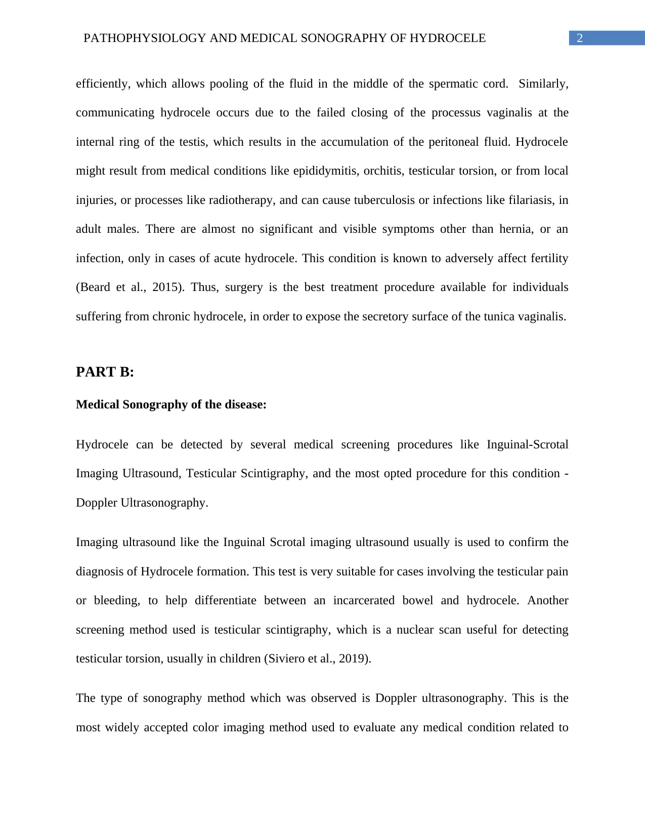
communicating hydrocele occurs due to the failed closing of the processus vaginalis at the
internal ring of the testis, which results in the accumulation of the peritoneal fluid. Hydrocele
might result from medical conditions like epididymitis, orchitis, testicular torsion, or from local
injuries, or processes like radiotherapy, and can cause tuberculosis or infections like filariasis, in
adult males. There are almost no significant and visible symptoms other than hernia, or an
infection, only in cases of acute hydrocele. This condition is known to adversely affect fertility
(Beard et al., 2015). Thus, surgery is the best treatment procedure available for individuals
suffering from chronic hydrocele, in order to expose the secretory surface of the tunica vaginalis.
PART B:
Medical Sonography of the disease:
Hydrocele can be detected by several medical screening procedures like Inguinal-Scrotal
Imaging Ultrasound, Testicular Scintigraphy, and the most opted procedure for this condition -
Doppler Ultrasonography.
Imaging ultrasound like the Inguinal Scrotal imaging ultrasound usually is used to confirm the
diagnosis of Hydrocele formation. This test is very suitable for cases involving the testicular pain
or bleeding, to help differentiate between an incarcerated bowel and hydrocele. Another
screening method used is testicular scintigraphy, which is a nuclear scan useful for detecting
testicular torsion, usually in children (Siviero et al., 2019).
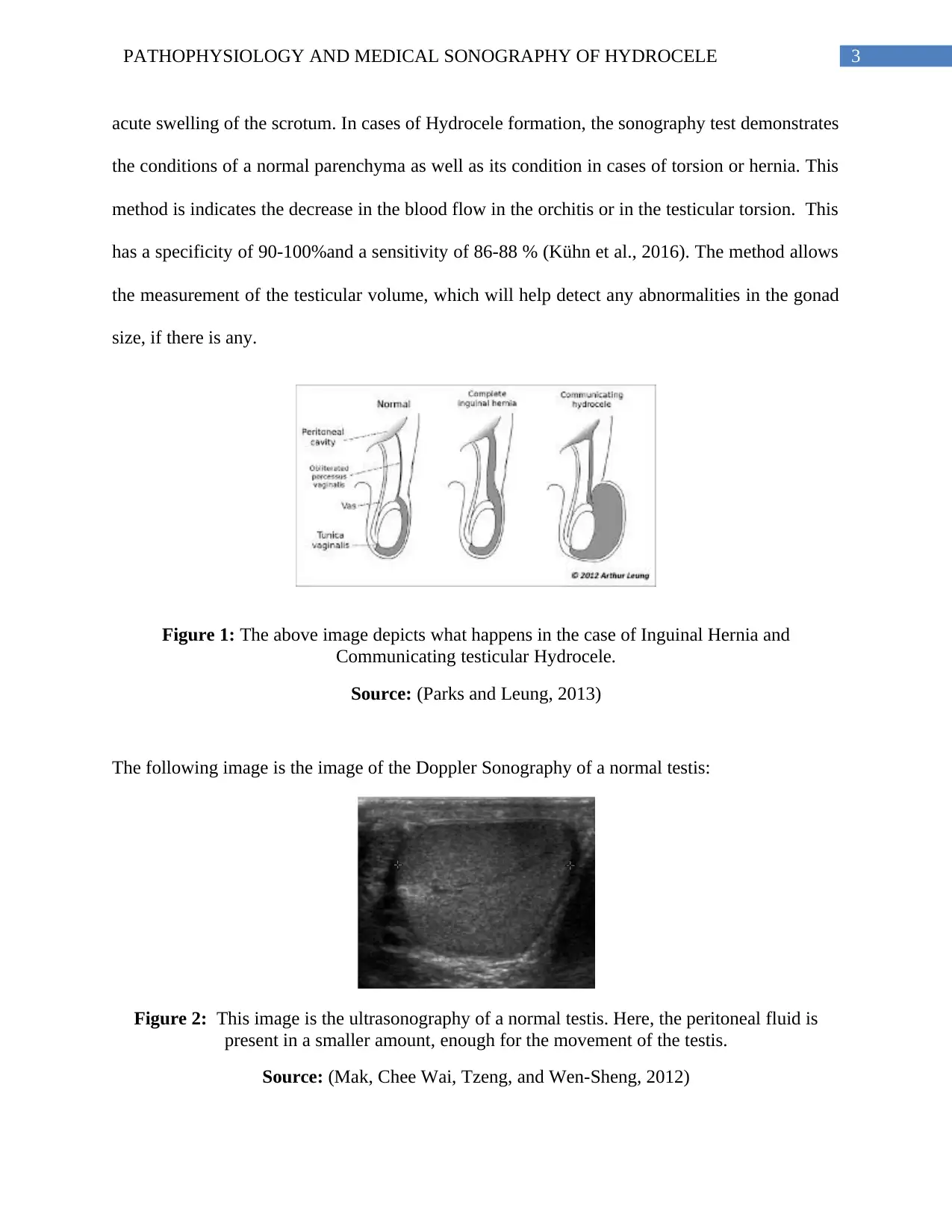
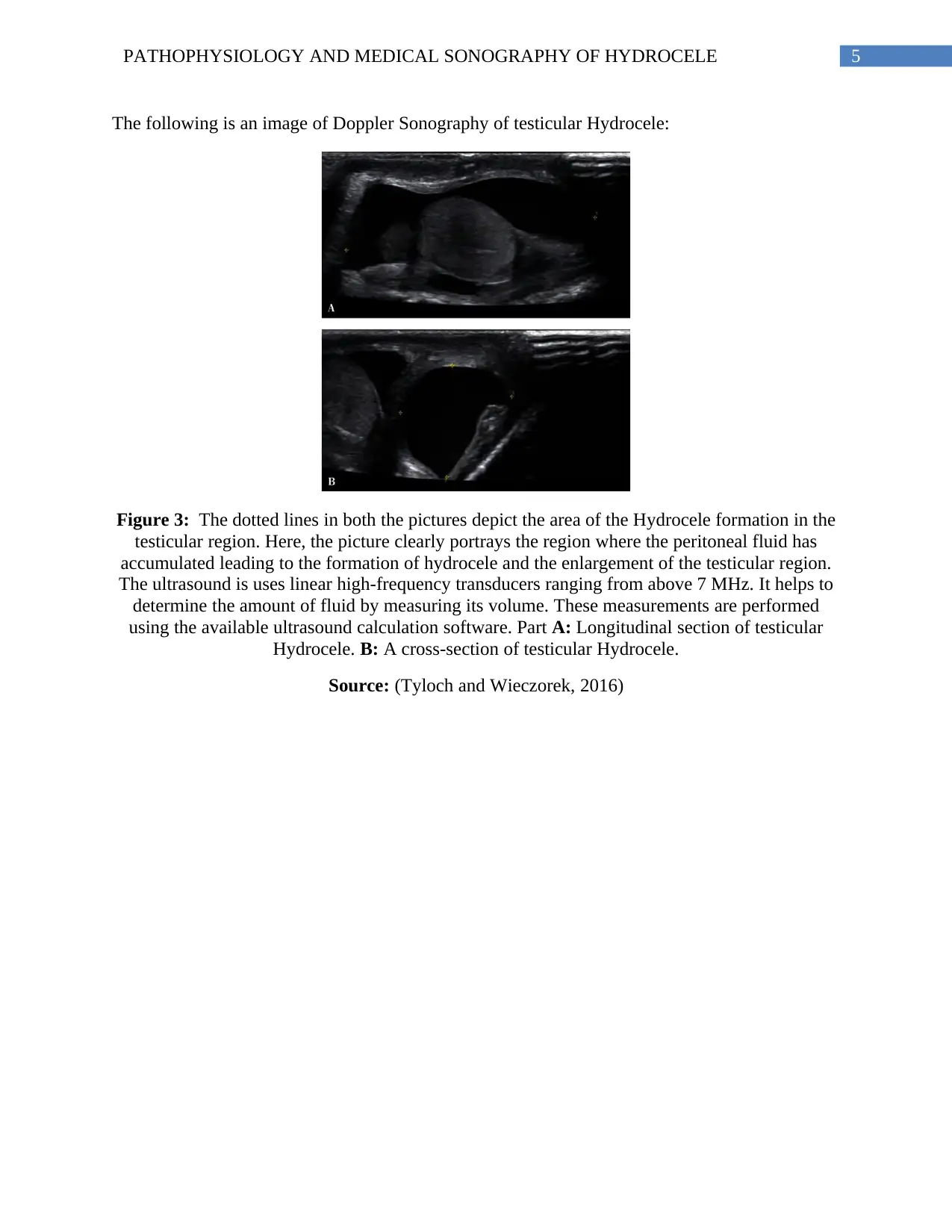
The type of sonography method which was observed is Doppler ultrasonography. This is the
most widely accepted color imaging method used to evaluate any medical condition related to
Pathophysiology and Medical Sonography of Hydrocele_3

End of preview

Want to access all the pages? Upload your documents or become a member.