Acute Pancreatitis: Pathophysiology, Lung Injury, and Treatment Report
VerifiedAdded on 2020/02/05
|9
|2224
|319
Report
AI Summary
This report provides a detailed analysis of the pathophysiology of acute pancreatitis, a condition characterized by acute inflammation of the pancreas. It explores the causes, ranging from gallstones and alcohol abuse to abdominal trauma. The report delves into the progression of the disease, including the development of acute lung injury and sepsis, and relates these complications to a case study of a patient named Betty Turner. It outlines expected physical assessment findings in patients with acute lung injury and sepsis, covering system-based findings, hemodynamic parameters, and acid-base balance. Furthermore, the report identifies and discusses evidence-based treatment goals and strategies, including sedation, oxygenation, hemodynamic management, and nutrition therapy, to prevent or minimize multi-organ failure. Diagnostic methods, including imaging and genetic testing, are also discussed, along with the importance of identifying disease severity for effective management. The report emphasizes the need for standardized nutritional support and evidence-based guidelines for patient care.
Running head: PATHOPHYSIOLOGY OF ACUTE PANCREATITIS 1
Pathophysiology Of Acute Pancreatitis
Student’s name
Institution
Pathophysiology Of Acute Pancreatitis
Student’s name
Institution
Paraphrase This Document
Need a fresh take? Get an instant paraphrase of this document with our AI Paraphraser
PATHOPHYSIOLOGY OF ACUTE PANCREATITIS 2
Pathophysiology of Acute Pancreatitis
1) Describe the pathophysiology of acute pancreatitis leading to acute lung injury and
respiratory failure, sepsis and septic shock and relate the pathophysiology of Betty’s
health problems to her deterioration in the Emergency Department.
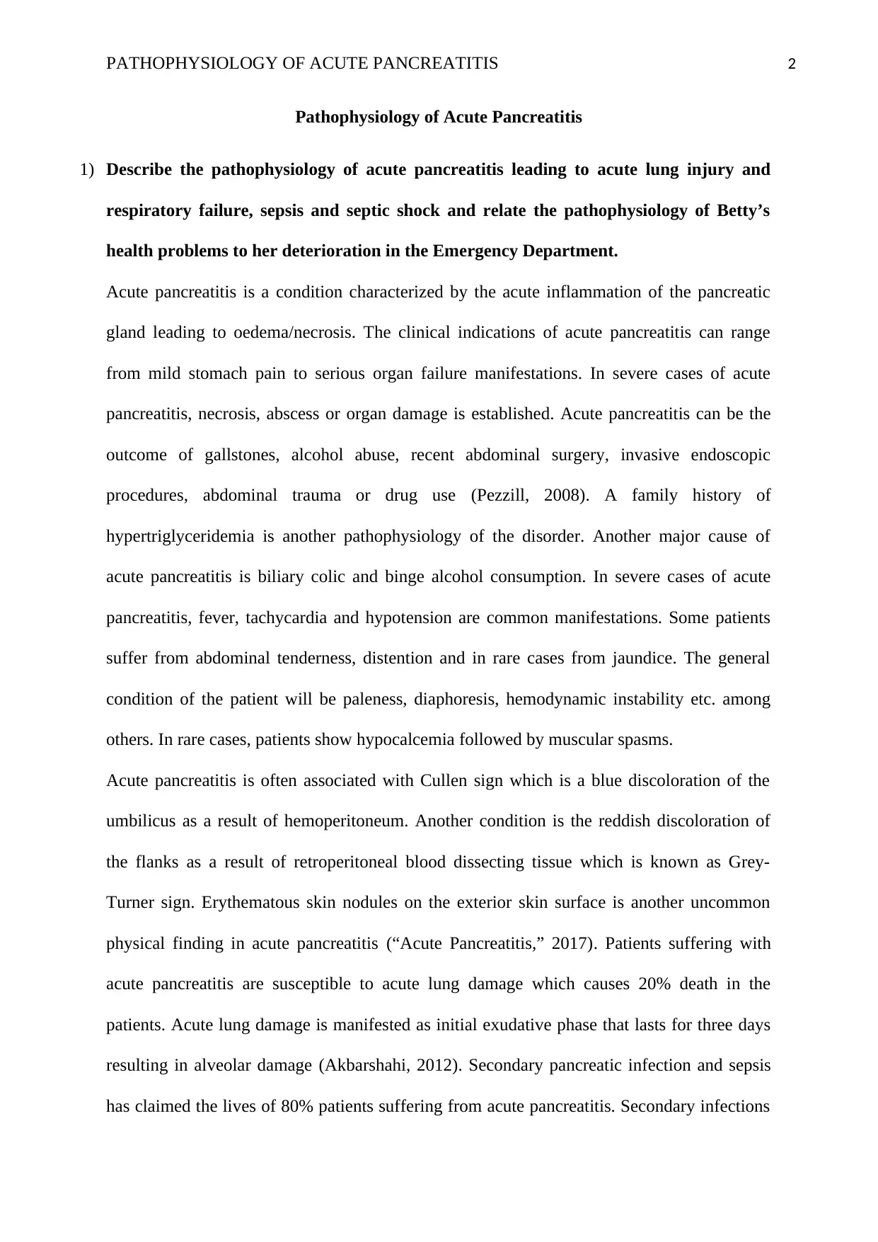
Acute pancreatitis is a condition characterized by the acute inflammation of the pancreatic
gland leading to oedema/necrosis. The clinical indications of acute pancreatitis can range
from mild stomach pain to serious organ failure manifestations. In severe cases of acute
pancreatitis, necrosis, abscess or organ damage is established. Acute pancreatitis can be the
outcome of gallstones, alcohol abuse, recent abdominal surgery, invasive endoscopic
procedures, abdominal trauma or drug use (Pezzill, 2008). A family history of
hypertriglyceridemia is another pathophysiology of the disorder. Another major cause of
acute pancreatitis is biliary colic and binge alcohol consumption. In severe cases of acute
pancreatitis, fever, tachycardia and hypotension are common manifestations. Some patients
suffer from abdominal tenderness, distention and in rare cases from jaundice. The general
condition of the patient will be paleness, diaphoresis, hemodynamic instability etc. among
others. In rare cases, patients show hypocalcemia followed by muscular spasms.
Acute pancreatitis is often associated with Cullen sign which is a blue discoloration of the
umbilicus as a result of hemoperitoneum. Another condition is the reddish discoloration of
the flanks as a result of retroperitoneal blood dissecting tissue which is known as Grey-
Turner sign. Erythematous skin nodules on the exterior skin surface is another uncommon
physical finding in acute pancreatitis (“Acute Pancreatitis,” 2017). Patients suffering with
acute pancreatitis are susceptible to acute lung damage which causes 20% death in the
patients. Acute lung damage is manifested as initial exudative phase that lasts for three days
resulting in alveolar damage (Akbarshahi, 2012). Secondary pancreatic infection and sepsis
has claimed the lives of 80% patients suffering from acute pancreatitis. Secondary infections
PATHOPHYSIOLOGY OF ACUTE PANCREATITIS 3
and sepsis develops due to bacterial contamination. To prevent the infection systemic
administration of antibiotics is the only available step (Mifkovic, 2006).
Betty Turner showed clinical symptoms such as nausea, vomiting and lethargy which are
characteristic of acute pancreatitis. In ED, Betty continued to deteriorate and her conscious
level decreased and she was intubated for airway management and ventilation support.
Respiratory failure and ventilation damage is a characteristic of severe case of pancreatitis.
2) Drawing upon the pathophysiology of acute pancreatitis and acute lung injury,
describe and rationalise what physical assessment findings you would expect to
see in a patient with acute lung injury and sepsis. In your description, you should
consider: a) expected system-based findings; b) haemodynamic parameters,
including right and left-sided heart pressures; and c) acid-base balance results,
serum biochemistry and haematology markers.
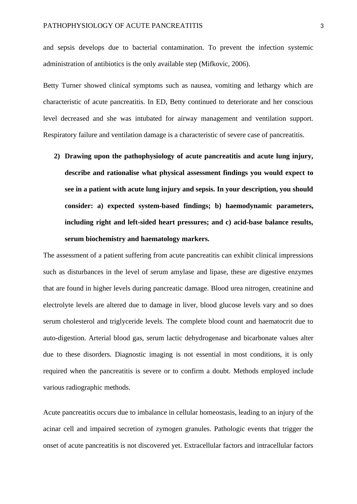
The assessment of a patient suffering from acute pancreatitis can exhibit clinical impressions
such as disturbances in the level of serum amylase and lipase, these are digestive enzymes
that are found in higher levels during pancreatic damage. Blood urea nitrogen, creatinine and
electrolyte levels are altered due to damage in liver, blood glucose levels vary and so does
serum cholesterol and triglyceride levels. The complete blood count and haematocrit due to
auto-digestion. Arterial blood gas, serum lactic dehydrogenase and bicarbonate values alter
due to these disorders. Diagnostic imaging is not essential in most conditions, it is only
required when the pancreatitis is severe or to confirm a doubt. Methods employed include
various radiographic methods.
Acute pancreatitis occurs due to imbalance in cellular homeostasis, leading to an injury of the
acinar cell and impaired secretion of zymogen granules. Pathologic events that trigger the
onset of acute pancreatitis is not discovered yet. Extracellular factors and intracellular factors
⊘ This is a preview!⊘
Do you want full access?
Subscribe today to unlock all pages.

Trusted by 1+ million students worldwide
PATHOPHYSIOLOGY OF ACUTE PANCREATITIS 4
(play a role. Also, acute pancreatitis can develop due to ductal cell injury. Onset of cellular
injury can lead the cellular membrane trafficking to become chaotic. The harmful effects are
Lysosomal and zymogen granule compartments fuse, enabling activation of trypsinogen to
trypsin, trypsin triggering the zymogen activation cascade, extrusion of secretory vesicles
across the basolateral membrane into the interstitium. These neutrophils then release
superoxide or proteolytic enzymes such as cathepsins, collagenase, and elastase etc. among
the others. Followed by macrophages releasing cytokines trigger the inflammatory responses.
Some of the mediators include tumor necrosis factor-alpha (TNF-α), interleukin etc.
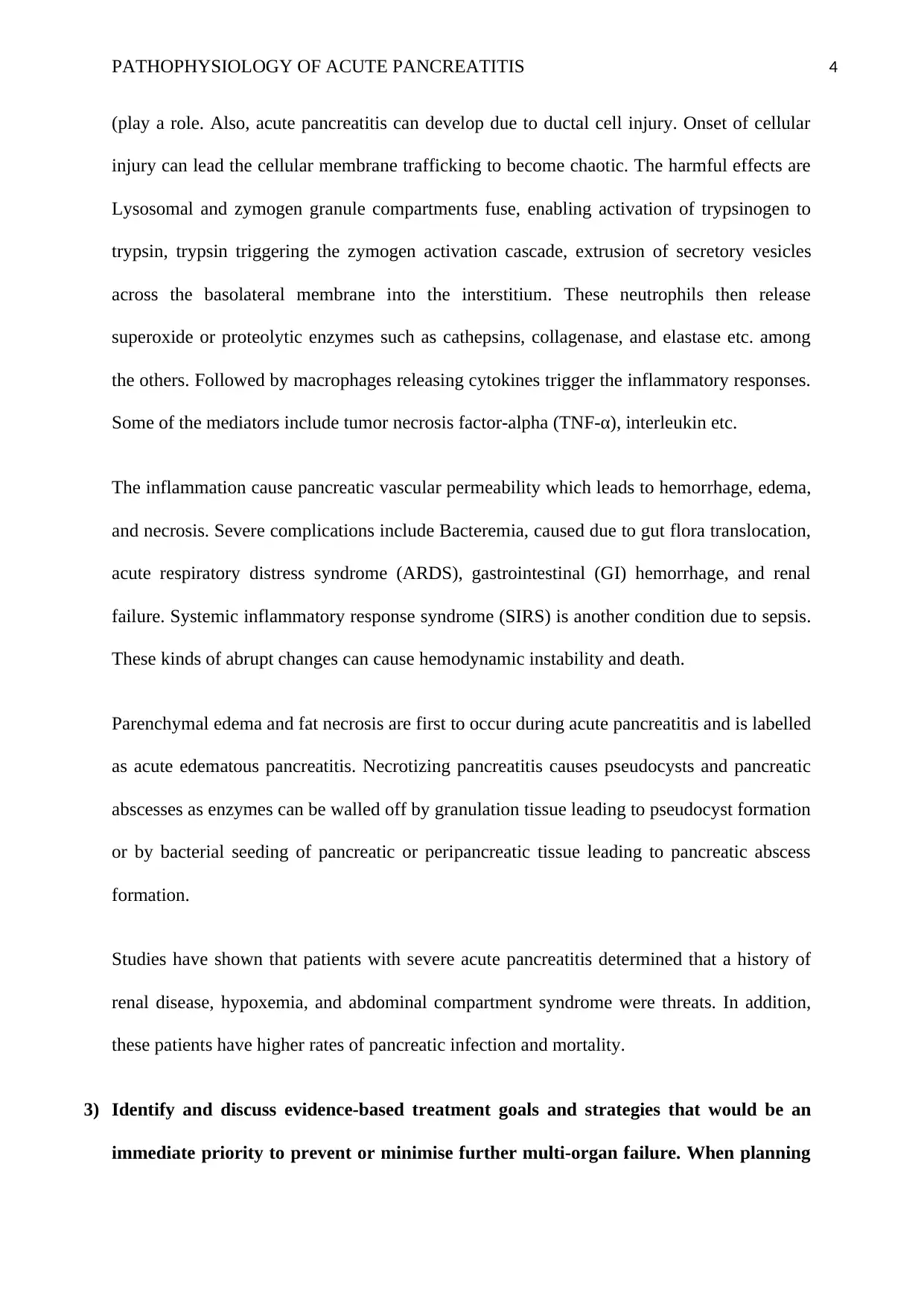
The inflammation cause pancreatic vascular permeability which leads to hemorrhage, edema,
and necrosis. Severe complications include Bacteremia, caused due to gut flora translocation,
acute respiratory distress syndrome (ARDS), gastrointestinal (GI) hemorrhage, and renal
failure. Systemic inflammatory response syndrome (SIRS) is another condition due to sepsis.
These kinds of abrupt changes can cause hemodynamic instability and death.
Parenchymal edema and fat necrosis are first to occur during acute pancreatitis and is labelled
as acute edematous pancreatitis. Necrotizing pancreatitis causes pseudocysts and pancreatic
abscesses as enzymes can be walled off by granulation tissue leading to pseudocyst formation
or by bacterial seeding of pancreatic or peripancreatic tissue leading to pancreatic abscess
formation.
Studies have shown that patients with severe acute pancreatitis determined that a history of
renal disease, hypoxemia, and abdominal compartment syndrome were threats. In addition,
these patients have higher rates of pancreatic infection and mortality.
3) Identify and discuss evidence-based treatment goals and strategies that would be an
immediate priority to prevent or minimise further multi-organ failure. When planning
Paraphrase This Document
Need a fresh take? Get an instant paraphrase of this document with our AI Paraphraser
PATHOPHYSIOLOGY OF ACUTE PANCREATITIS 5
treatment goals and strategies, you should consider the following: a) Sedation and pain
management; b) Oxygenation and ventilation; c) Haemodynamic management,
including fluid and medication therapy; and d) Nutrition therapy
Management of acute pancreatitis is a standardized method; although, patients with severe
acute pancreatitis require extensive care. The initial steps of supportive care include Fluid
resuscitation and Nutritional therapy. Another major treatment is Antibiotic therapy which
employs Antibiotics such as imipenem which is used in case of pancreatitis and infected
pancreatic necrosis. However, such drugs cannot be given for fever and initial care. This kind
of antibiotic therapy is not currently recommended. In patients, hospitalized due to severe
pancreatitis it is recommended to employ fluid therapy.
Another important method is Surgical intervention when other minimally non-invasive or
non-invasive methods are not available. Commonly used procedures in pancreatitis include
the removal of Gallstone pancreatitis which is known as Cholecystectomy, Image-guided
percutaneous placement of a collection tube into the fluid collection which is referred to as
pancreatic duct disruption. Surgeries also involve the removal of pseudocysts which is not
quite necessary in most cases, image-guided aspiration of infected pancreatic necrosis and
pancreatic abscess. Sedation is a form of pain management that can be achieved by general
anaesthesia during the interventional surgeries.
Abdominal radiography is a test to detect the free air in the kidney-ureters-bladder of an
upright patient. Abdominal ultrasonography is a test to detect gallstones and is one of the first
tests usually employed. Endoscopic ultrasonography is another test where microlithiasis and
periampullary lesions are detected which are not otherwise detected by other tests.
Endoscopic retrograde cholangiopancreatography is not used commonly and if used, should
be with caution. Magnetic resonance cholangiopancreatography is safer and non-invasive as
PATHOPHYSIOLOGY OF ACUTE PANCREATITIS 6
compared to endoscopic retrograde cholangiopancreatography but is not as sensitive. CT-
guided aspiration and drainage and genetic testing are two more methods to diagnose acute
pancreatitis.
Patients with biliary pancreatitis tend to have a higher mortality than patients with alcoholic
pancreatitis. Due to improvements in supportive care the rates of mortality has decreased over
the years. Type 2 diabetes mellitus contributes to higher mortality in patients with acute
pancreatitis. The patients with pancreatic necrosis who do not suffer from organ failure show
the mortality of zero. Most deaths result from multiorgan system failure in acute pancreatitis
patients. Bacterial infections do play a major role, but organ failure contributes to a major
rate of mortality. Acute respiratory distress syndrome (ARDS), acute renal failure, cardiac
depression, hemorrhage, and hypotensive shock are all severe manifestations of acute
pancreatitis.
Identification of patients based on greatest need of aggressive medical treatment by
understanding the disease severity as mild or severe is highly recommended. Strategies have
been employed to identify the severity of acute pancreatitis and predict its outcome. Ranson
criteria, Glasgow, Imrie are several clinical scoring systems that are available. The best
validated scoring system is The APACHE II. Biological markers and Genetic markers have
also been used for this purpose.
The specificity of Peritoneal lavage is a 93% but it has a low sensitivity of about 54%.
Dynamic CT scanning is widely used to predict acute pancreatitis of the abdomen. The
Balthazar criteria shows a sensitivity of 87% and specificity of 88%. Studies have shown that
the prognostic value of the neutrophil-lymphocyte ratio in 146 consecutive patients with
acute pancreatitis is elevated during the first 48 hours of treatment. The NLR is provides an
idea about inflammation in the abdomen. Data from 822 patients hospitalized with acute
⊘ This is a preview!⊘
Do you want full access?
Subscribe today to unlock all pages.

Trusted by 1+ million students worldwide
PATHOPHYSIOLOGY OF ACUTE PANCREATITIS 7
pancreatitis, showed that patients who had non-alcoholic fatty liver showed higher incidence
of moderately severe and severe acute pancreatitis than those without non-alcoholic fatty
liver. These patients were also seen to have higher C-reactive protein and showed an
APACHE II. These patients also exhibited CT scan severity index with organ failure and
local complications. (“Acute Pancreatitis: Practice Essentials, Background,
Pathophysiology,” 2016).
Standardized parenteral nutrition for providing nutrients to patients with severe acute
pancreatitis is in place. The concept behind this strategy was to avoid stimulation of exocrine
pancreatic secretory responses in order to ensure rest to the pancreas and in order to improve
the nutritional status. Nasojejunal feeding was compared with total parenteral nutrition and
no difference on the outcome was seen but the cost for enteral nutrition was four times lower.
In another group of data, comparison of intravenous feeding with no nutritional support
demonstrated that intravenous nutrition did not alter the outcome of patients with mild-to-
moderate pancreatitis otherwise contemplated by hospitalization, medications etc. However,
patients receiving total parenteral nutrition showed an increase in catheter-related infections.
The study suggests that parenteral nutrition can have certain associated shortcomings. Not
only is the increased risk of catheter-related sepsis but severe hyperglycemia has also been
reported. Hence, overfeeding is a chief risk factor for difficulties in patients receiving
parenteral nutrition. It is another concern that parenteral nutrition affects gut barrier function.
However, there is no evidence to support this claim.
In conclusion, nutritional support for severe and mild cases of acute pancreatitis should be
different and standardized across the globe. In the case of mild pancreatitis, no evidence is
present to prove that enteral or parenteral nutrition has a positive effect. Patients with pre-
existing malnutrition do not support the theory. Nutritional therapy is best for patients
Paraphrase This Document
Need a fresh take? Get an instant paraphrase of this document with our AI Paraphraser
PATHOPHYSIOLOGY OF ACUTE PANCREATITIS 8
suffering from severe pancreatitis. The mode of nutrient delivery can be parenteral or enteral
in such cases and the decision should be made depending on patient tolerance. If possible,
enteral is best attempted in all patients. The medical practitioner should carefully intervene
the necessary nutritional support of a patient. Some patients may also be needed to be
administered with enteral and parenteral nutrition. These statements are a result of
physiological studies and are not supported by evidence from cohort or cross-sectional
studies. The available controlled data should be utilized to plan and implement evidence
based guidelines and policies. (Meier, 2002).
PATHOPHYSIOLOGY OF ACUTE PANCREATITIS 9
References:
Acute Pancreatitis: Practice Essentials, Background, Pathophysiology. (2016). Retrieved May
6, 2017, from http://emedicine.medscape.com/article/181364-overview#a3
Acute Pancreatitis: Practice Essentials, Background, Pathophysiology. (2017). Retrieved
from http://emedicine.medscape.com/article/181364-overview
Akbarshahi. H. (2012). Acute lung injury in acute pancreatitis – Awaiting the big leap.
respiratoryMedicine
Mifkovic A. (2006). Septic complications of acute pancreatitis. - PubMed - NCBI. Retrieved
May 6, 2017, from https://www.ncbi.nlm.nih.gov/pubmed/17125065
Meier, R. (2002). CONSENSUS STATEMENT: ESPEN guidelines on nutrition in acute
pancreatitis - Acutepancreatitis.pdf. Retrieved May 6, 2017, from
http://espen.info/documents/Acutepancreatitis.pdf
Pezzill, R. (2008). Acute Pancreatitis: Pathophysiology, Clinical Aspects, Diagnosis of
Treatment.
⊘ This is a preview!⊘
Do you want full access?
Subscribe today to unlock all pages.

Trusted by 1+ million students worldwide
1 out of 9
Related Documents
Your All-in-One AI-Powered Toolkit for Academic Success.
+13062052269
info@desklib.com
Available 24*7 on WhatsApp / Email
![[object Object]](/_next/static/media/star-bottom.7253800d.svg)
Unlock your academic potential
Copyright © 2020–2025 A2Z Services. All Rights Reserved. Developed and managed by ZUCOL.


