This document discusses the PCR amplification and detection of malic enzyme. It covers the materials used, the method followed, the preparation of restriction enzymes, the preparation of agarose gel, running the gel, and the results obtained. The document does not mention the course code, course name, or college/university.
Contribute Materials
Your contribution can guide someone’s learning journey. Share your
documents today.
PCR amplification and detection of malic enzyme1 PCR AMPLIFICATION AND DETECTION OF MALIC ENZYME Course Professors Name Institution Location Date
Secure Best Marks with AI Grader
Need help grading? Try our AI Grader for instant feedback on your assignments.
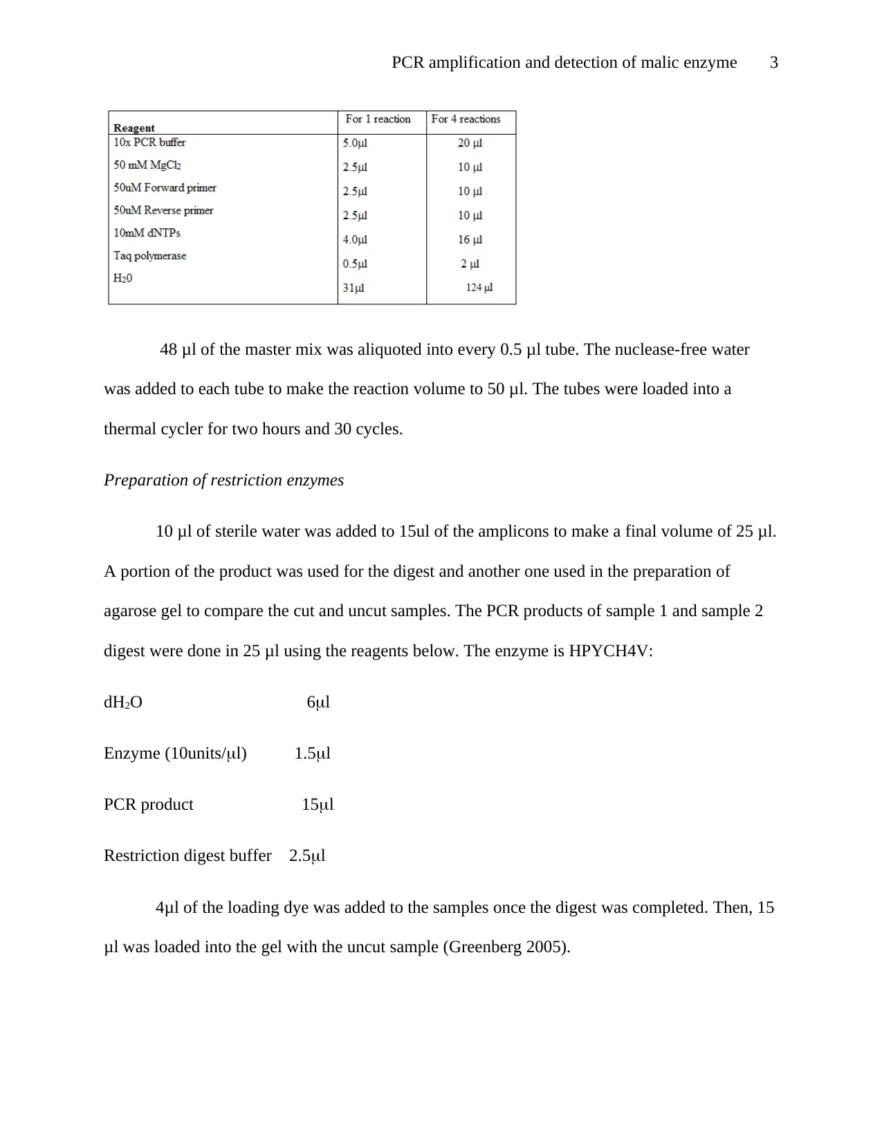
PCR amplification and detection of malic enzyme2 Introduction The Polymerase chain reaction amplifies DNA target region for detection by gel electrophoresis. Malic enzyme is a genome-coded mitochondrial enzyme which converts malate to pyruvate (Lenzenet al. 2005). The PCR process requires an enzyme known as Taq Polymerase, which helps in elongations and primers. The experiment aimed to amplify malic enzyme and visualize amplicons using agarose gel to determine the availability of alleles (Thermo Fisher Scientific 2019). Materials used 10mM dNTPs 10x PCR buffer Control sample Sterile deionized water Forward & reverse primers ME2rs585344R25’-GAAACATGGGTGTTGGAGACCC-3’ ME2rs585344F25’-GACTTCCCCGCTGTAAGCTG-3’ 25 mM MgCl2 Taq DNA polymerase Method An experiment had three tubes labeled from one to three. The negative control has sterile water, DNA template 1 and DNA template 2. The master mix was prepared using the calculations below.
PCR amplification and detection of malic enzyme3 48 μl of the master mix was aliquoted into every 0.5 μl tube. The nuclease-free water was added to each tube to make the reaction volume to 50 μl. The tubes were loaded into a thermal cycler for two hours and 30 cycles. Preparation of restriction enzymes 10 μl of sterile water was added to 15ul of the amplicons to make a final volume of 25 μl. A portion of the product was used for the digest and another one used in the preparation of agarose gel to compare the cut and uncut samples. The PCR products of sample 1 and sample 2 digest were done in 25 μl using the reagents below. The enzyme is HPYCH4V: dH2O6l Enzyme (10units/l)1.5l PCR product15l Restriction digest buffer2.5l 4μl of the loading dye was added to the samples once the digest was completed. Then, 15 μl was loaded into the gel with the uncut sample (Greenberg 2005).
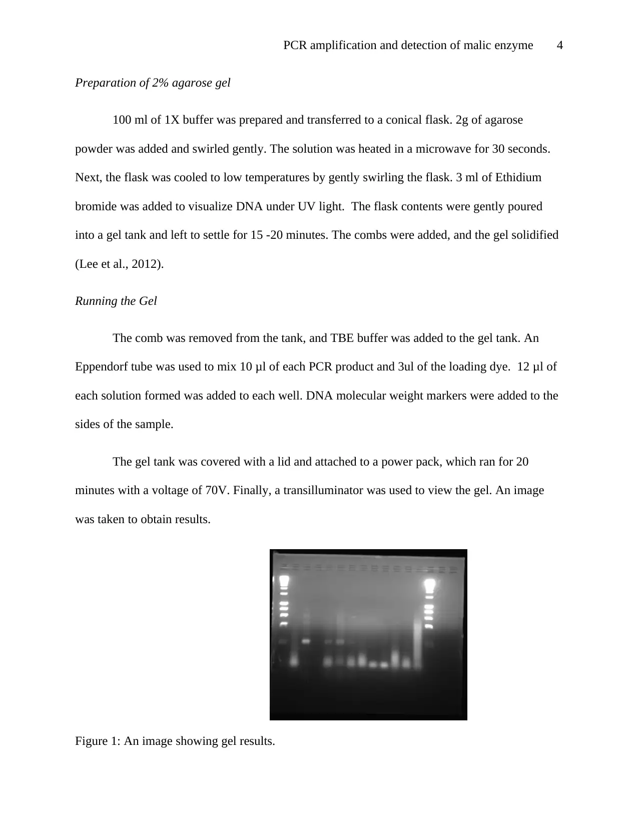
PCR amplification and detection of malic enzyme4 Preparation of 2% agarose gel 100 ml of 1X buffer was prepared and transferred to a conical flask. 2g of agarose powder was added and swirled gently. The solution was heated in a microwave for 30 seconds. Next, the flask was cooled to low temperatures by gently swirling the flask. 3 ml of Ethidium bromide was added to visualize DNA under UV light. The flask contents were gently poured into a gel tank and left to settle for 15 -20 minutes. The combs were added, and the gel solidified (Lee et al., 2012). Running the Gel The comb was removed from the tank, and TBE buffer was added to the gel tank. An Eppendorf tube was used to mix 10 μl of each PCR product and 3ul of the loading dye. 12 μl of each solution formed was added to each well. DNA molecular weight markers were added to the sides of the sample. The gel tank was covered with a lid and attached to a power pack, which ran for 20 minutes with a voltage of 70V. Finally, a transilluminator was used to view the gel. An image was taken to obtain results. Figure 1: An image showing gel results.
Paraphrase This Document
Need a fresh take? Get an instant paraphrase of this document with our AI Paraphraser
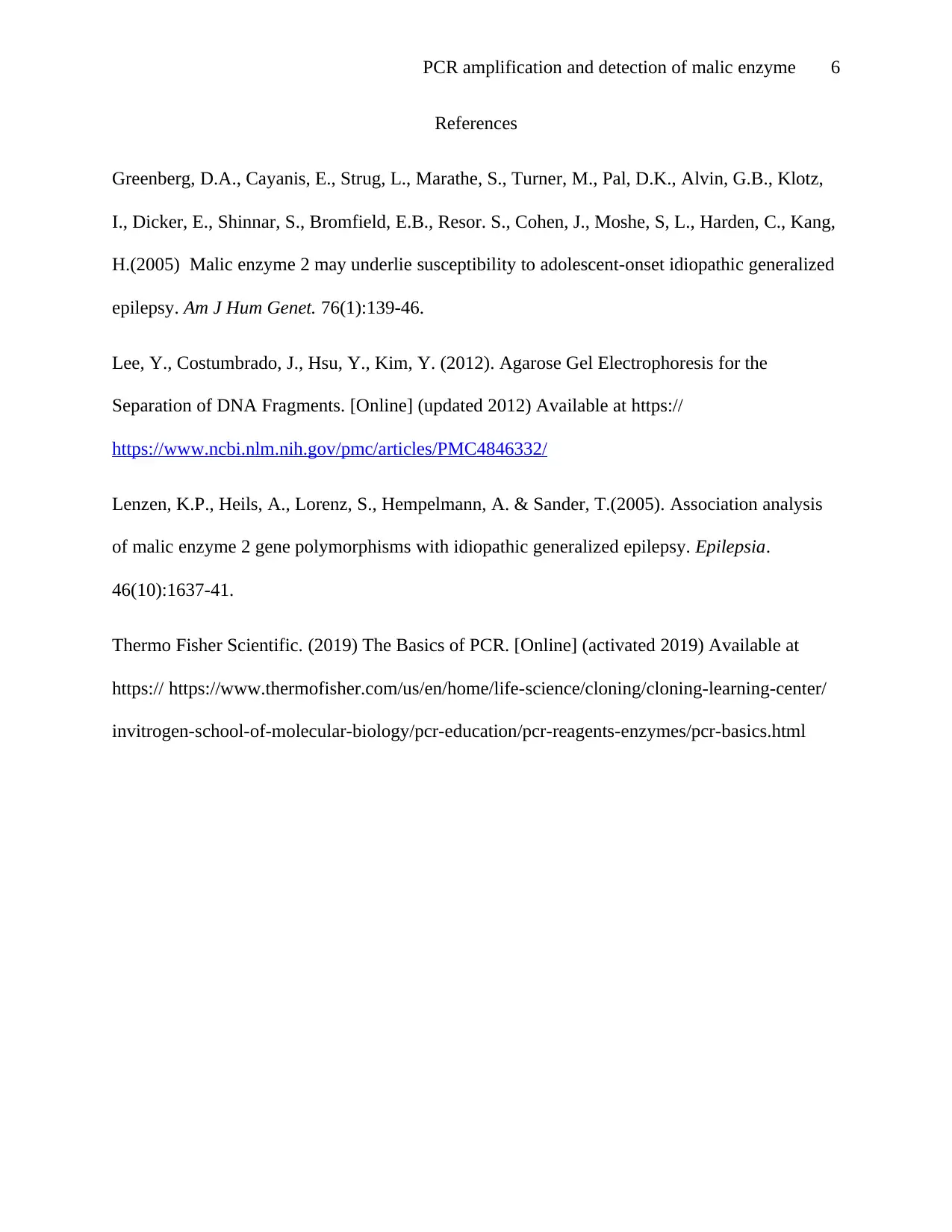
PCR amplification and detection of malic enzyme5 Figure 2: An image showing Gel digest. Discussion The experiment produced successful amplification at 200 bp. The result indicated that one homozygous gene was present in the band. However, all the wells did not provide good bands, due to either contamination or poor pipetting skills(Lee et al. 2012). Conclusion. The experiment aimed to amplify and detect malic enzyme. The aim was partly archived. However, the gel results indicated the presence of contamination. Therefore, the results were not satisfactory, and another experiment was to be done.
PCR amplification and detection of malic enzyme6 References Greenberg, D.A., Cayanis, E., Strug, L., Marathe, S., Turner, M., Pal, D.K., Alvin, G.B., Klotz, I., Dicker, E., Shinnar, S., Bromfield, E.B., Resor. S., Cohen, J., Moshe, S, L., Harden, C., Kang, H.(2005)Malicenzyme2may underlie susceptibility to adolescent-onset idiopathic generalized epilepsy.Am J Hum Genet.76(1):139-46. Lee, Y., Costumbrado, J., Hsu, Y., Kim, Y. (2012). Agarose Gel Electrophoresis for the Separation of DNA Fragments. [Online] (updated 2012) Available at https:// https://www.ncbi.nlm.nih.gov/pmc/articles/PMC4846332/ Lenzen, K.P., Heils, A., Lorenz, S., Hempelmann, A. & Sander, T.(2005).Association analysis ofmalicenzyme2gene polymorphisms with idiopathic generalizedepilepsy.Epilepsia. 46(10):1637-41. Thermo Fisher Scientific. (2019) The Basics of PCR. [Online] (activated 2019) Available at https://https://www.thermofisher.com/us/en/home/life-science/cloning/cloning-learning-center/ invitrogen-school-of-molecular-biology/pcr-education/pcr-reagents-enzymes/pcr-basics.html