Physics of Human Eye | Assignment
VerifiedAdded on 2022/09/06
|6
|1366
|24
AI Summary
Contribute Materials
Your contribution can guide someone’s learning journey. Share your
documents today.
Running head: PHYSICS OF HUMAN EYE 1
Physics of Human Eye
Name
Institutional Affiliation
Physics of Human Eye
Name
Institutional Affiliation
Secure Best Marks with AI Grader
Need help grading? Try our AI Grader for instant feedback on your assignments.
PHYSICS OF HUMAN EYE 2
Physics of Human Eye
The eye is an important part of every human as most if the information regarding the
environment is gathered through seeing. Therefore, vision is very important, and is achieved
when light enters the human eye. Light refers to an electromagnetic wav, and when it enters
the eye, the retina of an eye converts it into electrical impulses. The impulses are then
transferred into the brain for analysis and interpretations (3). Consequently, an individual is
able to perceive what he or she has seen.
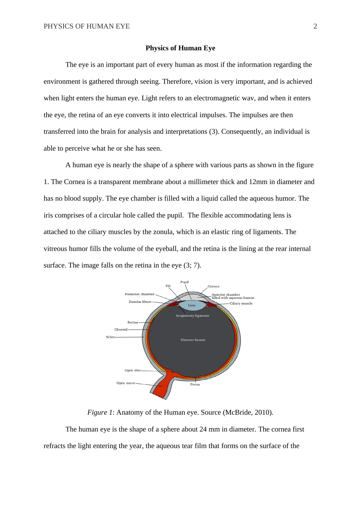
A human eye is nearly the shape of a sphere with various parts as shown in the figure
1. The Cornea is a transparent membrane about a millimeter thick and 12mm in diameter and
has no blood supply. The eye chamber is filled with a liquid called the aqueous humor. The
iris comprises of a circular hole called the pupil. The flexible accommodating lens is
attached to the ciliary muscles by the zonula, which is an elastic ring of ligaments. The
vitreous humor fills the volume of the eyeball, and the retina is the lining at the rear internal
surface. The image falls on the retina in the eye (3; 7).
Figure 1: Anatomy of the Human eye. Source (McBride, 2010).
The human eye is the shape of a sphere about 24 mm in diameter. The cornea first
refracts the light entering the year, the aqueous tear film that forms on the surface of the
PHYSICS OF HUMAN EYE 3
cornea ensures the smoothness of the first optical surface; hence providing the best possible
quality of the image. The role of the pupil is to limits the light entering the eye by controlling
the retinal illumination. The size of the pupil changes to at least 2 mm, in diameter when in
bright light and 8mm in the dark. The light enters in the eye, the cornea and crystalline lens to
form the retinal image. Once the light enters the eye, the light is refracted by the lens and it
enters the posterior chamber, through the vitreous humor and reaches the retina. The fovea is
the ventral point in the retina, which contains densely packed photo receptors (1).
The process of refraction and reflection plays a critical role in the functioning of the
eye. Refraction refers to the bending of light after it passes through a surface, while reflection
refers to the bouncing back of light in a different direction from the original ray. The
processes occur when lights hits a surface or passes through media such as air and the cornea.
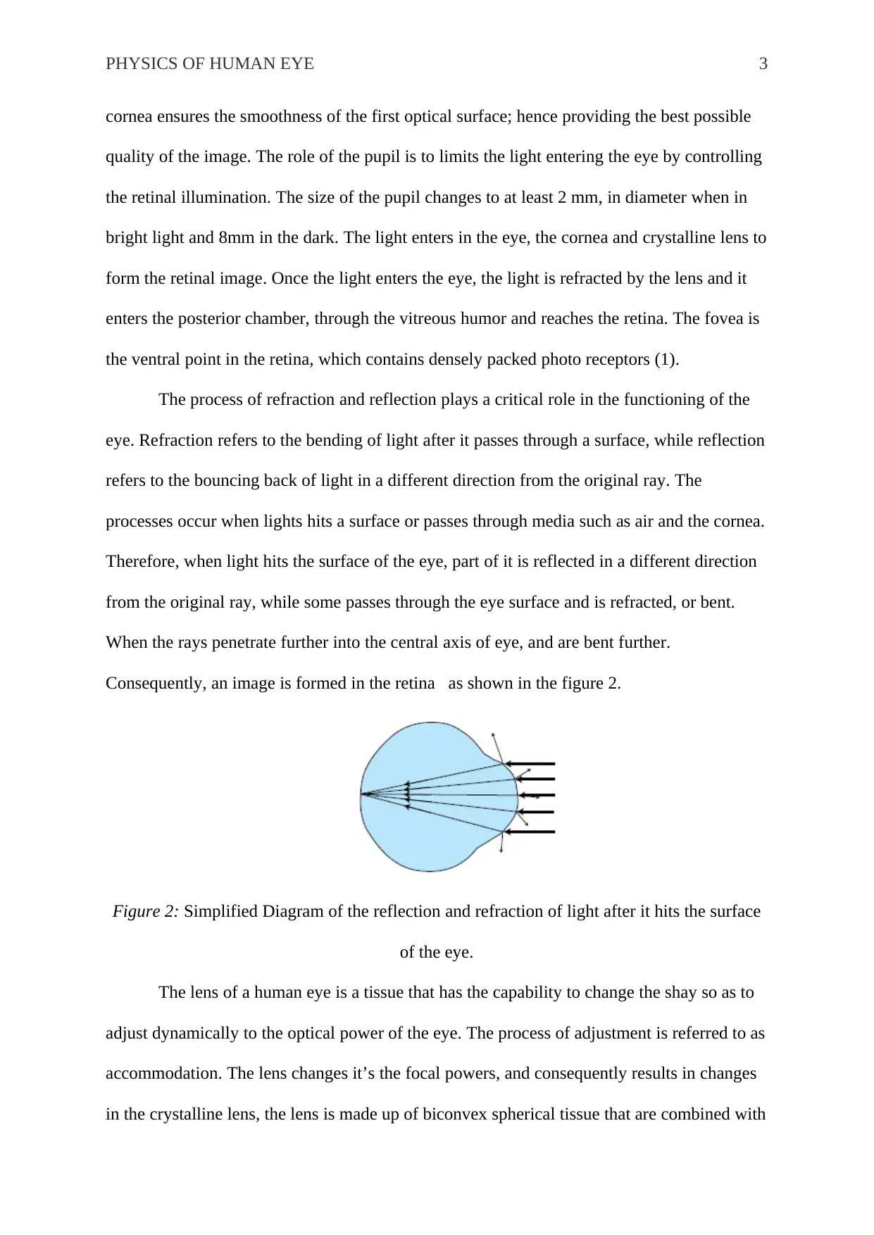
Therefore, when light hits the surface of the eye, part of it is reflected in a different direction
from the original ray, while some passes through the eye surface and is refracted, or bent.
When the rays penetrate further into the central axis of eye, and are bent further.
Consequently, an image is formed in the retina as shown in the figure 2.
Figure 2: Simplified Diagram of the reflection and refraction of light after it hits the surface
of the eye.
The lens of a human eye is a tissue that has the capability to change the shay so as to
adjust dynamically to the optical power of the eye. The process of adjustment is referred to as
accommodation. The lens changes it’s the focal powers, and consequently results in changes
in the crystalline lens, the lens is made up of biconvex spherical tissue that are combined with
PHYSICS OF HUMAN EYE 4
elastic outer membrane that varies in thickness. During the process of accommodation, the
external forces moves into the internal mass of the lens tissue, causing the anterior-posterior
of the lens to thicken (2).
Refractive errors are the major cause of eye defects among both children and adults.
Myopia is one such defect that is also referred to as nearsightedness. It refers to lack of the
ability to view distant objects, but no difficulties in viewing nearby objects. The condition
occurs when light is refracted more than the required level due to various eye conditions such
as bulging of the cornea, or developing an elongated eyeball. A bulged cornea means that it
has curved more that the customary requirement; hence refracts; light more than the usual.
Therefore, distant objects tend to form in front of the retina. In the case of horizontally
elongated eyeball, the distance between the retina and the cornea and lens systems is longer
than the custom; hence the images are also formed in front of the retina. Therefore, the nerves
at the fovea are not reached by the image and instead detect a blurry image of the distant
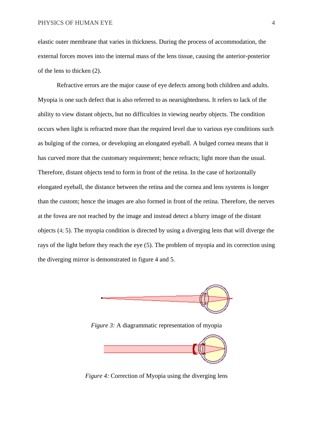
objects (4; 5). The myopia condition is directed by using a diverging lens that will diverge the
rays of the light before they reach the eye (5). The problem of myopia and its correction using
the diverging mirror is demonstrated in figure 4 and 5.
Figure 3: A diagrammatic representation of myopia
Figure 4: Correction of Myopia using the diverging lens
Secure Best Marks with AI Grader
Need help grading? Try our AI Grader for instant feedback on your assignments.
PHYSICS OF HUMAN EYE 5
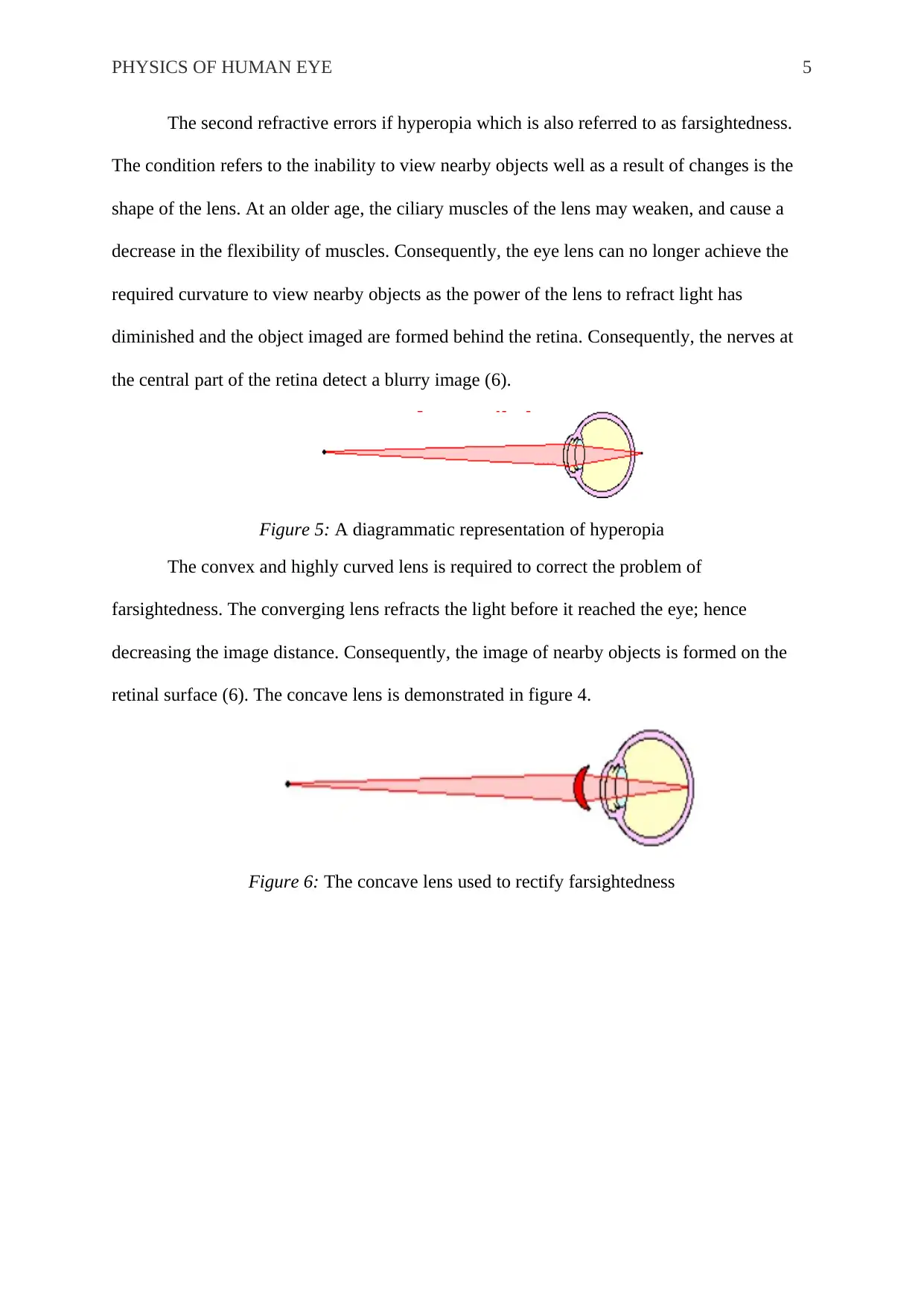
The second refractive errors if hyperopia which is also referred to as farsightedness.
The condition refers to the inability to view nearby objects well as a result of changes is the
shape of the lens. At an older age, the ciliary muscles of the lens may weaken, and cause a
decrease in the flexibility of muscles. Consequently, the eye lens can no longer achieve the
required curvature to view nearby objects as the power of the lens to refract light has
diminished and the object imaged are formed behind the retina. Consequently, the nerves at
the central part of the retina detect a blurry image (6).
Figure 5: A diagrammatic representation of hyperopia
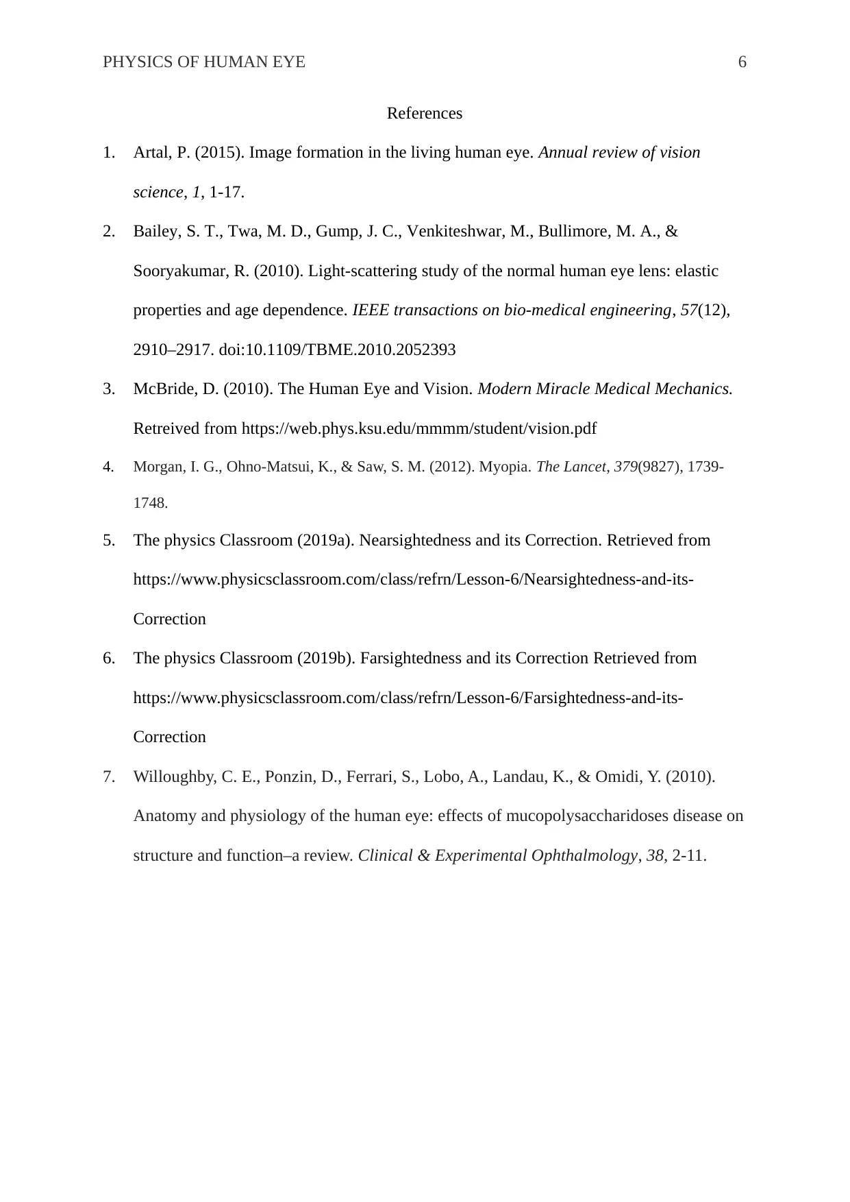
The convex and highly curved lens is required to correct the problem of
farsightedness. The converging lens refracts the light before it reached the eye; hence
decreasing the image distance. Consequently, the image of nearby objects is formed on the
retinal surface (6). The concave lens is demonstrated in figure 4.
Figure 6: The concave lens used to rectify farsightedness
PHYSICS OF HUMAN EYE 6
References
1. Artal, P. (2015). Image formation in the living human eye. Annual review of vision
science, 1, 1-17.
2. Bailey, S. T., Twa, M. D., Gump, J. C., Venkiteshwar, M., Bullimore, M. A., &
Sooryakumar, R. (2010). Light-scattering study of the normal human eye lens: elastic
properties and age dependence. IEEE transactions on bio-medical engineering, 57(12),
2910–2917. doi:10.1109/TBME.2010.2052393
3. McBride, D. (2010). The Human Eye and Vision. Modern Miracle Medical Mechanics.
Retreived from https://web.phys.ksu.edu/mmmm/student/vision.pdf
4. Morgan, I. G., Ohno-Matsui, K., & Saw, S. M. (2012). Myopia. The Lancet, 379(9827), 1739-
1748.
5. The physics Classroom (2019a). Nearsightedness and its Correction. Retrieved from
https://www.physicsclassroom.com/class/refrn/Lesson-6/Nearsightedness-and-its-
Correction
6. The physics Classroom (2019b). Farsightedness and its Correction Retrieved from
https://www.physicsclassroom.com/class/refrn/Lesson-6/Farsightedness-and-its-
Correction
7. Willoughby, C. E., Ponzin, D., Ferrari, S., Lobo, A., Landau, K., & Omidi, Y. (2010).
Anatomy and physiology of the human eye: effects of mucopolysaccharidoses disease on
structure and function–a review. Clinical & Experimental Ophthalmology, 38, 2-11.
1 out of 6
![[object Object]](/_next/static/media/star-bottom.7253800d.svg)


