Physiological basis of wound observations, sources of contamination and antibiotic rationale for Mary's wound
Verified
Added on 2023/06/04
|6
|1991
|218
AI Summary
This article explains the physiological basis of wound observations, sources of contamination and antibiotic rationale for Mary's wound. It also discusses the process by which Mary's wound will heal.
Contribute Materials
Your contribution can guide someone’s learning journey. Share your
documents today.
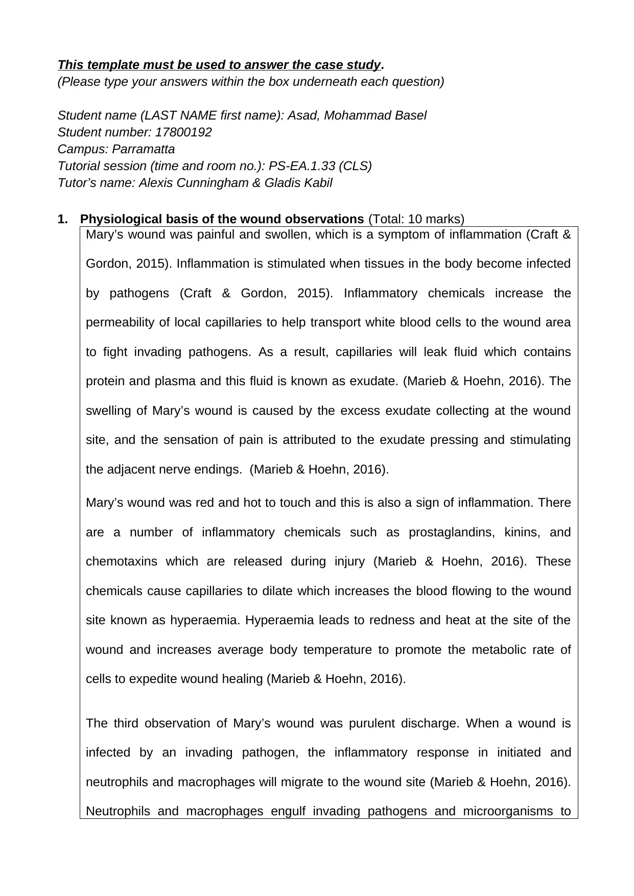
This template must be used to answer the case study. (Please type your answers within the box underneath each question) Student name (LAST NAME first name): Asad, Mohammad Basel Student number: 17800192 Campus: Parramatta Tutorial session (time and room no.): PS-EA.1.33 (CLS) Tutor’s name: Alexis Cunningham & Gladis Kabil 1.Physiological basis of the wound observations(Total: 10 marks) Mary’s wound was painful and swollen, which is a symptom of inflammation (Craft & Gordon, 2015). Inflammation is stimulated when tissues in the body become infected bypathogens(Craft&Gordon,2015).Inflammatorychemicalsincreasethe permeability of local capillaries to help transport white blood cells to the wound area to fight invading pathogens. As a result, capillaries will leak fluid which contains protein and plasma and this fluid is known as exudate. (Marieb & Hoehn, 2016). The swelling of Mary’s wound is caused by the excess exudate collecting at the wound site, and the sensation of pain is attributed to the exudate pressing and stimulating the adjacent nerve endings. (Marieb & Hoehn, 2016). Mary’s wound was red and hot to touch and this is also a sign of inflammation. There areanumberofinflammatorychemicalssuchasprostaglandins,kinins,and chemotaxinswhicharereleasedduringinjury(Marieb&Hoehn,2016).These chemicals cause capillaries to dilate which increases the blood flowing to the wound site known as hyperaemia. Hyperaemia leads to redness and heat at the site of the wound and increases average body temperature to promote the metabolic rate of cells to expedite wound healing (Marieb & Hoehn, 2016). The third observation of Mary’s wound was purulent discharge. When a wound is infectedbyaninvadingpathogen,theinflammatoryresponseininitiatedand neutrophils and macrophages will migrate to the wound site (Marieb & Hoehn, 2016). Neutrophils and macrophages engulf invading pathogens and microorganisms to
Secure Best Marks with AI Grader
Need help grading? Try our AI Grader for instant feedback on your assignments.
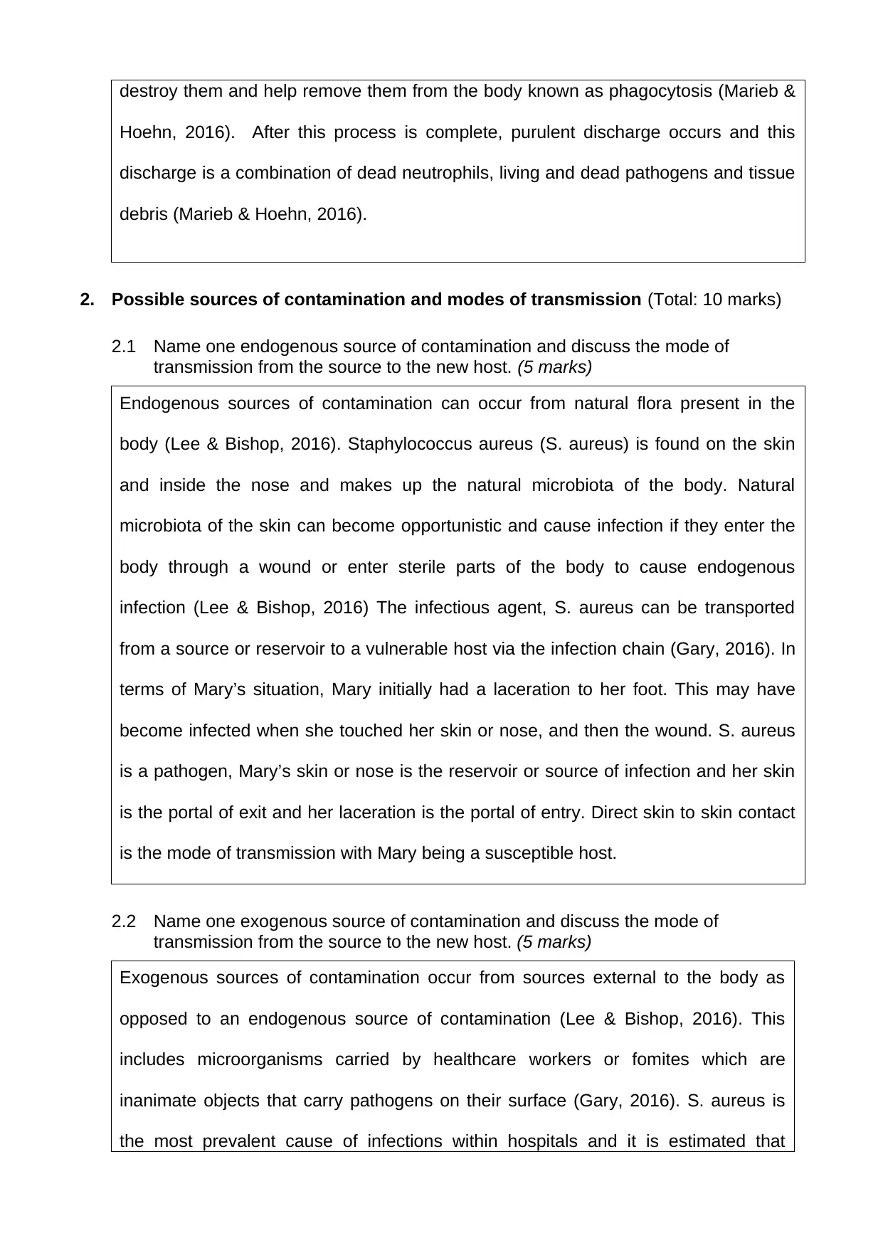
destroy them and help remove them from the body known as phagocytosis (Marieb & Hoehn, 2016).After this process is complete, purulent discharge occurs and this discharge is a combination of dead neutrophils, living and dead pathogens and tissue debris (Marieb & Hoehn, 2016). 2.Possible sources of contamination and modes of transmission(Total: 10 marks) 2.1Name one endogenous source of contamination and discuss the mode of transmission from the source to the new host.(5 marks) Endogenous sources of contamination can occur from natural flora present in the body (Lee & Bishop, 2016). Staphylococcus aureus (S. aureus) is found on the skin and inside the nose and makes up the natural microbiota of the body. Natural microbiota of the skin can become opportunistic and cause infection if they enter the body through a wound or enter sterile parts of the body to cause endogenous infection (Lee & Bishop, 2016) The infectious agent, S. aureus can be transported from a source or reservoir to a vulnerable host via the infection chain (Gary, 2016). In terms of Mary’s situation, Mary initially had a laceration to her foot. This may have become infected when she touched her skin or nose, and then the wound. S. aureus is a pathogen, Mary’s skin or nose is the reservoir or source of infection and her skin is the portal of exit and her laceration is the portal of entry. Direct skin to skin contact is the mode of transmission with Mary being a susceptible host. 2.2Name one exogenous source of contamination and discuss the mode of transmission from the source to the new host.(5 marks) Exogenous sources of contamination occur from sources external to the body as opposed to an endogenous source of contamination (Lee & Bishop, 2016). This includesmicroorganismscarriedbyhealthcareworkersorfomiteswhichare inanimate objects that carry pathogens on their surface (Gary, 2016). S. aureus is the most prevalent cause of infections within hospitals and it is estimated that
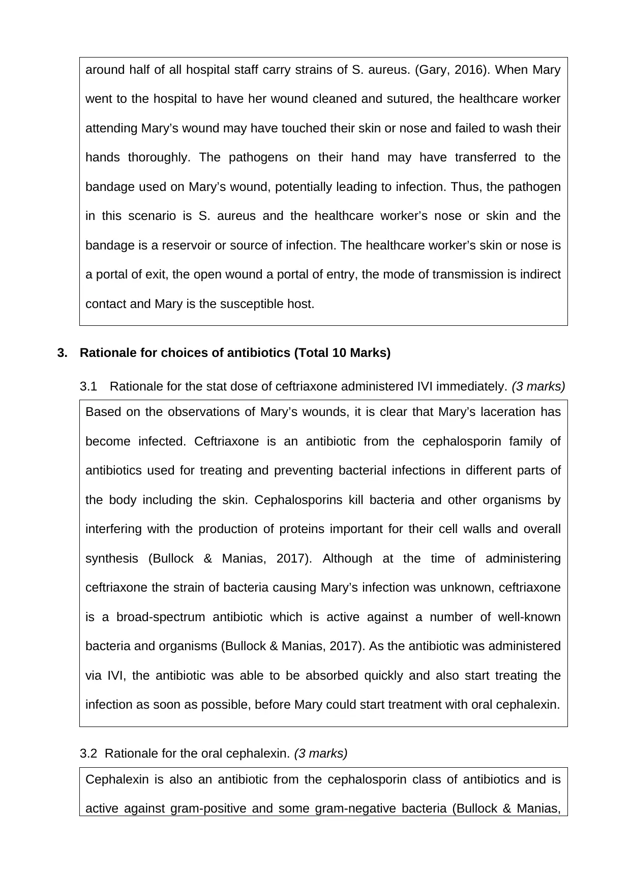
around half of all hospital staff carry strains of S. aureus. (Gary, 2016). When Mary went to the hospital to have her wound cleaned and sutured, the healthcare worker attending Mary’s wound may have touched their skin or nose and failed to wash their handsthoroughly.Thepathogensontheirhandmayhavetransferredtothe bandage used on Mary’s wound, potentially leading to infection. Thus, the pathogen in this scenario is S. aureus and the healthcare worker’s nose or skin and the bandage is a reservoir or source of infection. The healthcare worker’s skin or nose is a portal of exit, the open wound a portal of entry, the mode of transmission is indirect contact and Mary is the susceptible host. 3.Rationale for choices of antibiotics (Total 10 Marks) 3.1Rationale for the stat dose of ceftriaxone administered IVI immediately.(3 marks) Based on the observations of Mary’s wounds, it is clear that Mary’s laceration has becomeinfected.Ceftriaxoneisanantibioticfromthecephalosporinfamilyof antibiotics used for treating and preventing bacterial infections in different parts of the body including the skin. Cephalosporins kill bacteria and other organisms by interfering with the production of proteins important for their cell walls and overall synthesis(Bullock&Manias,2017).Althoughatthetimeofadministering ceftriaxone the strain of bacteria causing Mary’s infection was unknown, ceftriaxone is a broad-spectrum antibiotic which is active against a number of well-known bacteria and organisms (Bullock & Manias, 2017). As the antibiotic was administered via IVI, the antibiotic was able to be absorbed quickly and also start treating the infection as soon as possible, before Mary could start treatment with oral cephalexin. 3.2Rationale for the oral cephalexin.(3 marks) Cephalexin is also an antibiotic from the cephalosporin class of antibiotics and is active against gram-positive and some gram-negative bacteria (Bullock & Manias,
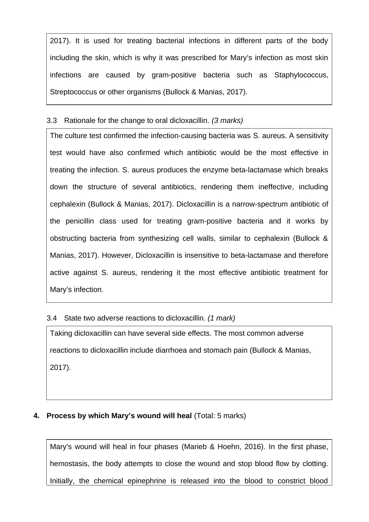
2017). It is used for treating bacterial infections in different parts of the body including the skin, which is why it was prescribed for Mary’s infection as most skin infectionsarecausedbygram-positivebacteriasuchasStaphylococcus, Streptococcus or other organisms (Bullock & Manias, 2017). 3.3Rationale for the change to oral dicloxacillin.(3 marks) The culture test confirmed the infection-causing bacteria wasS. aureus. A sensitivity test would have also confirmedwhich antibiotic would be the most effective in treating the infection.S. aureus produces the enzyme beta-lactamase which breaks downthestructureofseveralantibiotics,renderingthemineffective,including cephalexin(Bullock & Manias, 2017).Dicloxacillin is a narrow-spectrum antibiotic of thepenicillinclassusedfortreatinggram-positivebacteriaanditworksby obstructing bacteria from synthesizing cell walls, similar to cephalexin (Bullock & Manias, 2017). However, Dicloxacillin is insensitive tobeta-lactamase and therefore active against S. aureus, rendering it the most effective antibiotic treatment for Mary’s infection. 3.4State two adverse reactions to dicloxacillin.(1 mark) Taking dicloxacillin can have several side effects. The most common adverse reactions to dicloxacillin include diarrhoea and stomach pain (Bullock & Manias, 2017). 4.Process by which Mary’s wound will heal(Total: 5 marks) Mary’s wound will heal in four phases(Marieb & Hoehn, 2016).In the first phase, hemostasis, the body attempts to close the wound and stop blood flow by clotting. Initially, the chemical epinephrine is releasedintothebloodto constrict blood
Paraphrase This Document
Need a fresh take? Get an instant paraphrase of this document with our AI Paraphraser
vessels and reduce blood flow (Marieb & Hoehn, 2016). Platelet cells will then travel to the site of injury and aggregate to seal the wound. Platelets also release key cytokines that signal other cells to participate in the next phases of wound healing (Marieb & Hoehn, 2016). The inflammatory phase is the next phase of wound healing and is characterized by swelling, pain, heat and redness at the site of injury and this phase helps prevent infection (Marieb & Hoehn, 2016). Mary’s injury was at the inflammatory stage when she arrived at the hospital. During this phase capillaries are dilated and permeability is increased to allow healing and repair cells such as white blood cells, growth factors, nutrients and enzymes to move to the wound site, and bacteria, pathogens and dead tissue are removed from the site (Marieb & Hoehn, 2016). The third phase of wound healing is the proliferative phase. During this phase, granulation tissue consisting of collagen and extracellular matrix is formed and begins to fill the wound (Marieb & Hoehn, 2016).The granulation tissue will gradually proliferate and fill the wound. In doing so, the edges of the wound will contract to reduce the wound’s size and to allow the surrounding epithelial cells to divide, migrate and seal the wound (Marieb & Hoehn, 2016). In the fourth and final phase, the maturation phase, collagen fibres in the granular tissue will be reorganized, remodeled and will mature to gain tensile strength. However, the tensile strength of the repaired tissue is typically 80% of unwounded tissue (Marieb & Hoehn, 2016).
References Bishop, P.(2016). Epidemiology: How diseases are spread. In G. Lee & P. Bishop (Eds.).Microbiology and infection control for health professionals(6thed., pp. 161-183). Melbourne, Australia: Pearson. Bullock, S., & Manias, E. (2017).Fundamentals of pharmacology(8th ed.). Melbourne, Australia: Pearson. Craft, J., & Gordon, C. (2015). Introduction to clinical science. InJ. A. Craft, C. J. Gordon, E. S. Huether, K. L. McCance, L. V. Brashers & N. S. Rote (Eds). Understanding pathophysiology(2nd Australian and New Zealand ed., pp. 4-19). Sydney, Australia: Elsevier. Gary, L. (2016). Infection control in healthcare facilities. In G. Lee & P. Bishop (Eds.). Microbiology and infection control for health professionals(6th ed., pp. 295-324). Melbourne, Australia: Pearson. Lee, G., & Bishop, P. (Eds.). (2016). Microbiology and infection control for health professionals (6thed., pp. 161-183). Melbourne, Australia: Pearson. Marieb, E. N. & Hoehn, K. (2016).Human anatomy and physiology(10thed.). Essex, England: Pearson.