BCHM3P02: Comprehensive Analysis of Protein Structure & Function
VerifiedAdded on 2023/06/11
|18
|4418
|474
Report
AI Summary
This report delves into the intricate relationship between protein structure and function, emphasizing the roles of amino acids and their arrangements in determining protein characteristics. It outlines the four basic protein structures—primary, secondary, tertiary, and quaternary—explaining how interactions at each level influence overall protein function. The report also discusses various types of proteins, including enzymes, hormones, and structural proteins, highlighting their specific functions within the human body. A case study involving Variable Lymphocyte Receptor B (VLRB) illustrates how protein structure impacts antigen recognition and immune response. The analysis uses a zebrafish protein as a model, examining how structural modifications can affect protein activity and its implications for biological processes. Desklib provides this document and many more to aid students in their studies.

Name of student
Institutional Affiliations:
Name of Course:
Course Code:
Name of professor:
Institutional Affiliations:
Name of Course:
Course Code:
Name of professor:
Paraphrase This Document
Need a fresh take? Get an instant paraphrase of this document with our AI Paraphraser
Introduction to protein structures
Like many structures, attention should be paid to detail. It is the components of a structure that
determine the functioning and this is no different in proteins. They are the basic building blocks
of the human body and as such, are defined as the dry weight of the individual cells (Bailey,
2017). They are therefore very essential for our bodies and serve a range of functions that are
determined by the individual structure and components. In our bodies proteins are the major
components of most of the organs and serve various functions such as neurotransmittance,
locomotion to support among others. This specialization is mainly achieved through the
customized structure of the protein.
The nature of the protein may furthermore determine the flexibility as well as the rigidity. The
structure of the protein determines the nature as well as the function served. The most rigid
proteins include the exoskeleton while the most flexible are the muscles. The most rigid proteins
therefore may act as support elements while the most flexible serve the function of connectivity.
Amino acids are the building blocks of proteins which means that when broken down, the results
of proteins are amino acids. However, there are only two types of structures that establish the
protein: the fibrous structure and the globular structure. The fibrous proteins have an elongated
shape and are insoluble. On the other hand, the globular proteins are usually spherical, soluble
and compact. Regardless, there are only 20 sets of amino acids in which these two sets of amino
acids may fall into (Smith, 2017). The specific design of a particular protein depends on the
alignment of amino acids which may mean that one particular protein may be reengineered to
another type.
Moreover, the functioning of a protein is determined by its interaction with the neighboring
molecules which is determined by its structure. The structure of the protein therefore determines
the versatility and as such determines the various roles played in the biological functioning.
Some of the functions of protein are: acting as catalysts, provision of mechanical support,
transportation as well as the storage of other molecules, for immune provision, generation of
locomotion, transmission of impulses, and acting as the control factor in cell growth and
specialization. They therefore serve very important functions in biological processes.
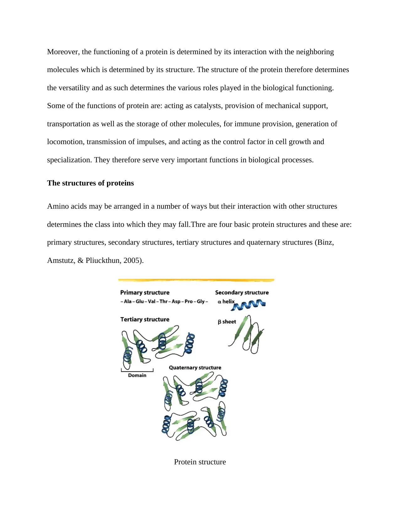
The structures of proteins
Amino acids may be arranged in a number of ways but their interaction with other structures
determines the class into which they may fall.Thre are four basic protein structures and these are:
primary structures, secondary structures, tertiary structures and quaternary structures (Binz,
Amstutz, & Pliuckthun, 2005).
Protein structure
molecules which is determined by its structure. The structure of the protein therefore determines
the versatility and as such determines the various roles played in the biological functioning.
Some of the functions of protein are: acting as catalysts, provision of mechanical support,
transportation as well as the storage of other molecules, for immune provision, generation of
locomotion, transmission of impulses, and acting as the control factor in cell growth and
specialization. They therefore serve very important functions in biological processes.
The structures of proteins
Amino acids may be arranged in a number of ways but their interaction with other structures
determines the class into which they may fall.Thre are four basic protein structures and these are:
primary structures, secondary structures, tertiary structures and quaternary structures (Binz,
Amstutz, & Pliuckthun, 2005).
Protein structure
⊘ This is a preview!⊘
Do you want full access?
Subscribe today to unlock all pages.
Trusted by 1+ million students worldwide
To begin with, the primary structure is made up of long chains of amino acids which is
determined by the arrangement of the 20 amino acids. The arrangement of the amino acids
determines the 3-D structure formed with each arrangement determining the function served.It is
the simplest arrangement of amino acids.
The second structure –secondary-is a bit more complex than the primary structure and usually
depend on the local interactions. Depending on the amino acids contained in the protein chain,
there are various interactions that may be formed which will consequently alter the 3-D structure.
The interaction in these structures as well as the alteration may be initiated by a number of
things. The alteration may be initiated by alcohols, carboxylic acids, carboamines among others
which may be directly linked to various parts of the sytructure.However,the, the two most likely
factors likely to lead to a change in the structure of the secondary structure are the alpha helix
and the beta pleated sheets (Smith, 2017). The former results to an alteration in the structure
because it forms a bond when the N-H interacts with the groups that are located in the
helix. It is this interaction of the background and the fore groups that results into the alteration in
the structure. On the other hand, the beta pleated sheet results to an alteration in the structure of
the protein because there is an interaction between the two groups, located in different strands,
each at the backbone of each strand.
The tertiary structure is formed as a result of the interaction in the secondary structure. However,
this is largely influenced by various components that are usually located in the protein structures.
Some of the structures that influence this change in structures, which means a change in
function, include the following: acidic, polar, non-polar groups among others.
Paraphrase This Document
Need a fresh take? Get an instant paraphrase of this document with our AI Paraphraser
Finally, there are quaternary protein structures. This is a more complex group, which, unlike the
primary secondary and tertiary structures, is made up of complex polypeptide chains. The
multiple sub units of the protein structure are arranged in different orientations which then
determines the function served. Some of the complex arrangements may result into different
folding which mat therefore result into different 3-D dimensions of the protein
sytructure.However,the, the interaction is not limited to other variables, there may also be
interaction between the different components such as macromolecules as well as interaction
between different parts of the protein. This complex interaction results into the formation of
advanced protein functions such as the replication of DNA (Cooper, et al., 2009).
Proteins and amino acids
As stated before, proteins are made up of amino acids. The basic composition of the amino acids
is that of a carbon atom, joined to other groups which include carboxyl and amino
groups .Moreover, they are connected to a hydrogen atom while the variable group, stated
before, varies from one amino acid to the next.A protein is therefore a 3 dimension element that
is formed when a number of polypeptide chains are twisted.
However, the process of protein formation is complex and is known as translation. Translation is
a process that involves the encoding of genes by both the DNA and RNA. The gene codes that
are contained in the DNA are transcripted into the RNA which are consequently transferred to
the peptides and with further modifications, result into proteins.
Types of proteins
The human body functions through the processes of various proteins. Some of the proteins that
the human body has include hormones, enzymes, and connective proteins among other. All these
are detailed below.
To begin with, one of the specialized proteins in the body are the antibodies. Their general
function is to prevent antigen from entering the body. As such, they are adapted for locomotion,
recognition as well as destruction. They are able to move through the blood vessels to locations
likely to have antigen and this is one of their major adaptations. As a matter of fact, they work by
ensuring that the antigen cannot move.
Actin and myosin are proteins which can be classified as contractile. They work by contraction
and relaxation which ensures that locomotion can be achieved. As such, they have been adapted
for flexibility. The most common type are the body muscles.
Thirdly, some proteins act as biochemical catalysts. These proteins are known as enzymes and
speed up the rate of biochemical reactions. The most common types of bodily enzymes are renin,
pepsin, lactase among others which usually speed up the rate of digestion. They determine the
rate at which the body digests the available food matter.
Hormones are proteins that control various body functioning. As such, they have been defined as
the body messengers which ensure that there is coordination between the various body functions.
Insulin, testosterone, oxytocin, estrogen are some of the hormones that ensure that there is
coordination between body activities. These activities include movement, reproduction among
others.
⊘ This is a preview!⊘
Do you want full access?
Subscribe today to unlock all pages.
Trusted by 1+ million students worldwide
As per the two classes under which proteins may fall, structural proteins fall into the fibrous
category. The structural proteins are designed in such a sense that they are generally stronger and
as such, they usually provide the body with support. Structural proteins include keratin and
elastin.
Another group of proteins is mainly concerned with transportation. One major example is
hemoglobin which usually transports oxygen from the lungs to the other body parts. Their
structural design is based on affinity and attachment. Once the substance to be transported has
been attracted, it cannot be displaced unless the protein releases it.
Finally, there are storage proteins that are designed for storage. The substances to be stored vary
both in structure as well as composition and as such, the proteins have to be adapted to a variety
of storage capabilities. Some examples of storage proteins include casein and ferritin among
others.
All the above stated, the protein activities are determined by the arrangement of the amino acids
and the side chains. Specialization is therefore an aspect of amino acid customization.
The structure of a protein and its function
The purpose of a protein is generally determined by its structure. Considering that some proteins
interact with each other such as in neurotransmittance, the general structure determines how the
two interact and bind. It is this interaction that ensures that the protein serves its purpose
adequately. Other proteins are used in providing a suitable environment for chemical reactions
and this is achieved by a pocket shape. The pocket is the environment in which the chemical
reactions are enhanced and this is very useful in enzymes as well as hormes.These are some of
the few notable instances that demonstrate that a protein structure is very useful in ensuring that
Paraphrase This Document
Need a fresh take? Get an instant paraphrase of this document with our AI Paraphraser
it serves its purpose.However,all this depends on the individual components of the protein
structure.
Various studies have been conducted to try and establish whether changing the structure may
result to a change in the functioning. All this is based on the fact that the structure of a protein
determines the purpose served as well as its functioning methodology.
The Study
Toll like receptor 5b, variable receptor b chimera
Introduction
The variable lymphocyte receptors are a group of immune proteins that have been discovered to
be very adaptive when controlling the antigen presence in a body (Cooper, Guo, Hirano, &
Herrin, 2009). They are therefore antibodies that do not use the conventional methods used by
other antibodies. As described before, antibodies function by attracting the antigen and
preventing their ability to move. It is this approach that enables them to manage and protect the
body from infections.
The ability of an antibody to detect the presence of an antigen in the body depends on the use of
immunoglobulin fold but this is not the process that is adopted by Variable Lymphocyte
receptors (Herrin & Cooper,, 2010).As such, it is fascinating how these proteins can recognize
antigens using adaptive strategies. The main strategy employed is based on the leucine rich
repeats (Kirchdoerfe, Herrin, Han, Cooper, & Wilson, 2012). The Leucine rich repeat is used in
determining the antigen which results into recognition.
The development of Leucine rich repeats forms the basis of antigen recognition and this been
particularly a very useful tool in the vertebrates whose immune system did not depend on
immunoglobulin. The adaptive response has ensures that all these vertebrates are well insured
against a variety of antigen. The VLR gene is responsible for producing these receptors and more
to this, there are three classes that may be produced: VLRA, VLRB and VLRC (Song, Jeon,
Namgung, hong, & yoon, 2017).However, the methodology employed by each receptor in
antigen recognition is totally unique but very effective.
VLRB
In this study, the concern is VLRB which detects the antigens by the process of agglutination and
binding. It will present a major breakthrough in the field of medicine by presenting an antibody
that adapts to the environment and can protect the body from an array of antigen.
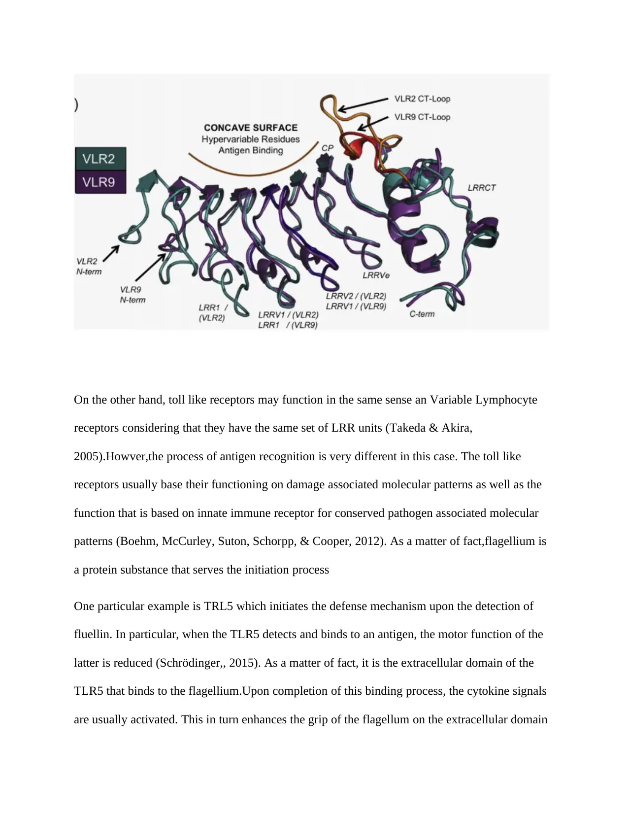
As per the shape of VLRB and the antigen binding sites, it has a crescent shape and some of the
other components of the protein are: A variety of LRR, an N-terminal leucine rich repeat cap,
leucine rich repeat,he peptide bond for connection, a leucine rich repeat which has a c-terminal
(Song, Jeon, Namgung, hong, & yoon, 2017). It is the concave shape of the VLRB that enables it
to bind the different antigens. The ability to bind to a variety of antigen has been brought about
by evolution over millions of years. Technology has also enabled researchers to isolate and
generate VLRBs from the protein.
particularly a very useful tool in the vertebrates whose immune system did not depend on
immunoglobulin. The adaptive response has ensures that all these vertebrates are well insured
against a variety of antigen. The VLR gene is responsible for producing these receptors and more
to this, there are three classes that may be produced: VLRA, VLRB and VLRC (Song, Jeon,
Namgung, hong, & yoon, 2017).However, the methodology employed by each receptor in
antigen recognition is totally unique but very effective.
VLRB
In this study, the concern is VLRB which detects the antigens by the process of agglutination and
binding. It will present a major breakthrough in the field of medicine by presenting an antibody
that adapts to the environment and can protect the body from an array of antigen.
As per the shape of VLRB and the antigen binding sites, it has a crescent shape and some of the
other components of the protein are: A variety of LRR, an N-terminal leucine rich repeat cap,
leucine rich repeat,he peptide bond for connection, a leucine rich repeat which has a c-terminal
(Song, Jeon, Namgung, hong, & yoon, 2017). It is the concave shape of the VLRB that enables it
to bind the different antigens. The ability to bind to a variety of antigen has been brought about
by evolution over millions of years. Technology has also enabled researchers to isolate and
generate VLRBs from the protein.
⊘ This is a preview!⊘
Do you want full access?
Subscribe today to unlock all pages.
Trusted by 1+ million students worldwide
On the other hand, toll like receptors may function in the same sense an Variable Lymphocyte
receptors considering that they have the same set of LRR units (Takeda & Akira,
2005).Howver,the process of antigen recognition is very different in this case. The toll like
receptors usually base their functioning on damage associated molecular patterns as well as the
function that is based on innate immune receptor for conserved pathogen associated molecular
patterns (Boehm, McCurley, Suton, Schorpp, & Cooper, 2012). As a matter of fact,flagellium is
a protein substance that serves the initiation process
One particular example is TRL5 which initiates the defense mechanism upon the detection of
fluellin. In particular, when the TLR5 detects and binds to an antigen, the motor function of the
latter is reduced (Schrödinger,, 2015). As a matter of fact, it is the extracellular domain of the
TLR5 that binds to the flagellium.Upon completion of this binding process, the cytokine signals
are usually activated. This in turn enhances the grip of the flagellum on the extracellular domain
Paraphrase This Document
Need a fresh take? Get an instant paraphrase of this document with our AI Paraphraser
of the TLR5.Therefore, the flagellum is the activator of the whole antigen binding process.
However, the high attraction rates between flagellum and TRL5 presents a major bottleneck in
the treatment of other pains and bacterial infections.
Considering that TRL5 and VLRB work in resemblance, the ability of the VLR to block the
signaling procedure that usually results into the binding process may be tested. The VLRB
antibodies may be attached to the extracellular domain of the TLR and therefore inhibiting the
process of binding (osterman, et al., 2012). However, these VLR have to be of very high affinity
and as such, the report presents some of the very effective, VLRs ranges which are VLR1, VLR2
and VLR9. Nevertheless, these three ranges have different rates of affinity and as such, have
different rates at which they can block the binding process that is usually the antigen
response.VLR1 has the least, followed by VLR2 and finally, VLR9 has the greatest effect on the
whole process (Gunn, Herrin , Acharya, Cooper, & Wilson, 2018) .The antigen used in the
research is obtained from zebrafish. Nevertheless, it is a truncated form that may present more
insight to the alteration of the functioning of a protein based on the structure.
Principles of changing the structure of VLR
The typical structure of VLR protein is that of a CT loop, LRR, an N-terminus and the c-terminal
cap. As such, there are a number of modifications that may be made to the structure to observe
the change in functioning. In this study, the main changes will be on the antigen binding sute.The
N-terminus that in this case is shifted from one position to another by the use of LRR.It
therefore shifts in position which will indicate the change in antigen binding performance of the
protein. The N-terminus in VLR2 and VLR9 are not in the same location with that of VLR9 7.0
A from the original position.
The results
To begin with, the screening process resulted into an array of VLR.These VLR will have to be
specific to the TLR process.However.it is important to understand that different levels and
ranges of antigen have varying effects on the response of the VLR.Very discernible antigens
such as bacteria, eukarotic cells and others usually have higher levels of influence and as such,
have very high levels of VLR mechanism employed..
The toll like receptor from zebra fish have to be reengineered to the specific requirement to the
variable lymphocyte receptor properties which will enhance the affinity levels. Engineering the
toll like receptor using human junket T cells was the strategy employed in order to increase the
response of the VLR receptor. By engineering the structure of the VRL and the toll like
receptors, the change in structure resulted into difference response. However, the various
differences have to be established which in turn enhances the sorting procediure.Sorting of these
antibodies can be done by the use of fluorescent or magnetization (Hong, Yoon, & Wilson,
2015). Furthermore, the sorting procedure may be done by determining the arrangement of the
amino acids on the polypeptide chain.
The three VLR clones indicated small differences in the sequence of amino acids in the peptide
chain and as such, presents variable responses to the antigen that is used. As a matter of fact, the
sequencing was about 80-70% similar but nevertheless, the motif of these three clones was the
same (Cooper, Guo, Hirano, & Herrin, 2009). Therefore, the difference in clone identity may be
used to establish he variable antigen recognition properties that are associated with a change in
the structure of the proteins.
⊘ This is a preview!⊘
Do you want full access?
Subscribe today to unlock all pages.
Trusted by 1+ million students worldwide
1 out of 18
Related Documents


Your All-in-One AI-Powered Toolkit for Academic Success.
+13062052269
info@desklib.com
Available 24*7 on WhatsApp / Email
![[object Object]](/_next/static/media/star-bottom.7253800d.svg)
Unlock your academic potential
Copyright © 2020–2026 A2Z Services. All Rights Reserved. Developed and managed by ZUCOL.