Medical Image Analysis: Dixon Sequence and NEMA Phantom Evaluation

Verified

Added on  2023/04/21

|4
|698
|292
Practical Assignment
AI Summary
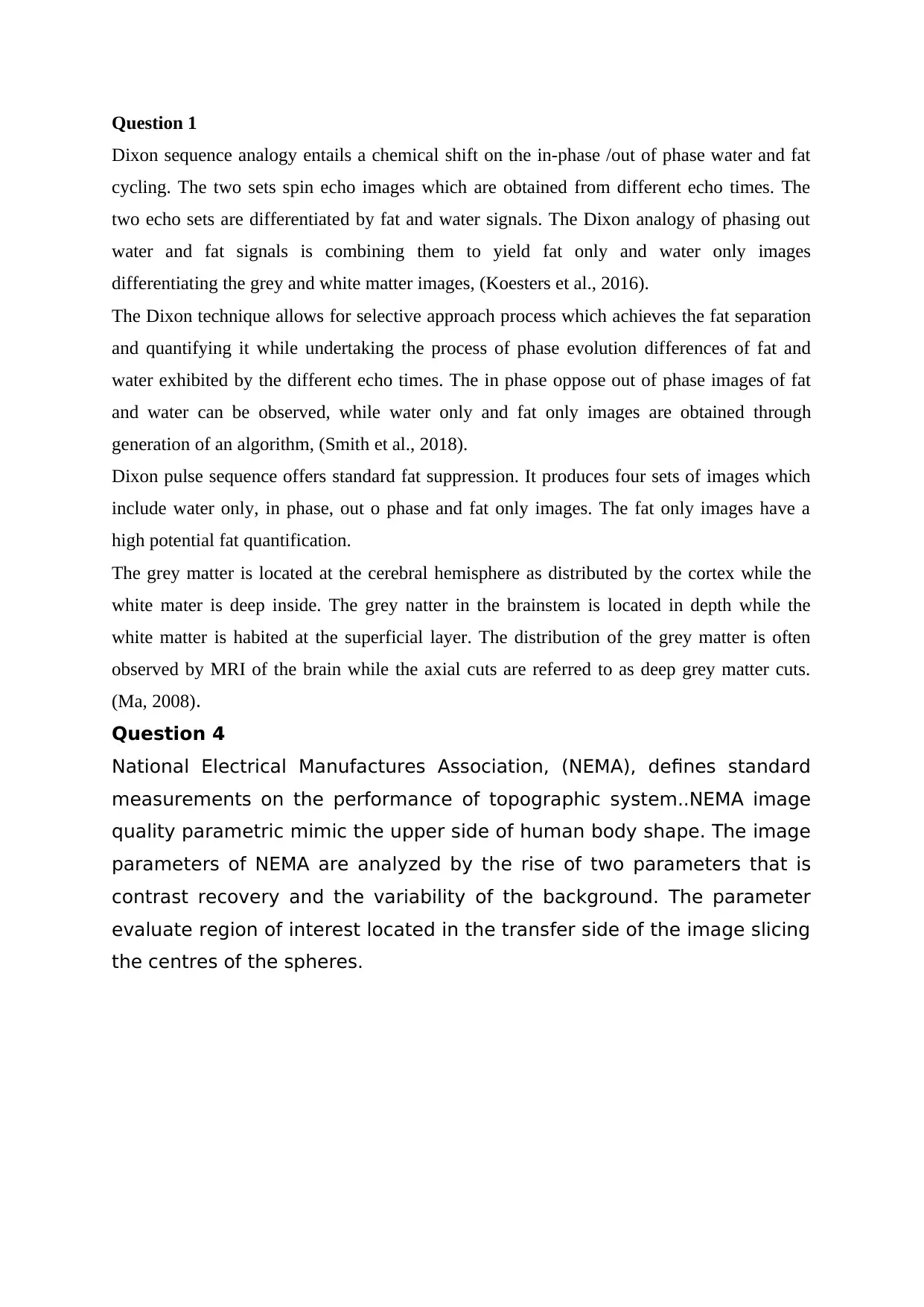
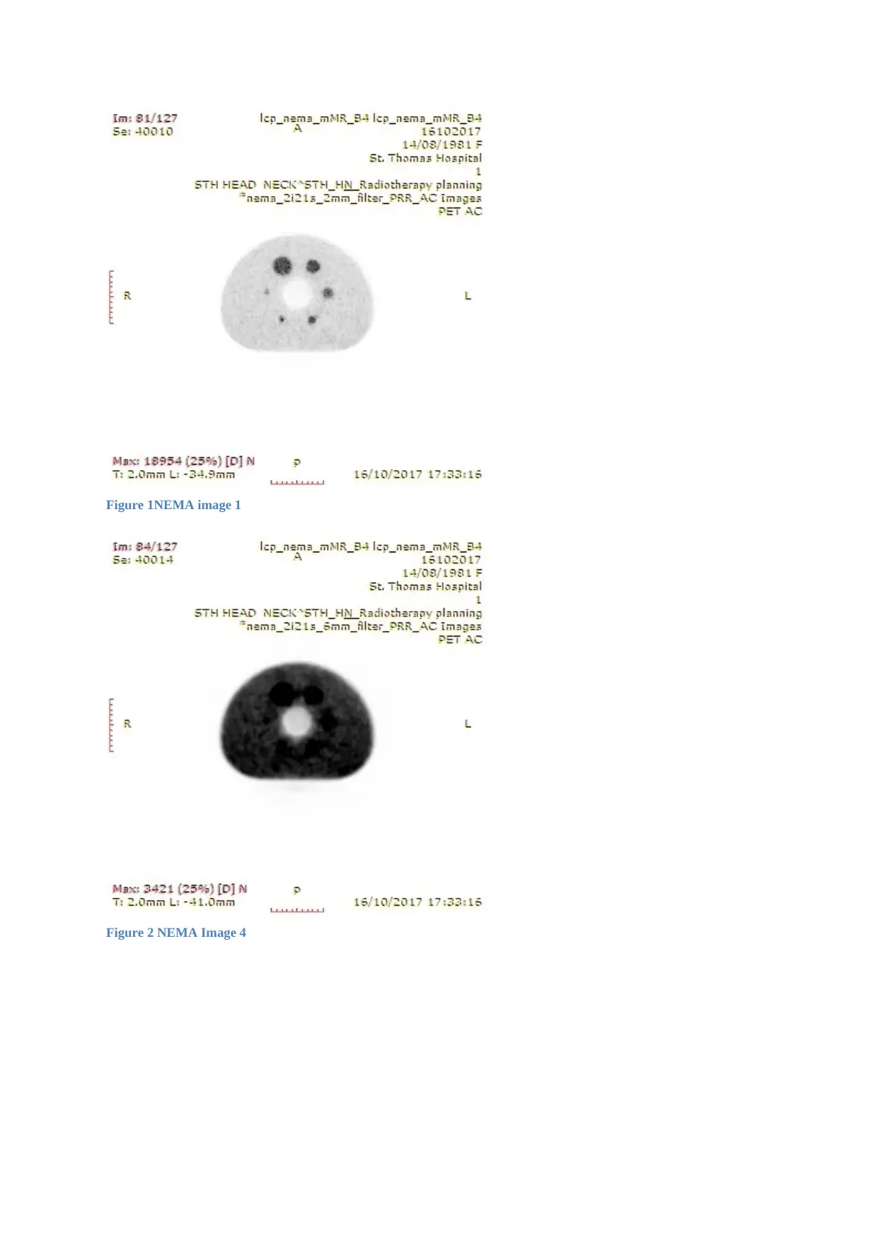
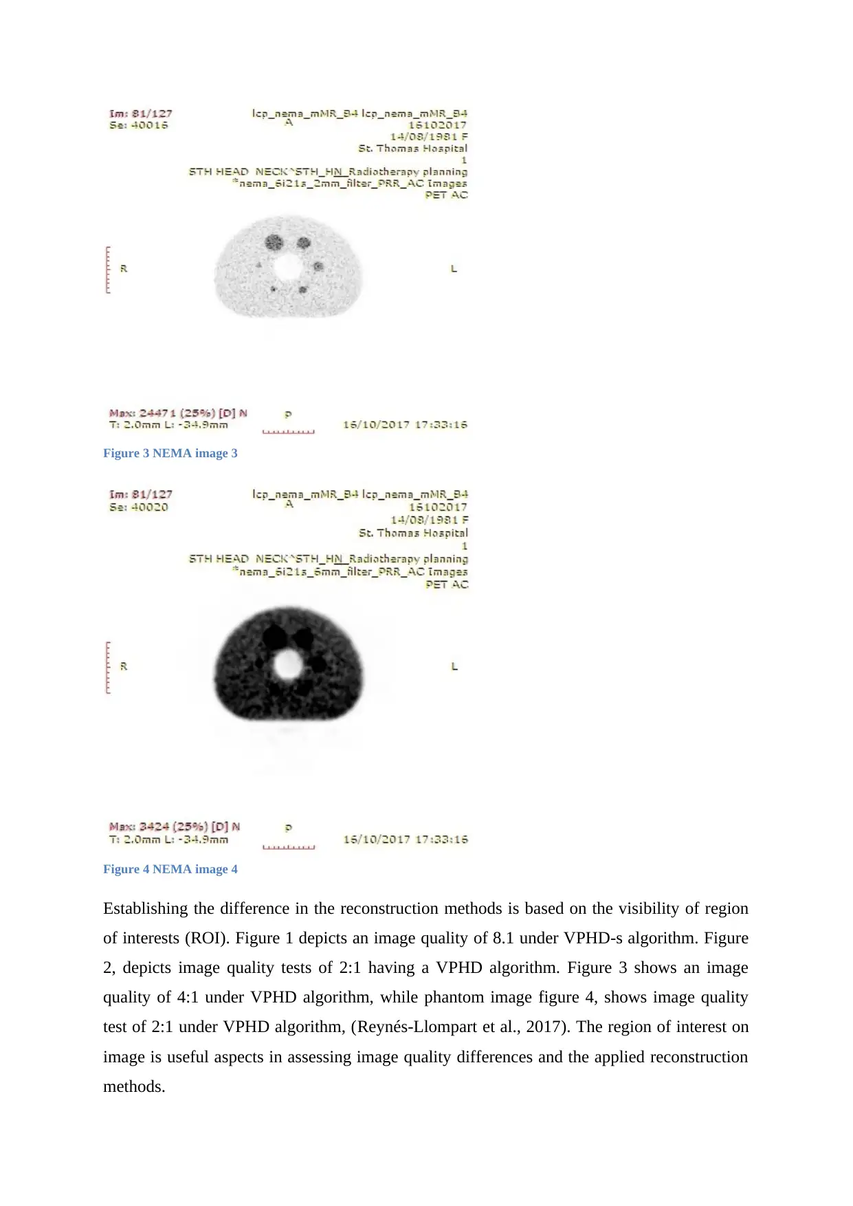
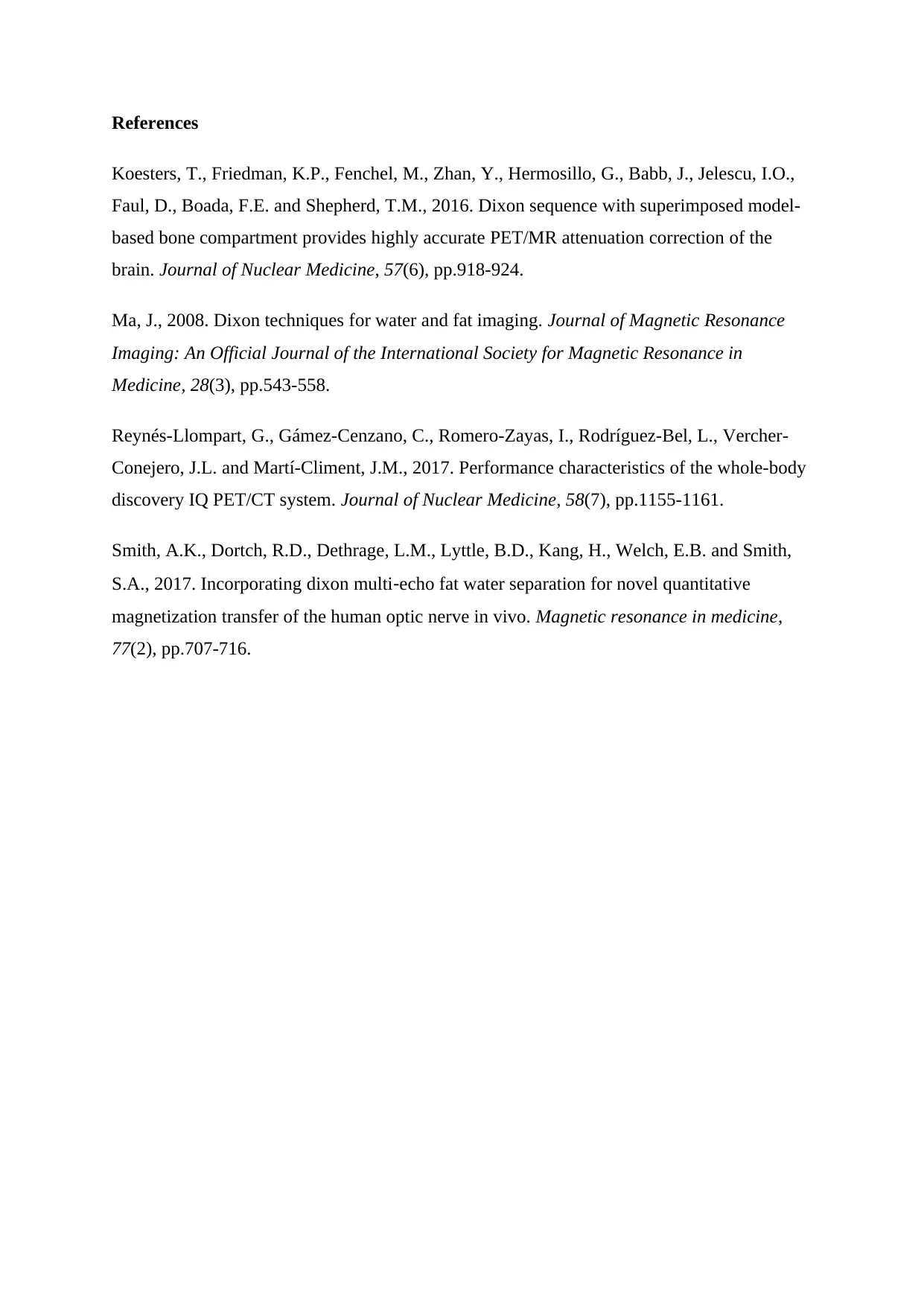
This assignment provides a detailed analysis of the Dixon sequence and NEMA phantom in the context of medical imaging. It explains the Dixon sequence analogy, focusing on chemical shifts and the generation of fat-only and water-only images, and discusses the differentiation of grey and white matter. Furthermore, the assignment examines NEMA image quality parameters, evaluating contrast recovery and background variability across different reconstruction methods. By visually comparing the reconstruction methods applied to the NEMA phantom, the assignment highlights the impact of each method on image quality and the visibility of regions of interest, supported by figures illustrating image quality under various algorithms. The document concludes with references to relevant research articles, enhancing the credibility and depth of the analysis. This resource, available on Desklib, provides students with a comprehensive understanding of these critical medical imaging techniques.
Document Page
Question 1
Dixon sequence analogy entails a chemical shift on the in-phase /out of phase water and fat
cycling. The two sets spin echo images which are obtained from different echo times. The
two echo sets are differentiated by fat and water signals. The Dixon analogy of phasing out
water and fat signals is combining them to yield fat only and water only images
differentiating the grey and white matter images, (Koesters et al., 2016).
The Dixon technique allows for selective approach process which achieves the fat separation
and quantifying it while undertaking the process of phase evolution differences of fat and
water exhibited by the different echo times. The in phase oppose out of phase images of fat
and water can be observed, while water only and fat only images are obtained through
generation of an algorithm, (Smith et al., 2018).
Dixon pulse sequence offers standard fat suppression. It produces four sets of images which
include water only, in phase, out o phase and fat only images. The fat only images have a
high potential fat quantification.
The grey matter is located at the cerebral hemisphere as distributed by the cortex while the
white mater is deep inside. The grey natter in the brainstem is located in depth while the
white matter is habited at the superficial layer. The distribution of the grey matter is often
observed by MRI of the brain while the axial cuts are referred to as deep grey matter cuts.
(Ma, 2008).
Question 4
National Electrical Manufactures Association, (NEMA), defines standard
measurements on the performance of topographic system..NEMA image
quality parametric mimic the upper side of human body shape. The image
parameters of NEMA are analyzed by the rise of two parameters that is
contrast recovery and the variability of the background. The parameter
evaluate region of interest located in the transfer side of the image slicing
the centres of the spheres.
tabler-icon-diamond-filled.svg

Paraphrase This Document

Need a fresh take? Get an instant paraphrase of this document with our AI Paraphraser
Document Page
Figure 1NEMA image 1
Figure 2 NEMA Image 4
Document Page
Figure 3 NEMA image 3
Figure 4 NEMA image 4
Establishing the difference in the reconstruction methods is based on the visibility of region
of interests (ROI). Figure 1 depicts an image quality of 8.1 under VPHD-s algorithm. Figure
2, depicts image quality tests of 2:1 having a VPHD algorithm. Figure 3 shows an image
quality of 4:1 under VPHD algorithm, while phantom image figure 4, shows image quality
test of 2:1 under VPHD algorithm, (Reynés-Llompart et al., 2017). The region of interest on
image is useful aspects in assessing image quality differences and the applied reconstruction
methods.
Document Page
References
Koesters, T., Friedman, K.P., Fenchel, M., Zhan, Y., Hermosillo, G., Babb, J., Jelescu, I.O.,
Faul, D., Boada, F.E. and Shepherd, T.M., 2016. Dixon sequence with superimposed model-
based bone compartment provides highly accurate PET/MR attenuation correction of the
brain. Journal of Nuclear Medicine, 57(6), pp.918-924.
Ma, J., 2008. Dixon techniques for water and fat imaging. Journal of Magnetic Resonance
Imaging: An Official Journal of the International Society for Magnetic Resonance in
Medicine, 28(3), pp.543-558.
Reynés-Llompart, G., Gámez-Cenzano, C., Romero-Zayas, I., Rodríguez-Bel, L., Vercher-
Conejero, J.L. and Martí-Climent, J.M., 2017. Performance characteristics of the whole-body
discovery IQ PET/CT system. Journal of Nuclear Medicine, 58(7), pp.1155-1161.
Smith, A.K., Dortch, R.D., Dethrage, L.M., Lyttle, B.D., Kang, H., Welch, E.B. and Smith,
S.A., 2017. Incorporating dixon multiecho fat water separation for novel quantitative
magnetization transfer of the human optic nerve in vivo. Magnetic resonance in medicine,
77(2), pp.707-716.
chevron_up_icon
1 out of 4
circle_padding
hide_on_mobile
zoom_out_icon
[object Object]