Radiation Physics: Decay Processes, Safety Regulations and Laser Use
VerifiedAdded on 2023/05/29
|7
|1771
|472
Homework Assignment
AI Summary
This assignment covers fundamental concepts in radiation physics, including radioactive decay processes such as alpha, beta, and gamma decay, explaining why beta particles lack discrete energy levels, and discussing photon attenuation mechanisms at different energy levels. It also delves into radiation safety, examining the Ionising Radiation Regulations 2017 (IRR17), controlled areas, optimization principles in radiation exposure, and methods for reducing fluoroscopy radiation. Furthermore, the assignment explores factors influencing laser choice in eye surgery, defines maximum permissible exposure and accessible emission limits, and outlines hazard control measures in laser eye surgery. Quantitative problems involving half-life calculations and equivalent dose are also addressed. The document concludes with relevant references, providing a comprehensive overview of key topics in radiation physics and safety.

University
Assignment
Student Name
Tutor
Assignment
Student Name
Tutor
Paraphrase This Document
Need a fresh take? Get an instant paraphrase of this document with our AI Paraphraser
Question 1
Part a
i) Alpha – changes in atomic mass occurs by 2
ii) Beta – occurs in the heavy nucleus as in stable nucleus does not lead to decay
iii) Beta decay process- the process takes place in the proton-rich nucleus
iv) Gamma decay process- yields emission of electromagnetic waves
Part b Beta decay process
Beta particles are often negatively charged electron which is emitted within the nucleus.
The electron mass is usually small, a tiny fraction of atomic mass. The nucleus mass often
undergoes decay process changed by small amounts; often the mass number is unchanged.
The nucleus does not have electrons but rather through beta decay process which occurs
after changes on the neutron into a proton in the nucleus. In a stable nucleus there is no
decay. A free neutron has an excess of neutrons, which can decay emitting beta particles.
Sharing the energy with the particle causes neutrino, which has little or no mass which does
not have any mass; it carries energy and momentum force. The source of energy in the
process is released into beta decay which is often explained by parent mass isotope which is
larger than the overall sum of masses in the decay process. The mass obtained is converted
into energy thus leaving no discrete quantity, (Eichholz, 2018).
Part c
Photon mass attenuation is essential in radiological activities. The photon can be displayed
in forms of X-rays, gamma-rays and beta rays. The attenuation mass is dependent on the
absorption and scattering of radiation caused by different mechanisms.
Photons interact with matter in varied ways depending on the available energy and material
nature. A photon with 1.5 MeV has high interaction due to more energy loaded with it
compared to Photon with MeV energy, (Elmahroug, Y., Tellili, B., & Souga, C. (2015).
Question 3
a. Medical ionizing radiation encompasses x-rays and nuclear can treatments such as
radiotherapy, which is often utilized in hospitals. Ionising and radiation regulations set the
holders with guidelines for radiation protection.
It guides the participants on minimizing unintended, and incorrect medical exposures,
ensures benefits which outweigh the risks associated on each exposure and keeping
diagnostics doses as low as possible for its optimization.
Regulation
- The ionizing Radiation regulations of 2017 and the Ionizing radiation regulation
amendment act of 2018.
Occupational radiation legal based are covered by the Ionising Radiation regulations of
2017 referred to IRR17. I replaced ionizations radiations IRR99 and implementing in work
safety places.
Employees under this act can work in ionizing radiations conditions through an approved
code of practice. The IRR17 Regulation applies to a large range of workplace where
radioactive substances emitting ionization used, (UK Government 2018).
b. Controlled area
Under the Ionising Radiation regulations of 2017 referred to IRR17, controlled area entails
rules and guidelines which guide through the restriction of exposure of ionizing radiation
should have minimum exposure to radiation. Further, any person working in the area should
receive an effective dose of greater than 6mSv or equivalent not more than 15 mSv for eye
lens or greater than 150mSv in a year for skin
Employers must designate areas which are controlled whereby
- It is necessary to keep the conditions under control
- Any individual likely to receive doses greater than 1 mSv or greater than 5 mSv from
the lens of greater than 50 mSv must be supervised, (UK Government 2018).
Part c
Optimization refers to the likelihood of increasing exposures, the number of people exposed
and the magnitude levels of those exposed, which should be kept as low as possible. This
thus refers to protection level which should be best be kept under the prevailing conditions,
which maximizing benefit over harm. In order to facilitate this, there should restrictions on
doses or risks of individuals.
Examples of optimization include;
- Optimizing x-ray beam which provides a balance between image quality and patient
exposure. The spectrum in the x-ray machine is defined by the filtration in the beam of
light. The kV values used and filtration is undertaken are optimized for specific types of
examinations.
- Usage of optimization during CT scan imaging is geared towards reducing the
exposures subjected to the patients.
- Optimization can be undertaken in dental radiology arena, (Ollila et al., 2018).
Part d
Reducing fluoroscopy radiation
1. Time of exposure
You're viewing a preview
Unlock full access by subscribing today!
Time exposure is the basic thing for reducing external exposure radiations. It is important
to limit the amount of time spent in the exposure room. This reduces further on effective
doses which limits the exposure level.
2. Radiation equipment notifier
In the fluoroscopy room use of radiation producing equipment, the notifier is essential in
understanding the different effects of radiation. This monitors the exposure levels to the
staff.
3. Fluoroscopic procedures
Healthcare staff should ensure that the patient is kept as low to the image intensifier side
of the fluoroscopic unit. Standing on the side of image intensifier reduces intensity
radiation.
4. Avoiding direct beam exposures
There is a need to avoid direct contact of the radiation beams and wearing of protective
gloves should be used with 0.25 millimeters equivalent, (Yi et al., 2017).
Question 6
Part a
Factors influencing laser choice in eye surgery
Laser eye surgery entails the correction of the vision poor cess which involves usage of
lasers which reshape the front surface of the eyes which enables focusing to be better. It is
essential for correcting short-sightedness, and stigmatization, (Jiménez, Alarcón, Anera &
del Barco, 2016). Key factors on the choice of laser include;
- Patient refractory index
- Available technology
- Techniques adopted by the surgeon
- Continuity of care
Part b
Maximum permissible exposure
This refers to the highest power density in W/cm2 J/cm2 which is the highest point of source
of energy which is regarded to be safe, that is the negligible probability of creating damage.
It is often doses of about 10% that is having 50 % chance of creating damage in worst cases,
(Frigerio & Biazzi, 2018).
Accessible emission limit
Accessible emission limit refers to the maximum level which is permitted at a particular
Paraphrase This Document
Need a fresh take? Get an instant paraphrase of this document with our AI Paraphraser
class for emission to occur. The aversion response in the exposure form the bright visible
light is often assumed to limit the exposure to a specific retinol are to around 0.25 seconds
or less, (Duncan, 2016).
Part c
Hazard control measures in eye laser surgery
Implementing control measures to reduce the hazards linked to laser devices is control.
Mitigation measures include beam hazards control, nonbeam hazards and biological
hazards. Control measures to ensure safety entail;
- Using interlocks on a protective house designed to prevent access to beams above the
maximum permissible limit
- Using remote interlock connectors which facilitate electrical connections to the
emergencies master switch.
- Engaging EH&S Laser Safety Officer to routinely conduct analysis in order to establish
safe nominal hazard zones.
- Using and installing beam stop attenuator which is able to prevent laser radiation which
is excess than the normal MPE Level, (Parker & Parker, 2017).
Question 2
Part a
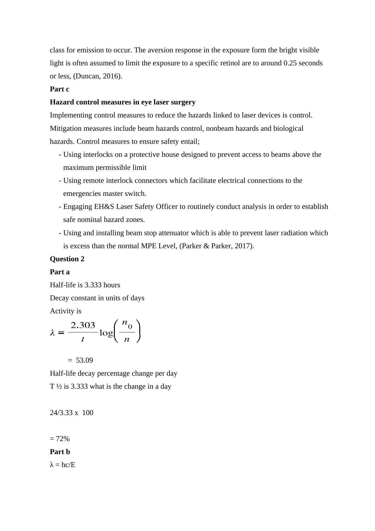
Half-life is 3.333 hours
Decay constant in units of days
Activity is
= 53.09
Half-life decay percentage change per day
T ½ is 3.333 what is the change in a day
24/3.33 x 100
= 72%
Part b
λ = hc/E
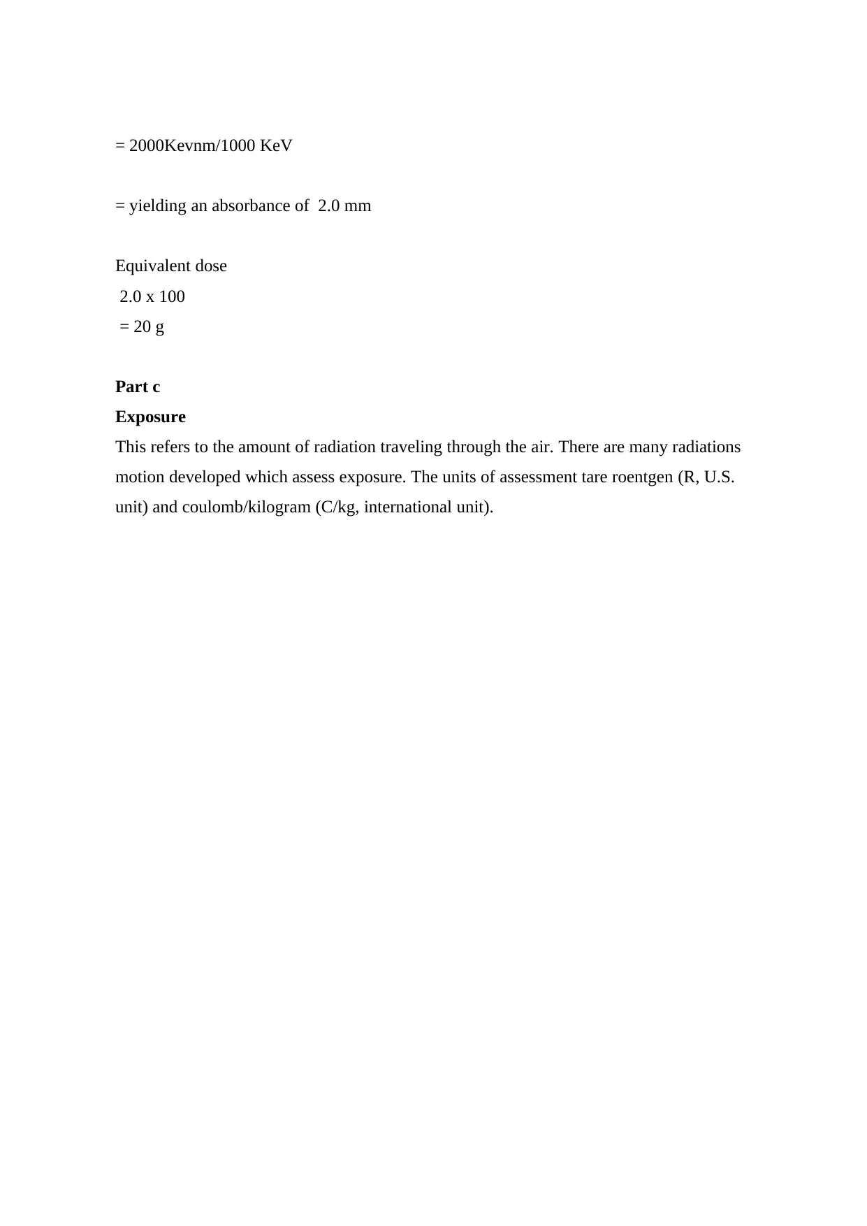
= 2000Kevnm/1000 KeV
= yielding an absorbance of 2.0 mm
Equivalent dose
2.0 x 100
= 20 g
Part c
Exposure
This refers to the amount of radiation traveling through the air. There are many radiations
motion developed which assess exposure. The units of assessment tare roentgen (R, U.S.
unit) and coulomb/kilogram (C/kg, international unit).
You're viewing a preview
Unlock full access by subscribing today!
References
Duncan, K. J. (2016, October). Laser based power transmission: Component selection and
laser hazard analysis. In Emerging Technologies: Wireless Power Transfer (WoW),
2016 IEEE PELS Workshop on (pp. 100-103). IEEE.
Eichholz, G. G. (2018). Principles of Nuclear Radiation Detection: 0. CRC Press.
Elmahroug, Y., Tellili, B., & Souga, C. (2015). Determination of total mass attenuation
coefficients, effective atomic numbers and electron densities for different shielding
materials. Annals of Nuclear Energy, 75, 268-274.
Frigerio, F., & Biazzi, L. (2018, June). Maximum permissible exposure in the laser display
shows. In 2018 IEEE International Conference on Environment and Electrical
Engineering and 2018 IEEE Industrial and Commercial Power Systems Europe
(EEEIC/I&CPS Europe) (pp. 1-7). IEEE.
Jiménez, J. R., Alarcón, A., Anera, R. G., & del Barco, L. J. (2016). Q-optimized algorithms:
Theoretical analysis of factors influencing visual quality after myopic corneal refractive
surgery. Journal of Refractive Surgery, 32(9), 612-617.
Ollila, S., Vainio, M., Peltola, J., Nord, J., Kuusela, E., Kauppinen, J., ... & Rusanen, M.
(2018). U.S. Patent Application No. 15/269,882.
Parker, P. J., & Parker, S. P. (2017). Laser Safety. In Lasers in Dentistry—Current Concepts
(pp. 87-106). Springer, Cham.
UK Government , (2018). Radiaion Legal Base. Accessed on 9/11/2012. Accessed at http;
http://www.hse.gov.uk/radiation/ionising/legalbase.htm
Yi, H. J., Sung, J. H., Lee, D. H., Kim, S. W., & Lee, S. W. (2017). Analysis of radiation
doses and dose reduction strategies during cerebral digital subtraction angiography.
World neurosurgery, 100, 216-223.
1 out of 7
Related Documents
Your All-in-One AI-Powered Toolkit for Academic Success.
+13062052269
info@desklib.com
Available 24*7 on WhatsApp / Email
![[object Object]](/_next/static/media/star-bottom.7253800d.svg)
Unlock your academic potential
© 2024 | Zucol Services PVT LTD | All rights reserved.


