MD7015 Orthopaedics: Scoliosis, Diagnosis & Treatment Strategies
VerifiedAdded on 2023/06/18
|15
|4057
|431
Report
AI Summary
This report provides a comprehensive review of scoliosis, including its definition, causes (idiopathic, congenital, neuromuscular, etc.), clinical features, biomechanics of progression, and diagnostic strategies. It evaluates the effectiveness of MRI and CT scans in diagnosing and managing scoliosis, discussing their roles in identifying underlying conditions and guiding surgical treatment. The report also covers the pathophysiology, clinical incidence, and different types of scoliosis, such as idiopathic, congenital, and neuromuscular, and it emphasizes the importance of understanding the three-dimensional nature of spinal curvature in scoliosis progression and management. The assessment was submitted in partial fulfillment of the requirements for the Orthopaedics module MD7015.

Assessment 2 Report
Paraphrase This Document
Need a fresh take? Get an instant paraphrase of this document with our AI Paraphraser
Table of Contents
INTRODUCTION...........................................................................................................................3
MAIN BODY...................................................................................................................................3
CONCLUSION................................................................................................................................3
REFERENCES................................................................................................................................4
INTRODUCTION
Scoliosis is refer as abnormal or deformity which consist of lateral curvature and rotation
movement of the spine. (Brochet, and Ruet, 2019) Generally, the usual spinal curve happens on
cervical of a person, thoracic and also the areas of body part which is well known as sagittal
plane of a body. During the movement, human's natural curves position and its location at the
head over the region of pelvis of an individual and can do activity as similar to as shock
absorbers in the body which can help with distribution of mechanical stress. Scoliosis is
frequently called as spinal curvature in the coronal of an individual's body or it also known as
frontal plane in an individual's body. Studies state that it is actually more complex, aslo the
curvature the degree of curvature is measured in coronal plane. (Burk,and Pasterkamp, 2019) It
is three dimensional complication that can include planes such as axial plane, coronal plane and
axial plane. There are several causes of scoliosis which several and are distinguished broadly,
such as, congenital, neuromuscular, related with syndrome, idiopathic and also spinal curvature.
Studies reported spinal curvature happens due to secondary reasons. The common cases
which generally encounter by the general practitioners are idiopathic scoliosis which are more
common in young individuals, whereas, in older people it has been the most common scoliosis
which is observed degenerative scoliosis. It has been observed it can also may occur secondary
to tumour, infection as well as trauma. (Butti, and Patten, 2019) In general cases it has been
observed that individual who are suffering from this complications debits asymmetry that are in
chest wall as well as in the back of an individual. This report review in respect of current
knowledge of the condition scoliosis and its fundamental prevailing principles and underpins on
understanding of causes of scoliosis and their effective diagnosis and treatment plan with aid of
RI and CT Scan.
Question to investigate
To determine understanding of scoliosis and its treatment measures, moreover if MRI and CT
scan are effective strategy for this spinal disease?
Current knowledge of the condition
Studies state that scoliosis described as the availability of one and more than one sidelong
bends in regards with vertebral section within coronal plane, but it is unusual bends may
influence spinal arrangement in each of the three measurements in which Radiography,
computed tomography (CT) and also magnetic resonance (MR) imaging, debits essential role in
⊘ This is a preview!⊘
Do you want full access?
Subscribe today to unlock all pages.
Trusted by 1+ million students worldwide
the process of evaluation of scoliosis as well as in the deciding its fundamental reason
(Casselman and Schauwvlieghe, 2021)
In spite of the fact, that scoliosis is as a usual (in 80% of cases) idiopathic, different
congenial or neurologic abnormalities might promote unusual lateral curvature of the
individual's spine cord as well. Also, determination of one of the most relevant imaging
methodology, to adress specific assessment might help through more familiar along with
imaging manifestations of knowledge in respect to the imaging signs of different causes of
scoliosis; moreover, this imagining process might be improved by an understanding principle,
progression as well as progression which lie in the process of development, progression as well
as management of scoliosis(Brochet, and Ruet, 2019)
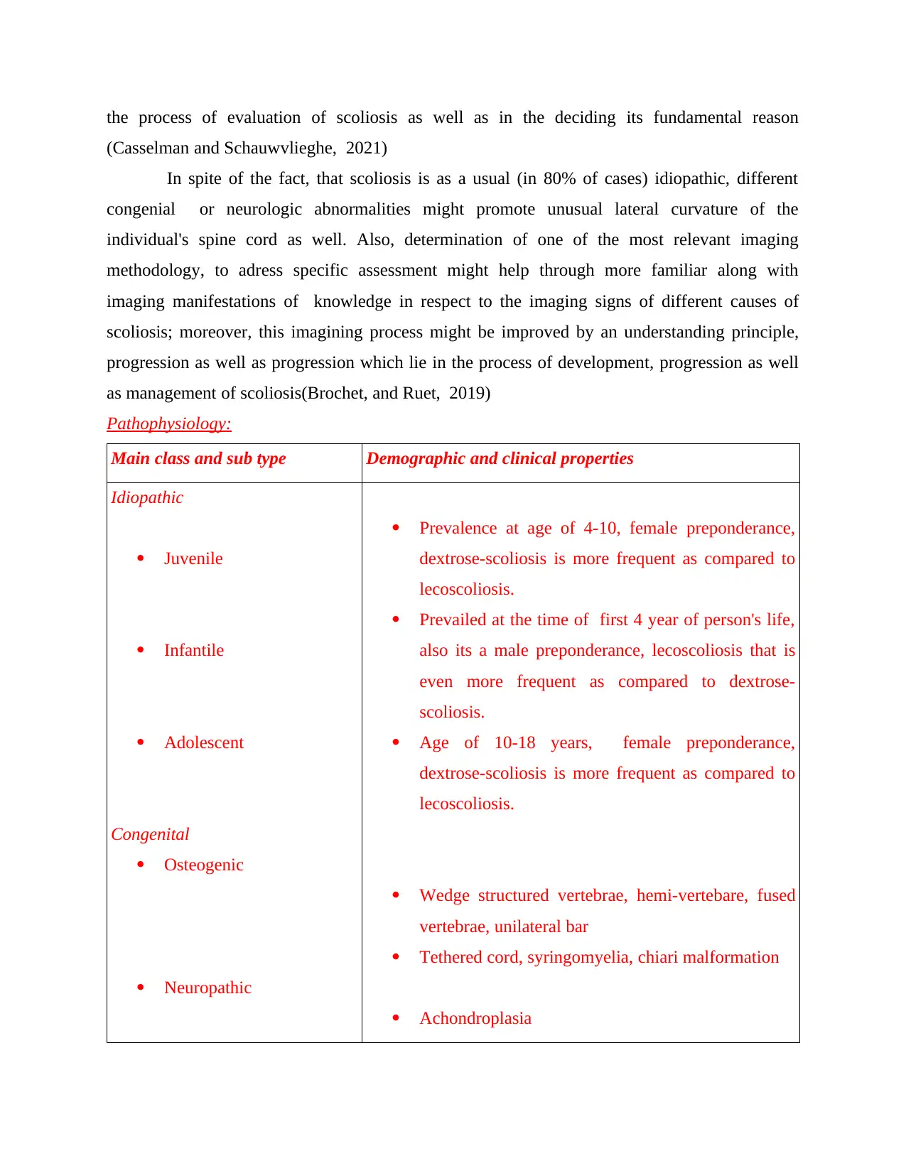
Pathophysiology:
Main class and sub type Demographic and clinical properties
Idiopathic
Juvenile
Infantile
Adolescent
Congenital
Osteogenic
Neuropathic
Prevalence at age of 4-10, female preponderance,
dextrose-scoliosis is more frequent as compared to
lecoscoliosis.
Prevailed at the time of first 4 year of person's life,
also its a male preponderance, lecoscoliosis that is
even more frequent as compared to dextrose-
scoliosis.
Age of 10-18 years, female preponderance,
dextrose-scoliosis is more frequent as compared to
lecoscoliosis.
Wedge structured vertebrae, hemi-vertebare, fused
vertebrae, unilateral bar
Tethered cord, syringomyelia, chiari malformation
Achondroplasia
(Casselman and Schauwvlieghe, 2021)
In spite of the fact, that scoliosis is as a usual (in 80% of cases) idiopathic, different
congenial or neurologic abnormalities might promote unusual lateral curvature of the
individual's spine cord as well. Also, determination of one of the most relevant imaging
methodology, to adress specific assessment might help through more familiar along with
imaging manifestations of knowledge in respect to the imaging signs of different causes of
scoliosis; moreover, this imagining process might be improved by an understanding principle,
progression as well as progression which lie in the process of development, progression as well
as management of scoliosis(Brochet, and Ruet, 2019)
Pathophysiology:
Main class and sub type Demographic and clinical properties
Idiopathic
Juvenile
Infantile
Adolescent
Congenital
Osteogenic
Neuropathic
Prevalence at age of 4-10, female preponderance,
dextrose-scoliosis is more frequent as compared to
lecoscoliosis.
Prevailed at the time of first 4 year of person's life,
also its a male preponderance, lecoscoliosis that is
even more frequent as compared to dextrose-
scoliosis.
Age of 10-18 years, female preponderance,
dextrose-scoliosis is more frequent as compared to
lecoscoliosis.
Wedge structured vertebrae, hemi-vertebare, fused
vertebrae, unilateral bar
Tethered cord, syringomyelia, chiari malformation
Achondroplasia
Paraphrase This Document
Need a fresh take? Get an instant paraphrase of this document with our AI Paraphraser
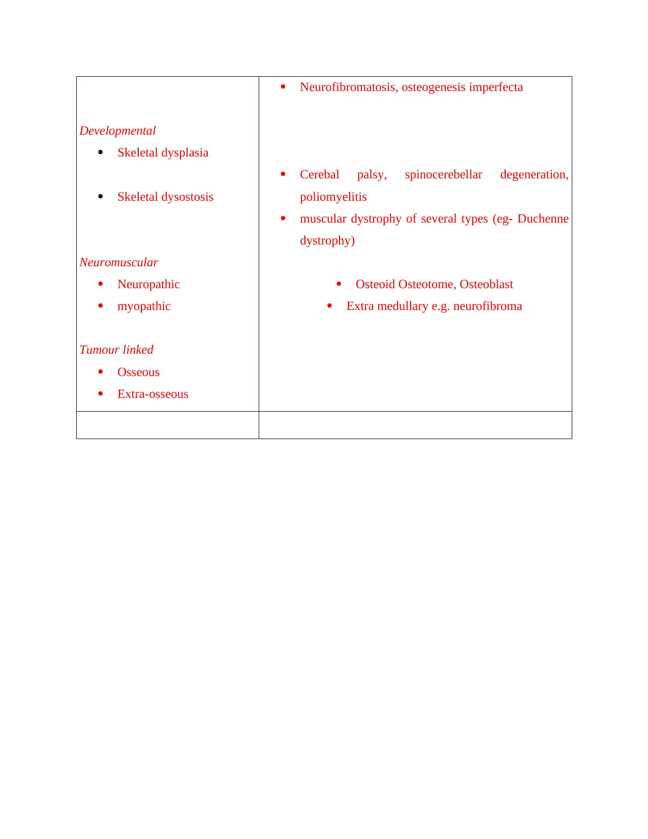
Developmental
Skeletal dysplasia
Skeletal dysostosis
Neuromuscular
Neuropathic
myopathic
Tumour linked
Osseous
Extra-osseous
Neurofibromatosis, osteogenesis imperfecta
Cerebal palsy, spinocerebellar degeneration,
poliomyelitis
muscular dystrophy of several types (eg- Duchenne
dystrophy)
Osteoid Osteotome, Osteoblast
Extra medullary e.g. neurofibroma
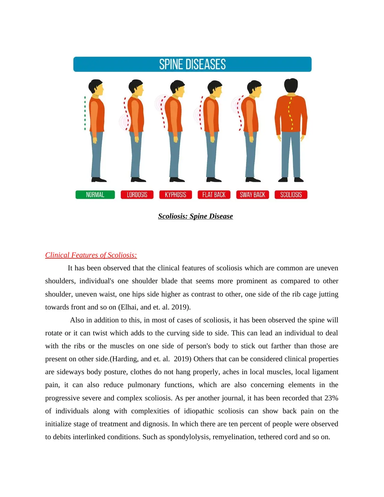
Scoliosis: Spine Disease
Clinical Features of Scoliosis:
It has been observed that the clinical features of scoliosis which are common are uneven
shoulders, individual's one shoulder blade that seems more prominent as compared to other
shoulder, uneven waist, one hips side higher as contrast to other, one side of the rib cage jutting
towards front and so on (Elhai, and et. al. 2019).
Also in addition to this, in most of cases of scoliosis, it has been observed the spine will
rotate or it can twist which adds to the curving side to side. This can lead an individual to deal
with the ribs or the muscles on one side of person's body to stick out farther than those are
present on other side.(Harding, and et. al. 2019) Others that can be considered clinical properties
are sideways body posture, clothes do not hang properly, aches in local muscles, local ligament
pain, it can also reduce pulmonary functions, which are also concerning elements in the
progressive severe and complex scoliosis. As per another journal, it has been recorded that 23%
of individuals along with complexities of idiopathic scoliosis can show back pain on the
initialize stage of treatment and dignosis. In which there are ten percent of people were observed
to debits interlinked conditions. Such as spondylolysis, remyelination, tethered cord and so on.
⊘ This is a preview!⊘
Do you want full access?
Subscribe today to unlock all pages.
Trusted by 1+ million students worldwide
Clinical incidence of Spinal disease
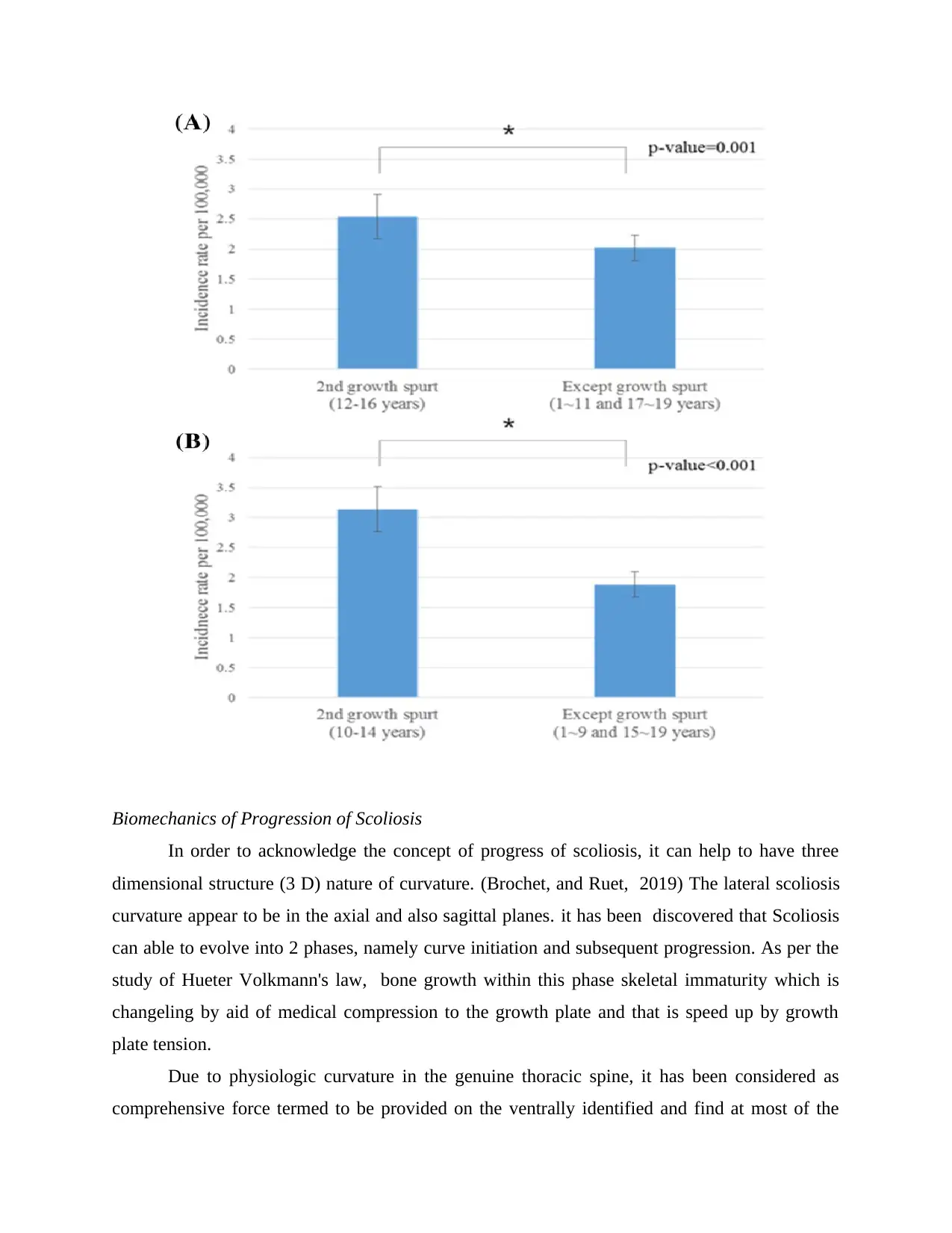
Studies concluded that Scoliosis affects 3% to 4% of the population, or it can also be
estimated to nine million people of in the UK. (Patsopoulos, 2018) It has tendency to develop in
early childhood development. It has been observed that the primary age for scoliosis is 10-15
years children, that can be prevailed equally in both genders. In which it has been recorded that
women are eight to nine time likely to get infected to curve magnitude which needs acute
treatment. (Lim, and et. al. 2018)
Furthermore, it has been observed each year, in UK, individual who are dealing with
scoliosis are able to attain more than 600, 000 visits to their personal or private healthcare
professionals, for estimation of 30,000 children are eligible to fit with stand and this stand is
known as brace for individuals who are suffering from sclerosis as well as 38,000 individuals can
subjected in the spinal fusion surgery as well. Idiopathic scoliosis is one of well-known kind and
estimated prevalence for 80% of scoliosis cases.
Congenital scoliosis that incorporates scoliosis caused, due to structural abnormalities of
bone and neural tissues of a person. Studies concluded that it is the second common type
representing 8 -20% of cases. Neuromuscular, developmental and cancer related scoliosis
combines establish the excess 10 -15%. (Lubetzki, and et. al. 2020) At present, degenerative
scoliosis and traumatic scoliosis are termed as likewise to be significant subcategories by those
associated board of the illness. Currently, in the management of the disease.
Paraphrase This Document
Need a fresh take? Get an instant paraphrase of this document with our AI Paraphraser
Biomechanics of Progression of Scoliosis
In order to acknowledge the concept of progress of scoliosis, it can help to have three
dimensional structure (3 D) nature of curvature. (Brochet, and Ruet, 2019) The lateral scoliosis
curvature appear to be in the axial and also sagittal planes. it has been discovered that Scoliosis
can able to evolve into 2 phases, namely curve initiation and subsequent progression. As per the
study of Hueter Volkmann's law, bone growth within this phase skeletal immaturity which is
changeling by aid of medical compression to the growth plate and that is speed up by growth
plate tension.
Due to physiologic curvature in the genuine thoracic spine, it has been considered as
comprehensive force termed to be provided on the ventrally identified and find at most of the
part of the vertebral column, in which the dis-tractive force is project on dorsally situate region.
Over time, it has been discrepancy manifests as alteration or alteration in the directivity of spinal
curvature, which can be ventrally fourth inside region of vertebral column that can termed to be
become the concave side as well as dorsally found at the part convex part (Withofs,and et. al.
2019).
Furthermore, it has been figured out that critical degree of curvature which have been
form, a considered fell mechanical cycle that can promotes the progress of scoliosis, that can
grows faster at the times of rapid spinal development. Resultant, considered impacts on both time
and 3D primary rotation should be considered while studying about scoliosis movement and
planning administration of this bone abnormalities.
In the process of evaluation, it is important for health practitioners to identify apex and
significant vertebrae. The essential signifying the curve type, choosing significant and relevant
approaching instrumentation framework that can be effective for the fusion. Surgical
methodologies depends on the surgeon. The apex is the vertebra or a round disk like structure
with the rotary motion from the focal point of the vertebral region. End vertebrae are considered
as those along with maximal slant which is toward the apex of the curvature and also they are
utilized to determine the Cobb angle. (Northrup,and et. al. 2021) Neutral vertebrae are those
that represent no proof of movement on vertical front facing ( poster-anterior or PA and ante-
posterior or AP radiographs, their pedicles are in the average as well as symmetric positions.
Studies state that neutral vertebrae always might be at similar even as end vertebrae, it
can be present at underneath distal to bend or curve, but also they are never close to apex than
end vertebrae. Whereas, the Stable vertebrae are the vertebrae uttermost cephalad that are
divided by the central sacral vertical line (CSVL) at a level beneath the end vertebra of the distal
curve. (Cerqueira, and et. al. 2018) The CSVL is a generally upward or vertical line that is drawn
opposite to an imaginary tangential line drawn across the top of the iliac crests on radiographs. It
separates with the help of sacrum.
Evaluation of a diagnostic strategy i.e. MRI and CT
Scoliosis is generally confirmed by performing physical examination of an individual, in
which, intervention like X-ray, spinal radiograph, CT scan and MRI plays a significant role. As
the curve of bones metric by the aid of Cobb method and it is diagnosed in the terms of
⊘ This is a preview!⊘
Do you want full access?
Subscribe today to unlock all pages.
Trusted by 1+ million students worldwide
complexities by the several of degrees (Patsopoulos, 2018) There are curves that can be exceed
45 to 50 degrees that are considered to be complex and also often more intensive treatment.
Studies confirmed that standard exam that are used in sometimes by paediatricians which
well known as Adam's Forward Bend Test. At the time of this test on children, individual leans
at front with his feet joining together and the bend perpendicular at the waist. (Brochet, and Ruet,
2019) From this angle, any abnormal spinal curvature of an individual can be easily observed.
Use of CT and MR Imaging in Scoliosis:
MRI and CT scan are considered as initial screening test in order to diagnose patient. It is
limited as they are unable to detect exact type of the deformity of patients. The main aim of
performing MRI or CT imaging in individual in order to identifying the main cause of the
problem. (Lim, and et. al. 2018) 2020) Also, the cross sectional imaging modalities are termed
to utilizing to guidance surgical treatment as evaluation postoperative complications.
Radiography is known as the mainstay within idiopathic scoliosis imaging. This is used
for confirming diagnosis and ruling out any underlying conditions. The presence of scoliosis in
spinal cord is a lateral rotatory curve within spine or coronal plane. This is characterised as side
to side deformity and also its 3 dimensional rotational deformity. There are various causes for
scoliosis which is known as diagnosis of exclusion. Scoliosis is the lateral spinal curvature
within Cobb angle of 10 degrees. This is known as abnormal curve and it can result for an
underlying congenital osseous or neurologic abnormality.
The imaging modalities includes radiography, magnetic resonance and imaging and it
plays pivotal roles in the diagnosis. There is treatment of primary role within the significance of
neurologic curve. There is need for managing the vertebrae or the curve type as it is known as
degree of angulation and it should be treated in less time. The aid of idiopathic scoliosis is the
governance for intensity and it is governed by the progression within likely usage during periods.
The planning of treatment as well as biomechanics include curve progression and it should well-
advised within the idiopathic scoliosis. The understanding of nomenclature includes method for
measurement and it is used for describing the scoliosis when it is essential for radiologists to
council spine experts. There is discussion with clinical implications for the individual vertebrae.
The ned vertebrae The measurement of include disk with the greatest rotation and developing the
significance for bisected the apex and vertical line for the perpendicular crests. The measurement
Paraphrase This Document
Need a fresh take? Get an instant paraphrase of this document with our AI Paraphraser
of Cobb angle is having certain limitations and it is performed by equally reliant upon
epidemiologic analysis.
There are other effective methods which are used for initial Scoliosis is referring as the
lateral spinal curvature which is associated with the angle of 10 degrees and more. Moreover,
this is also analysing that the abnormal curvature has shown the outcome of congenital or
developmental osseous or the neurologic abnormalities. As per this, the most the cases are
unknown when the identification of their causes is signify (Withofs,and et. al. 2019). Moreover,
the imaging modalities such as radiography, computed tomography is being used in with the
additional support of magnetic resonance MR imaging play key role in the development of
diagnosis, monitoring and the management of the disease which is firmly started with the
abnormal curvature of the body. As per this the radiography play a functional role and with the
CT and MR Imaging which show the indication of the presence which is based in underlying
process of osseous or the neurological cause which has been suspected. It is important to
indent8ify that the significance of the various bone and vertebra in or near the curved segment
which may include apex, end vertebra, neutral vertebra and stable vertebra. Moreover, the degree
of angulation is defined as the measured with the help of cobb method.
The degree of vertebral rotation which is show the measurement with the help of Nash-
Moe method. Moreover, the longitudinal extend of the spinal have some of the deviation which
has been taken in such a way which can render the spinal cord and their functional in the body.
This disease is progressive in nature and start with the factor of body with the last zone of spinal
cord and change the curvature of body and shown the factor of abnormal. (Brochet, and Ruet,
2019). With all the idiopathic scoliosis and their progression is defined with context of period of
rapid growth, and the optimal flow is taken which show the skeletally immature patient which
the short duration of 4 months. Also, the skeletal maturity is gained the curve of the bone is
more than the 30 degree form the 10 degree which is monitored with the help to MRI and CT
scan.
Medication and Treatment
There are different drugs which can be used in the case of scoliosis and can have high
impact on individual with such type of diseases. There are various treatment options for scoliosis
which are available in the market. These includes scoliosis braces, physical therapy, scoliosis
surgery, psychosocial support, scoliosis medications which includes analgesics and other
medication. These are the one which allow to provide relief from this diseases. Analgesics are
effective and allow to provide relaxation from severe pain. (Brochet, and Ruet, 2019) This is
effective in order to managing pain for some time period. This includes acetaminophen or
ibuprofen or aspirin. There is involvement of prescription when there is severe pain with
advanced disc degeneration in opiates with extreme cases.
Other medication like neuromuscular scoliosis or syndrome related scoliosis refers to
secondary effects on other condition like cerebral palsy, neurofibromatosis, Duchene muscular
dystrophy, myelodysplasia. These are some of the condition which can be treated with specific
medication. There are some of the best medication for the treatment for the scoliosis. This can be
deliberating and can cause severe pain within individual. Some prescribed drugs can be effective
like ibuprofen, acetaminophen or NSAID's in rare cases. (Williamsand et. al. 2020) There is
description of some of the drugs which may help to provide better relief in this condition.
Tylenol is the analgesics which is provided oral in tablet form with doses in every 4 to 6 hours.
This can cause nausea, stomach pain or loss of appetite as common side effects.
Motrin is the NSAID which also provides orally and can be taken as table at every 4 hours.
Stomach pain, nausea and upset stomach can be side effect. Diclofenac is the prescription
NSAID which is given orally and should be given in table form 2 doses daily. Constipation,
diarrhoea and abdominal pain is common side effect. (Lubetzki, and et. al. 2020)
CONCLUSION
Overall it can be concluded as Sclerosis is a spine disease. Although this disease can be
found to be usual in most of the cases. It can promote number of complication to an individual to
deal with illness. The clinical features of this disease that can serious such as even shoulders,
uneven waist, clothes may appear to be handing on an individual and so on. It is characterised as
both MR imaging and CT can be suggested by health practitioner in order to determine the
inherent reason of sclerosis. CT can provide guidance preoperative planning for intervention they
can provide effective results. As it has been seeming that it is deformities in bones which can
allow to form curvature in an individual body. Though CT scanning the observation which can
be undermine are osseous causes and also in some of the cases in regards with conjunction with
MR imaging. MR imaging are important if it is a neuropathic cause can be analysed. Through the
⊘ This is a preview!⊘
Do you want full access?
Subscribe today to unlock all pages.
Trusted by 1+ million students worldwide
1 out of 15
Your All-in-One AI-Powered Toolkit for Academic Success.
+13062052269
info@desklib.com
Available 24*7 on WhatsApp / Email
![[object Object]](/_next/static/media/star-bottom.7253800d.svg)
Unlock your academic potential
Copyright © 2020–2026 A2Z Services. All Rights Reserved. Developed and managed by ZUCOL.