Analysis of Sepsis Case Study: Pathogenesis, Nursing, ABGs
VerifiedAdded on 2023/01/11
|7
|1846
|76
Report
AI Summary
This report presents a comprehensive analysis of a sepsis case study involving a 75-year-old male patient, Mr. Kirkman, admitted with a urinary tract infection that progressed to sepsis. The report begins by dissecting the pathogenesis of sepsis in relation to the patient's clinical manifestations, including the role of gram-negative bacteria, endotoxins, and the body's inflammatory response. It then identifies and justifies an appropriate nursing strategy, specifically intravenous fluids, based on the latest research and clinical guidelines. Finally, the report critically examines the patient's arterial blood gas (ABG) results, relating them to the underlying pathophysiology and explaining the observed deviations from normal ranges, such as low partial pressure of oxygen, altered carbon dioxide levels, elevated lactate, and pH imbalances. The analysis integrates current medical literature to support all conclusions.

Running head: SEPSIS 1
Sepsis
Name
Institution
Tutor
Date:
Sepsis
Name
Institution
Tutor
Date:
Paraphrase This Document
Need a fresh take? Get an instant paraphrase of this document with our AI Paraphraser
SEPSIS 2
1. Analyze the pathogenesis in relation to the clinical manifestation in the case study.
Sepsis is a condition that arise as the immune system of the body fight back against an
infection. Such infection includes Urinary tract infection like it was the case of Mr.Kirkmann.
The World Health Organization defined a urinary tract infection as any form of disease that
affects any part of the urinary system. The anatomy of the urinary system comprises of bladder,
urethra and ureter. They are made in a manner that makes it quite difficult for any pathogen to
invade. In case a bacteria invades the urinary system which is mainly through unprotected sexual
intercourse or insertion of catheter under hygienic condition, they replicate rapidly in the bladder
leading to inflammation. During this period, the patient would experience a burning sensation
while urinating. Furthermore, the patient will also experience sharp abdominal pain that radiates
from the right flank (Craft, Gordon, Huether, McCance, & Brashers, 2015). This is exactly what
Mr.Kirkmann in the case study was experiencing. At this juncture, the best nursing intervention
is treatment using antibiotics. However, thorough microbiology tests ought to be conducted to
find out the bacteria responsible for the underlying urinary tract infections. This is later followed
up by sensitivity tests to find out the best drug or antibiotic. If the Urinary tract infection is not
combated properly, it progresses and leads to a condition known as sepsis.
Studies indicate that Urinary Tract infections contributes to 20-30% cases of sepsis. In
this case study for example, the patient, Mr. Kirkmann, has a history of Urinary tract infection.
The patient had a catheter inserted and this could have been the source of the gram negative
bacteria (Gotts & Matthay, 2016). Once bacteria colonize the urethra, they lead to Urinary tract
infections. According to studies, gram negative bacteria have endotoxins such as
lipopolysaccharides in their cell walls. Such endotoxins are the ones that contribute to the
manifestation of sepsis (Kumar, 2017). The endotoxin stimulate both inflammatory, coagulation
SEPSIS 3
and the complement systems that in return stimulate monocytes, macrophages, neutrophils and
finally dendritic cells. The monocytes then stimulate production of both interlukin-1 and tumor
necrosis factor alpha that mediate sepsis. The immune response results to inflammation that leads
to fever. This why Mr.Kirkmann had a high temperature of 39 degree Celsius. The immune
responds by injuring vital organs and the body responds by producing more coagulation agents.
This is why there were high platelets, WBCs in the diagnosis. Furthermore, injury to the vital
body organs like heart and lungs lead to respiratory failure and this subsequently lead to tissue
perfusion. Tissue perfusion leads to metabolic acidosis and this explains the discrepancy in the
arterial blood gases. Furthermore, respiratory failure means the patient cannot breathe well and
as a result he will experience high heart and breathing rates of above 20 and 90 beats per minute.
2. Select one appropriate nursing strategy and explain the underpinning evidence base for
this rationale.
Intravenous fluids is the recommended management in patients with sepsis and would
also be used in this case. Several studies have been conducted and point out that antibiotics alone
cannot be used to effectively clear sepsis (Levi & Van der Poll, 2017). The same studies propose
fluids in the form of crystalloids or colloids. The fluids are administered through IV so that the
nurse is able to monitor and keep track on the quantity of the fluids being administered. In 2002,
the sepsis campaign was set up with the sole objective of coming up with the best strategies to
prevent sepsis (Levy, Evans, & Rhodes, 2018). After extensive research in 2004, it was agreed
that fluid resuscitation is the best management approach of sepsis. This can be done either by
colloids or crystalloids. In 2012, after a surge of research to establish which fluids are the best, it
was recommended that crystalloids should be the first choice (Curtis, Ramsden, 2016). It is also
recommended that albumin should be used as the adjuvant.
⊘ This is a preview!⊘
Do you want full access?
Subscribe today to unlock all pages.

Trusted by 1+ million students worldwide
SEPSIS 4
The IV fluids are divided on the basis of their composition. They are either colloids or
crystalloids. Colloids are fluids that do not easily cross the cell membranes as compared to the
crystalloids. It is for this reason that research recommend crystalloids as first choice (Limongi,
D'Agostini, & Ciotti, 2016). Since colloids do not diffuse through cellular membranes easily,
they tend to remain in the intravascular space for quite some time. Studies also indicate that
when colloids are administered to a patient with sepsis, they work through osmosis thus retaining
fluids in the intravascular compartments. Crystalloids on the other hand are composed of soluble
substances that diffuse easily through the cellular membrane and are divided into either balanced
or non-balanced formulas (Simpson, 2016). An Example of a non-balanced fluid is normal saline
which is isotonic to blood plasma but a different electrolyte composition. Examples of the
balanced crystalloids on the other hand include Lactate, Ringers Acetate and the Plasmalyte.
Unlike the non-balanced crystalloids, these fluids have an electrolyte composition similar to
plasma (Singer et al., 2016). Since sepsis is associated with low blood pressure, the rationale of
this nursing intervention is to provide extra fluids so as to prevent blood pressure from
decreasing continuously.
3. Critically analyze the arterial blood gas results, and relate them to underlying
pathogenesis.
One of the diagnostics for the patient in the case study was arterial blood gases (ABGs)
and the results were as follows. For partial pressure of carbon dioxide and oxygen, the results
were 32mmHg and 75 mmHg respectively. Both the parameters were below the normal ranges
since the normal range for partial pressure of oxygen is between 80 and 100 mmHg while that of
carbon dioxide is between 35 and 45 mmHg. The results for lactate were 3.2 mmol/L and this
were higher than the normal level of between 0.3 and 0.8 mmol/L. The PH was 7.25 and was
Paraphrase This Document
Need a fresh take? Get an instant paraphrase of this document with our AI Paraphraser
SEPSIS 5
also below the normal ranges of between 7.35 and 7.45. The results for HCO3‾ was 15 mmol/L.
This results were below the normal levels of between 22 and 32 mmol/L. Those of Base Excess
(BE) was -6 mmol/L while the normal range is between -2 and 2 mmol/L. The variations in the
blood gases results is as a result of the effect of the sepsis.
Recent studies indicate that Lactate is one of the sepsis markers. This is because during
sepsis, there is an altered microcirculation and this also leads to impaired exchange of oxygen
and more of lactate is produced. This is why in this case, Lactate was high than the normal
values. Since sepsis is simply the inflammatory response of the body to infections that lead to
hemodynamic changes as well as respiratory failure (Van der Poll, Van de Veerdonk, Scicluna,
& Netea, 2017). This two processes eventually lead to decreased oxygenation in the tissues and
this is the reason for low partial oxygen pressure in the patient. Partial carbon dioxide in the case
study was lower than the normal ranges. This is because during sepsis, there is poor tissue
perfusion and tis leads to the buildup of serum lactate leading to metabolic acidosis and patients
therefore increase minute ventilation and this eventually blows off carbon dioxide (Vincent,
Mira, & Antonelli, 2016). This is the same reason for the low HCO3‾ and the Base Excess. The
PH on the other was higher than the normal range. This is due to impaired excretion of Hydrogen
ions in the urine due to the UTI.
SEPSIS 6
References
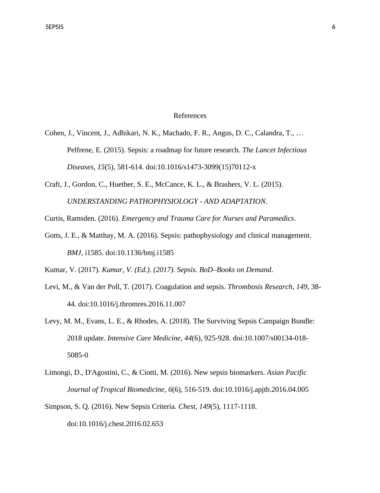
Cohen, J., Vincent, J., Adhikari, N. K., Machado, F. R., Angus, D. C., Calandra, T., …
Pelfrene, E. (2015). Sepsis: a roadmap for future research. The Lancet Infectious
Diseases, 15(5), 581-614. doi:10.1016/s1473-3099(15)70112-x
Craft, J., Gordon, C., Huether, S. E., McCance, K. L., & Brashers, V. L. (2015).
UNDERSTANDING PATHOPHYSIOLOGY - AND ADAPTATION.
Curtis, Ramsden. (2016). Emergency and Trauma Care for Nurses and Paramedics.
Gotts, J. E., & Matthay, M. A. (2016). Sepsis: pathophysiology and clinical management.
BMJ, i1585. doi:10.1136/bmj.i1585
Kumar, V. (2017). Kumar, V. (Ed.). (2017). Sepsis. BoD–Books on Demand.
Levi, M., & Van der Poll, T. (2017). Coagulation and sepsis. Thrombosis Research, 149, 38-
44. doi:10.1016/j.thromres.2016.11.007
Levy, M. M., Evans, L. E., & Rhodes, A. (2018). The Surviving Sepsis Campaign Bundle:
2018 update. Intensive Care Medicine, 44(6), 925-928. doi:10.1007/s00134-018-
5085-0
Limongi, D., D'Agostini, C., & Ciotti, M. (2016). New sepsis biomarkers. Asian Pacific
Journal of Tropical Biomedicine, 6(6), 516-519. doi:10.1016/j.apjtb.2016.04.005
Simpson, S. Q. (2016). New Sepsis Criteria. Chest, 149(5), 1117-1118.
doi:10.1016/j.chest.2016.02.653
⊘ This is a preview!⊘
Do you want full access?
Subscribe today to unlock all pages.

Trusted by 1+ million students worldwide
SEPSIS 7
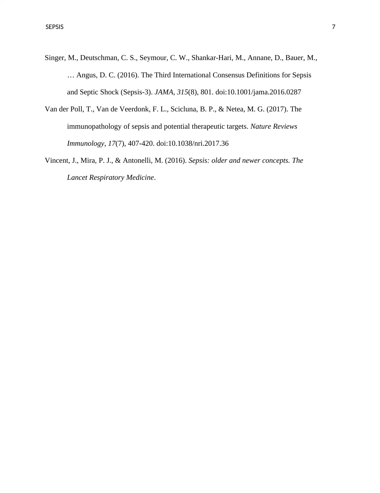
Singer, M., Deutschman, C. S., Seymour, C. W., Shankar-Hari, M., Annane, D., Bauer, M.,
… Angus, D. C. (2016). The Third International Consensus Definitions for Sepsis
and Septic Shock (Sepsis-3). JAMA, 315(8), 801. doi:10.1001/jama.2016.0287
Van der Poll, T., Van de Veerdonk, F. L., Scicluna, B. P., & Netea, M. G. (2017). The
immunopathology of sepsis and potential therapeutic targets. Nature Reviews
Immunology, 17(7), 407-420. doi:10.1038/nri.2017.36
Vincent, J., Mira, P. J., & Antonelli, M. (2016). Sepsis: older and newer concepts. The
Lancet Respiratory Medicine.
1 out of 7
Related Documents
Your All-in-One AI-Powered Toolkit for Academic Success.
+13062052269
info@desklib.com
Available 24*7 on WhatsApp / Email
![[object Object]](/_next/static/media/star-bottom.7253800d.svg)
Unlock your academic potential
Copyright © 2020–2025 A2Z Services. All Rights Reserved. Developed and managed by ZUCOL.


