Treating Peptic Ulcers: A Case Study
VerifiedAdded on 2020/03/16
|11
|2929
|76
AI Summary
This assignment focuses on the treatment of a patient with peptic ulcers. It outlines various medical interventions, including proton pump inhibitors, cytoprotective agents, and antibiotics for H. pylori eradication. The case also emphasizes behavioral modifications like smoking cessation and dietary adjustments to aid in ulcer healing.
Contribute Materials
Your contribution can guide someone’s learning journey. Share your
documents today.
Running Head: SARS 1
Severe Acute Respiratory Syndrome (SARS)
Name of Student
Institutional Affiliation
Severe Acute Respiratory Syndrome (SARS)
Name of Student
Institutional Affiliation
Secure Best Marks with AI Grader
Need help grading? Try our AI Grader for instant feedback on your assignments.
SARS 2
SARS
Case Study 1
Q1. Epidemiology, Epidemic, and Pandemic
Epidemiology refers to the study of the spreading and causes of events or cases
associated with health and the use of the same study to regulate the infection and other
challenges of health. Epidemiological investigations use methods such as descriptive studies
and surveillance to study the spread of diseases. Also, it uses analytical studies to study the
causes. The epidemic is the sudden increase of disease cases above the normal expectations
in a particular population of a specific area. On the other hand, a pandemic is an epidemic
that has propagated over various continents or countries, normally affecting a lot of people.
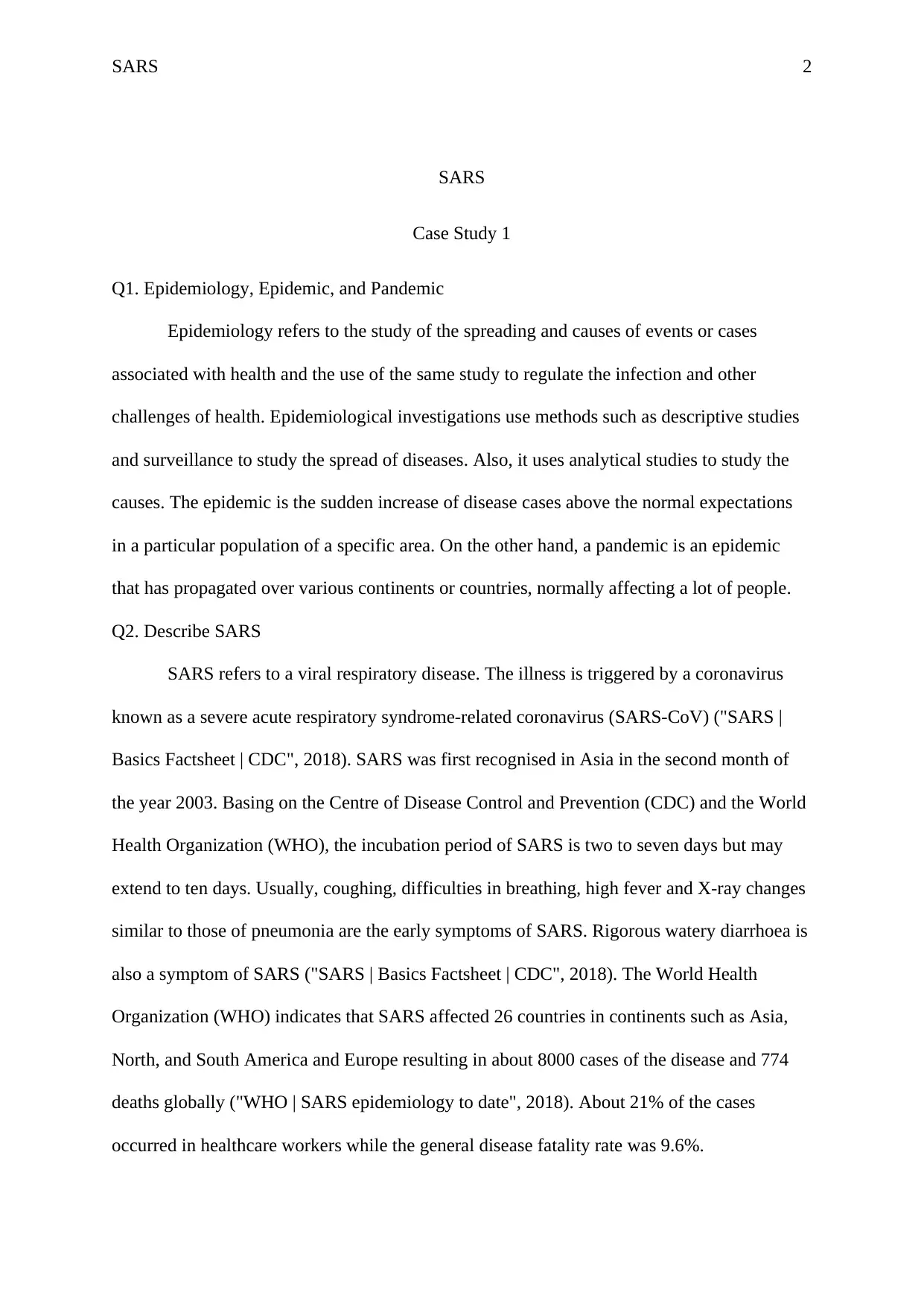
Q2. Describe SARS
SARS refers to a viral respiratory disease. The illness is triggered by a coronavirus
known as a severe acute respiratory syndrome-related coronavirus (SARS-CoV) ("SARS |
Basics Factsheet | CDC", 2018). SARS was first recognised in Asia in the second month of
the year 2003. Basing on the Centre of Disease Control and Prevention (CDC) and the World
Health Organization (WHO), the incubation period of SARS is two to seven days but may
extend to ten days. Usually, coughing, difficulties in breathing, high fever and X-ray changes
similar to those of pneumonia are the early symptoms of SARS. Rigorous watery diarrhoea is
also a symptom of SARS ("SARS | Basics Factsheet | CDC", 2018). The World Health
Organization (WHO) indicates that SARS affected 26 countries in continents such as Asia,
North, and South America and Europe resulting in about 8000 cases of the disease and 774
deaths globally ("WHO | SARS epidemiology to date", 2018). About 21% of the cases
occurred in healthcare workers while the general disease fatality rate was 9.6%.
SARS 3
Q3. Source of the infection
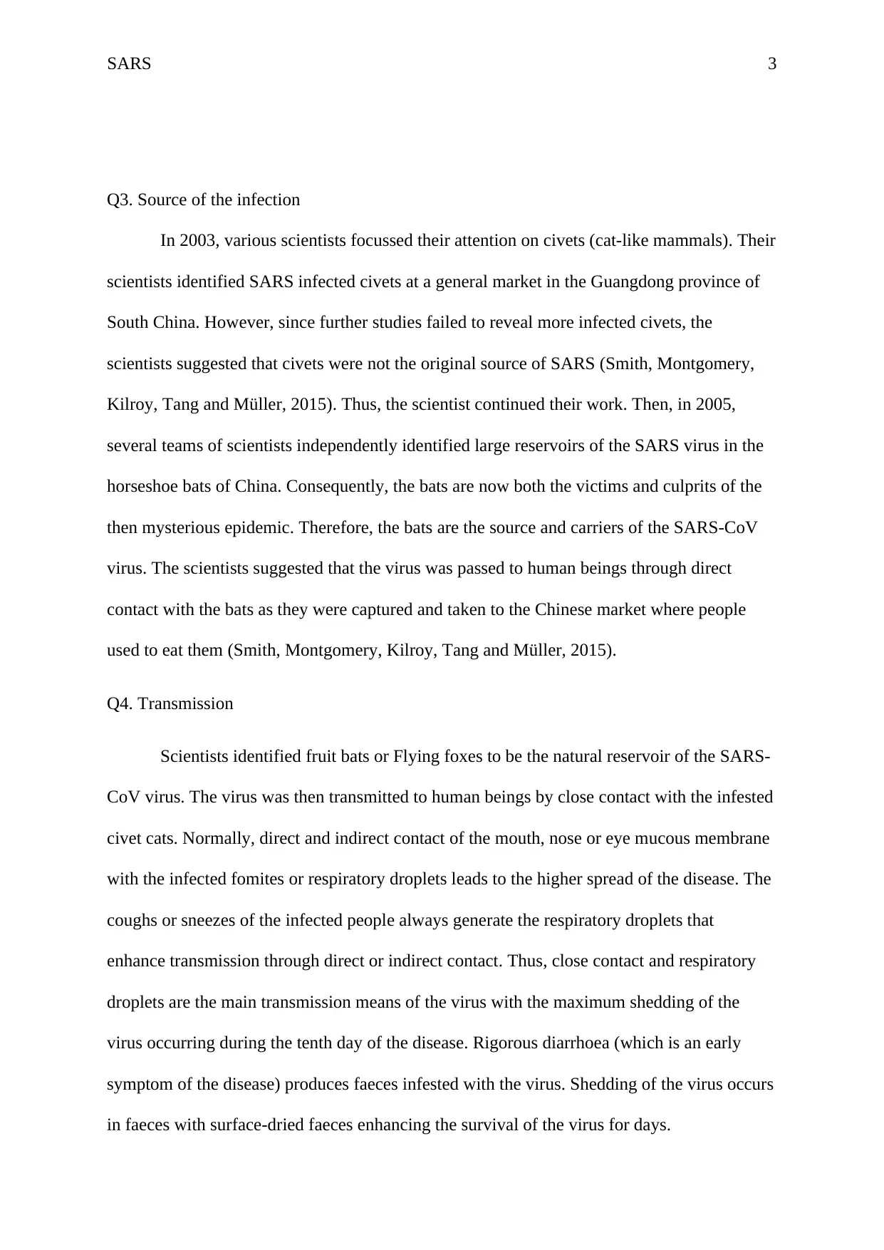
In 2003, various scientists focussed their attention on civets (cat-like mammals). Their
scientists identified SARS infected civets at a general market in the Guangdong province of
South China. However, since further studies failed to reveal more infected civets, the
scientists suggested that civets were not the original source of SARS (Smith, Montgomery,
Kilroy, Tang and Müller, 2015). Thus, the scientist continued their work. Then, in 2005,
several teams of scientists independently identified large reservoirs of the SARS virus in the
horseshoe bats of China. Consequently, the bats are now both the victims and culprits of the
then mysterious epidemic. Therefore, the bats are the source and carriers of the SARS-CoV
virus. The scientists suggested that the virus was passed to human beings through direct
contact with the bats as they were captured and taken to the Chinese market where people
used to eat them (Smith, Montgomery, Kilroy, Tang and Müller, 2015).
Q4. Transmission
Scientists identified fruit bats or Flying foxes to be the natural reservoir of the SARS-
CoV virus. The virus was then transmitted to human beings by close contact with the infested
civet cats. Normally, direct and indirect contact of the mouth, nose or eye mucous membrane
with the infected fomites or respiratory droplets leads to the higher spread of the disease. The
coughs or sneezes of the infected people always generate the respiratory droplets that
enhance transmission through direct or indirect contact. Thus, close contact and respiratory
droplets are the main transmission means of the virus with the maximum shedding of the
virus occurring during the tenth day of the disease. Rigorous diarrhoea (which is an early
symptom of the disease) produces faeces infested with the virus. Shedding of the virus occurs
in faeces with surface-dried faeces enhancing the survival of the virus for days.
SARS 4
Q5. How SARS was contained in 2003
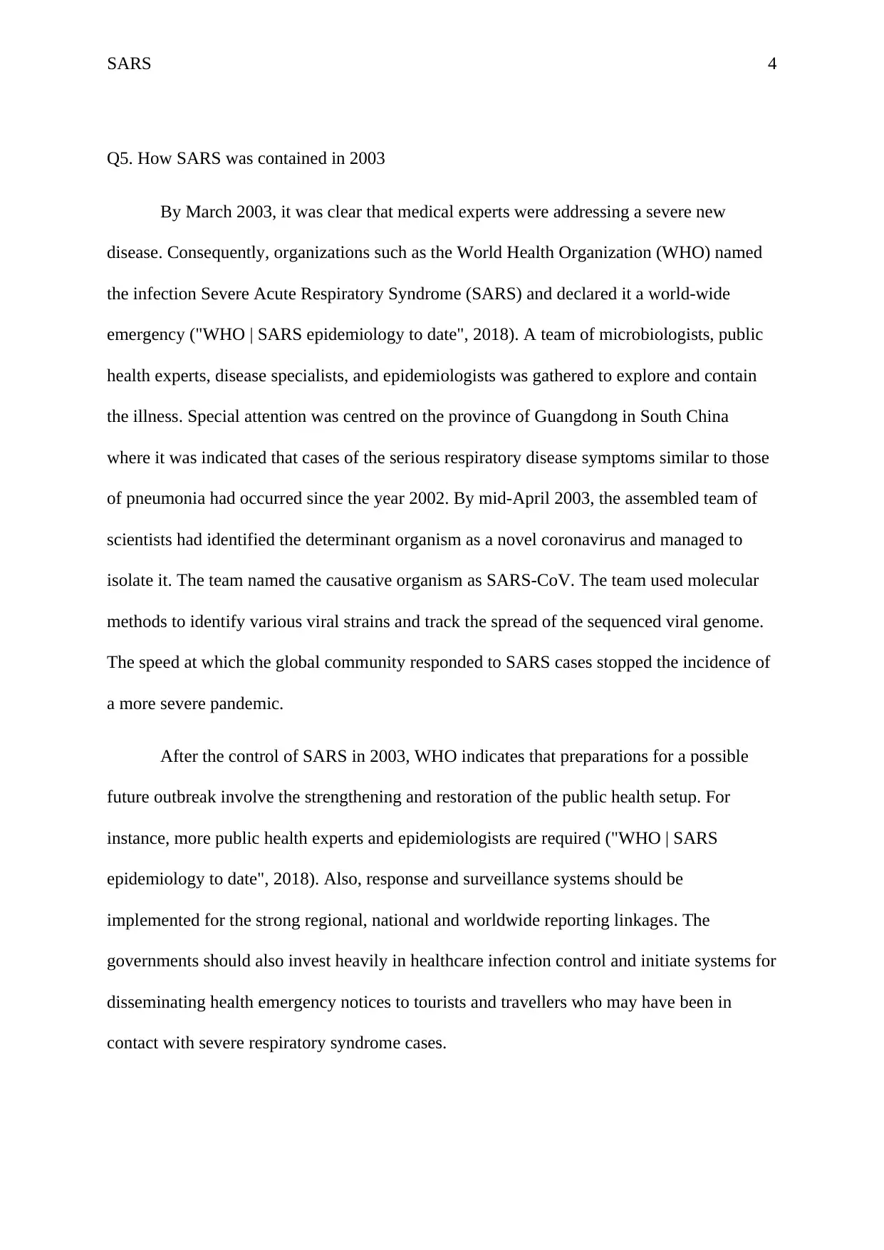
By March 2003, it was clear that medical experts were addressing a severe new
disease. Consequently, organizations such as the World Health Organization (WHO) named
the infection Severe Acute Respiratory Syndrome (SARS) and declared it a world-wide
emergency ("WHO | SARS epidemiology to date", 2018). A team of microbiologists, public
health experts, disease specialists, and epidemiologists was gathered to explore and contain
the illness. Special attention was centred on the province of Guangdong in South China
where it was indicated that cases of the serious respiratory disease symptoms similar to those
of pneumonia had occurred since the year 2002. By mid-April 2003, the assembled team of
scientists had identified the determinant organism as a novel coronavirus and managed to
isolate it. The team named the causative organism as SARS-CoV. The team used molecular
methods to identify various viral strains and track the spread of the sequenced viral genome.
The speed at which the global community responded to SARS cases stopped the incidence of
a more severe pandemic.
After the control of SARS in 2003, WHO indicates that preparations for a possible
future outbreak involve the strengthening and restoration of the public health setup. For
instance, more public health experts and epidemiologists are required ("WHO | SARS
epidemiology to date", 2018). Also, response and surveillance systems should be
implemented for the strong regional, national and worldwide reporting linkages. The
governments should also invest heavily in healthcare infection control and initiate systems for
disseminating health emergency notices to tourists and travellers who may have been in
contact with severe respiratory syndrome cases.
Secure Best Marks with AI Grader
Need help grading? Try our AI Grader for instant feedback on your assignments.
SARS 5
Case Study 2
Q1. Blood pathology
Results from the blood examination provide a proper picture for the
pathophysiological analysis. First off, the hemoglobin levels are lower than normal indicating
an anemic state. The abnormality in hemoglobin levels is coupled with low ferritin levels and
low transferrin saturation. Reduced ferritin levels and transferrin saturation also signify an
anemic condition (Medlineplus.gov., 2018). Low ferritin levels often result from a heavy
blood loss due to injury, bleeding in the gastrointestinal tract, poor absorption of iron in the
GIT, or chronic intake of food with poor iron concentrations (Medlineplus.gov., 2018). The
low transferrin saturation indicates a prolonged bleeding condition in the digestive tract, poor
iron absorption because of some underlying conditions in the gastrointestinal tract, or a poor
supply of dietary iron.
The blood test also reveals that the patient presents with a corpuscular volume of low
mean. The low mean corpuscular volume indicates microcytic anemia, which is confirmed by
the blood slide examination. The blood slide exam also shows that the red blood cells are
hypochromic hence microcytic hypochromic anemia. Microcytic hypochromic anemia is
highly associated with iron deficiency anemia but in this case, the iron levels in the blood and
the total iron binding capacity are normal (Chaudhry and Kasarla, 2017). Other than iron
deficiency anemia, the microcytic hypochromic state of the red blood cells can be attributed
to lead poisoning, chronic disease, and acute or chronic loss of blood due to various
conditions (Chaudhry and Kasarla, 2017). The white blood cell count exists within the
normal range hence showing that the patient is not suffering from acute bacterial or parasitic
infection. With conditions such as ancylostomiasis, the blood cell investigation would include
SARS 6
a measure of the eosinophils, which in a positive case would present with eosinophilia. The
platelet count is also normal thus signifying a normal clotting process unless in conditions
where an ulcerated surface encounters persistent irritation.
Q2. Other noteworthy issues
The mid-central pain that occurs in the abdomen is an important diagnostic factor as it
raises suspicion over anomalies in the gastrointestinal tract and other organs in the region
such as the liver and spleen. The etiology of the pain is however gastrointestinal due to the
nature of its presentation. The pains occur mostly and intensely during the night which gives
a clue of a possible ulceration of the GIT mucosa. The soothing character of the antacids
confirms the presence of ulcers in a region of the gastrointestinal tract where acidic
substances are the main secretion. Acids are known to irritate the lining of the gastrointestinal
tract especially in situations where the mucosal protection is eroded. Ingestion of food or
antacids is known to reduce the effects of acidic digestive juices on the gastrointestinal lining
with eroded mucosal lining.
Another important observation from the presentation is the 42-year-old’s smoking
behavior. The blood examination indicates a higher than normal red blood cell count, which
is common in habitual smokers. The smoking behavior and the current state of low
hemoglobin could be useful in explaining the state of fatigue. Smoking is also related to the
occurrence of stomach ulcers and esophageal regions due to reflux of acidic content and a
probable association with H.pylori proliferation as suggested by Li et al. (2014).
Q3. Most Probable Diagonosis
The clinical presentation and laboratory evidence directs the diagnosis to a possible
existence of peptic ulcers. Peptic ulcers occur in either the stomach lining or in the duodenum
hence explaining the abdominal pain. The pains mainly occur at night and in conditions
SARS 7
where the stomach is empty and is soothed by intake of non-acidic food or treatment with
antacids. The ulceration can cause hemorrhage or interfere with iron intake thus justifying the
low hemoglobin levels, low ferritin levels, low transferrin concentration, and the resultant
microcytic hypochromic condition of the red blood cells. Peptic ulcers can be attributed to
many causes but the most prominent one is a chronic infection by the bacteria Helicobacter
pylori (H. pylori).
Q4. Other diagonosis tests
Peptic ulcers can be confirmed through an array or tests ranging from imaging to
laboratory tests where stool and blood samples are specimens of choice. To note the presence,
condition, and severity of the ulcers, an endoscopy procedure would be appropriate.
Endoscopy will provide the most suitable evidence for the existence of ulcers or any other
structural anomaly in the stomach and the extent of severity.
A stool test for fecal occult blood, ova and cyst, H.pylori antigen, and routine macro-
and microscopic examination will also be essential. The fecal occult blood (FOB) is
important in indicating the occurrence of hemorrhages in the gastrointestinal tract especially
in cases of unexplained low hemoglobin levels. In the case of peptic ulcers, the FOB test
would be positive. The H.pylori antigen test is critical in this case as it confirms the presence
of an infection with the bacteria hence not only does it ratify the presence of peptic ulcers, but
also gives its etiology and a baseline for the treatment protocol (Anand, 2017). The
macroscopic examination checks for the presence of any parasite, while the microscopic
examination is suitable for identification of any ova or cyst of pathogenic parasites such as
Necator americanus or Ancylostoma duodenale, which are also known to cause anemia and
ulcerations on the luminal walls of the small intestines. The microscopic ova and cyst test
helps to rule out parasitic infection.
Paraphrase This Document
Need a fresh take? Get an instant paraphrase of this document with our AI Paraphraser
SARS 8
Q5. Pathophysiology and the treatment protocol
Pathophysiology
Peptic ulcers are defects in the duodenum and mucosal stomach lining leading to
irritation of the underlining epithelial cells by the acidic nature of fluids in the two regions. In
normal conditions, the mucosa offers primary protection to the cells of the region thus
preventing the scouring action of the digestion fluids produced in the stomach. An
interruption of the mucosa exposes the vulnerable epithelial cells beneath leading to
ulcerations. Chronic ulcerations are thought to result from the persistent action of
Helicobacter pylori, which feeds on the mucosa and irritate the epithelial cells developing
sores.
The occurrence of peptic ulcers results from an imbalance between the aggressive
factors and the defensive factors. The aggressive factors are known to destroy the
gastrointestinal mucosa and such include endogenic factors such as peptic secretions, which
are known to have high acidity. Exogenous factors known to disrupt the mucosa include
chronic H.pylori infection, medications such as the nonsteroidal anti-inflammatory drugs
(NSAIDs), excessive smoking, chronic alcoholism and prolonged exposure to stress (Anand,
2017). Of the exogenous aggressive factors, chronic H.pylori infection and detrimental
effects of NSAIDs cause greater damage to the gastrointestinal mucosa compared to the
others (Anand, 2017). Factors responsible for the defense mechanism against the action of
acid and pepsin are of great physiological importance even with the presence of aggressive
factors. The defense mechanism can be divided into the pre-epithelial defense, which
comprises of the secretion of bicarbonates and mucus, the epithelial defense including the
epithelial restitution, replication, and secretion of bicarbonates, the extracellular buffers such
as bicarbonates, and the post-epithelial defense resulting from mucosal microcirculation and
SARS 9
the tissue acid-base balance. An imbalance of the two factors in favor of the aggressive
factors, therefore, results in the occurrence of ulcerations due to a compromised defense
mechanism.
Presence of sores leads to gastrointestinal bleeding that can be detected in the stool
through a fecal occult blood test and through a blood examination for complete blood count
and peripheral blood film examination. The blood examination in cases of chronic peptic
ulcers reveals anemic conditions. In normal conditions, the ulcerated surfaces are expected to
recover through the physiological healing processes but in conditions where the aggressive
factors such as an intense chronic H.pylori infection are persistent, the peptic ulcers disease
becomes chronic.
Treatment
The treatment protocol for peptic ulcers disease follows various criteria depending on
the clinical presentation. In the presented case, the treatment process can start by reducing or
countering the effects of pepsin and acids by prescribing proton pump inhibitors to reduce
acid production and allow the ulcers to heal (Mayoclinic.org, 2018). Cytoprotective agents
such as sucralfate and misoprostol can accompany the proton pump inhibitors as additional
prescriptions for the protection of gastrointestinal lining (Mayoclinic.org, 2018). In presence
of a positive result for the presence of H.pylori infection, the condition should be treated
using antibodies. The treatment of H.pylori is effected through a combination of antibiotics
and the prescribed dose should be followed to prevent resistance.
Behavioral factors such as smoking and alcoholism are known to cause or enhance the
occurrence of peptic ulcers. Therapeutic processes including abstinence from alcohol and
cigarette smoking would be effective in helping to treat the ulcers as Li et al. (2014) confirms
that cigarette smoking is highly associated with the occurrence of peptic ulcers and other

SARS 10
gastrointestinal disorders. The patient should also modify his foods to avoid those with high
acidity and instead take foods that are known to contain very little or no acidity. He should
also avoid skipping meals as such will aggravate the condition by allowing the pepsin and the
peptic acids to work on the stomach and duodenal walls.
Secure Best Marks with AI Grader
Need help grading? Try our AI Grader for instant feedback on your assignments.
SARS 11
References
Anand, B. (2017). Peptic Ulcer Disease: Background, Anatomy, Pathophysiology. Retrieved
from https://emedicine.medscape.com/article/181753-overview
Chaudhry HS, Kasarla MR. Anemia, Microcytic Hypochromic. [Updated 2017 Dec 4]. In:
StatPearls [Internet]. Treasure Island (FL): StatPearls Publishing; 2018
Jan-. Available from: https://www.ncbi.nlm.nih.gov/books/NBK470252/
Li, L., Chan, R., Lu, L., Shen, J., Zhang, L., & Wu, W. et al. (2014). Cigarette smoking and
gastrointestinal diseases: The causal relationship and underlying molecular
mechanisms (Review). International Journal Of Molecular Medicine, 34(2), 372-380.
doi: 10.3892/ijmm.2014.1786
Mayoclinic.org. (2018). Peptic ulcer - Diagnosis and treatment - Mayo Clinic. Retrieved from
https://www.mayoclinic.org/diseases-conditions/peptic-ulcer/diagnosis-treatment/drc-
20354229
Medlineplus.gov. (2018). Ferritin blood test: MedlinePlus Medical Encyclopedia. Retrieved
from https://medlineplus.gov/ency/article/003490.htm
SARS | Basics Factsheet | CDC. (2018). Retrieved from https://www.cdc.gov/sars/about/fs-
sars.html
Smith, R., Montgomery, M., Kilroy, G., Tang, S., & Müller, S. (2015). Tropical low
formation during the Australian monsoon: the events of January 2013 (paper updated
July 2016). Australian Meteorological And Oceanographic Journal, 65(3/4), 318-341.
doi: 10.22499/2.6503.003
WHO | SARS epidemiology to date. (2018). Retrieved from
http://www.who.int/csr/sars/epi2003_04_11/en/
1 out of 11
Related Documents
Your All-in-One AI-Powered Toolkit for Academic Success.
+13062052269
info@desklib.com
Available 24*7 on WhatsApp / Email
![[object Object]](/_next/static/media/star-bottom.7253800d.svg)
Unlock your academic potential
© 2024 | Zucol Services PVT LTD | All rights reserved.


