Skeleton and Muscles: Components, Functions, and Effects on the Body | Desklib
VerifiedAdded on 2022/11/16
|11
|2922
|154
AI Summary
This document covers topics such as the skull, pectoral girdle, elbow joint components, muscle types, and the effects of osteoporosis. Suitable for students studying anatomy, physiology, and related courses.
Contribute Materials
Your contribution can guide someone’s learning journey. Share your
documents today.
Running head: SKELETON AND MUSCLES 1
SKELETON AND MUSCLES
Student’s Name
Institutional Affiliation
Course
Instructor
Date
SKELETON AND MUSCLES
Student’s Name
Institutional Affiliation
Course
Instructor
Date
Secure Best Marks with AI Grader
Need help grading? Try our AI Grader for instant feedback on your assignments.
SKELETON AND MUSCLES 2
SECTION A
Assignment Criteria 1.1 & 1.2
Question 1: The Skull
The skull is an essential component of the axial skeleton and it’s a bony structure
forming the head. Its primary function is to support the facial structures and protect the brain
cavity. The skull has a total of 22 bones which are categorised into two major parts with
different embryological origin. The parts include the viscerocranium and neurocranium. The
neurocranium is the major component of the crania cavity whose primary roles are
surrounding and protecting the brainstem and the brain as a whole. Additionally, the skull has
sinuses which are filled with air to lessen its weight minimally reducing the strength. The
sinuses also contribute to voice resonance and assists in moistening and warming of the
inhaled air (Roberts, 2014).
Question 2: The pectoral Girdle
The pectoral girdle is a component of the appendicular skeleton consisting of the
clavicle and scapulae whose role is to link the upper limbs to the central body core or the
axial skeleton. The connection between the pectoral girdle and the upper limbs result in the
formation of joints or articulations. The pectoral girdle has a loose scapulae attachment to the
axial skeleton which makes it move in multiple directions with the aid of muscular activity.
There is usually no point of articulation between the vertebral column and the pelvic girdle
those enhancing a wide range of motion for the arm (Roberts, 2014).
SECTION B
Assignment Criteria 1.3, 1.4 & 2.2
Components of the Elbow Joint
Component 1-Ligament
SKELETON AND MUSCLES 3
Component 2-Articular Cartilage
Component 3-Joint Capsule
Component 4-Muscle
Component 5-Synovial Cavity with Synovial fluid
Component 6-Synovial membrane (Synovium)
Component 7-Tendon
The movement of the elbow joint is facilitated by two major muscles namely triceps
and biceps. The two muscles have antagonistic movements during flexion and extension of
the elbow joint. The elbow joint has seven key muscles which are responsible for its
extension, flexion, and rotation of the forearm. The muscles are broadly categorised into
extensor and flexor groups. The flexor groups consist of biceps brachii, brachialis, and
brachioradialis while the extensor group consists of triceps brachii and the anconeus. The
mechanisms involved in the antagonistic movements of the muscles are contraction and
relaxation (Roberts, 2014).
The biceps are located at the anterior aspect of the forearm while the triceps are at the
posterior aspect of the forearm. For instance, when raising the forearm, the biceps tend to
contract while the triceps relax. The reverse happens when lowing the forearm whereby the
biceps relax while the triceps contract. The various components of the elbow joint have roles
which significantly contribute to its movement. The ligaments provide lateral and medial
strength to the joint capsule of the joint thus ensuring stability and resistance to mechanical
stresses during flexion and extension movements (Standring, 2015).
The articular cartilages provide coverage to the ends of the bones forming the elbow
joint to prevent injuries during movement. The muscles facilitate the antagonistic movement
SKELETON AND MUSCLES 4
of the elbow joint through contraction and relaxation during extension, flexion, and the
rotation of the forearm. The joint capsule surrounds the elbow joint while providing
lubrication and strength for easy movement. The synovial cavity has the synovial fluid which
acts as a lubricant to reduce friction at the cartilages during motion/ movement. The synovial
membrane is a connective tissue which produces the synovial fluid for lubrication of the
surfaces of the joint to allow smooth movement. The tendon is connective tissue which is
highly organized whose function is to join the muscles to bones and resist high tensile forces
during the transmission of forces from muscles to the bones (Rahman, Cil, & Stylianou,
2016).
SECTION C
Assignment Criterion 2.1
Question 1: Striated, Smooth, and Cardiac Muscles
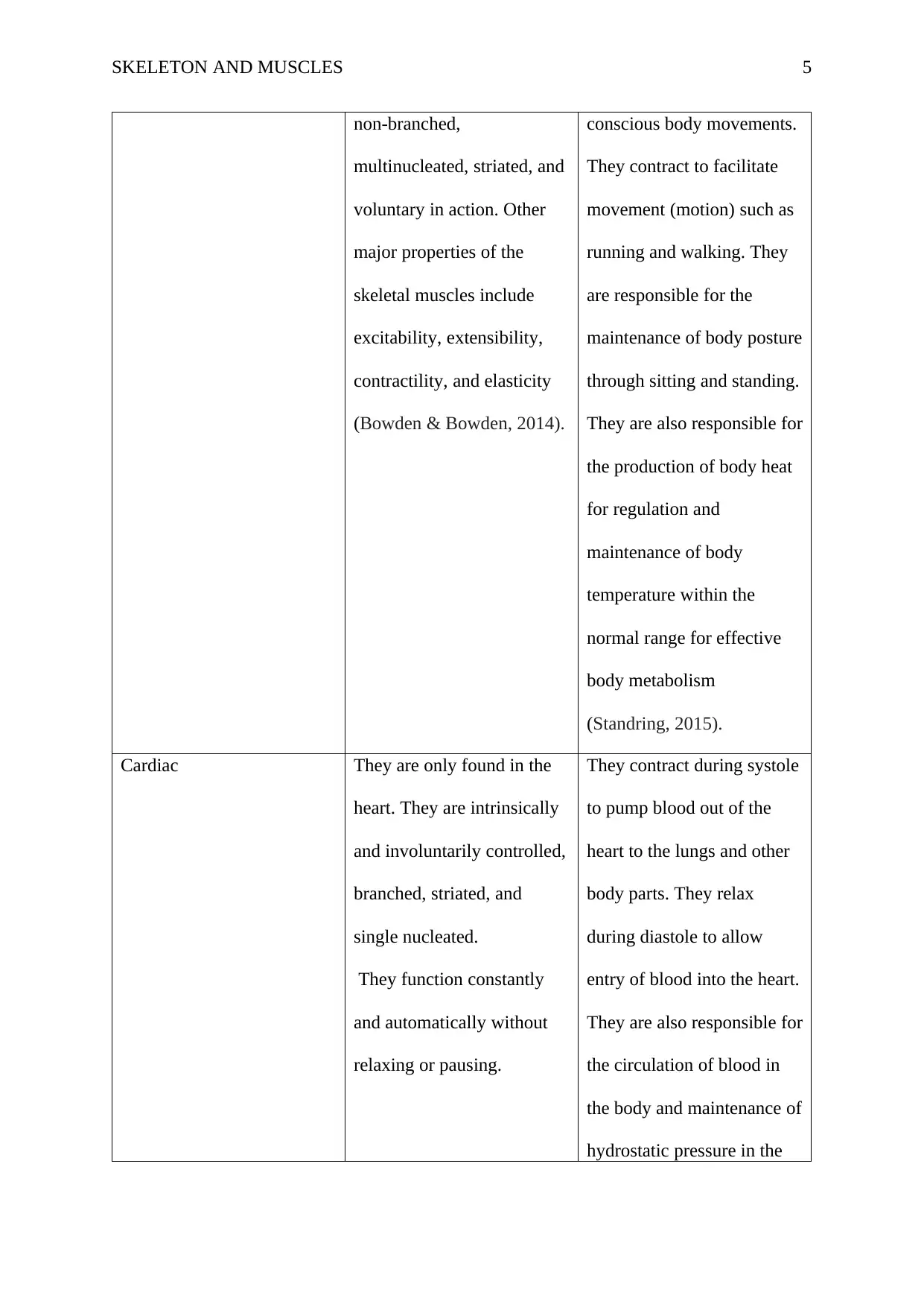
Muscle Type Characteristics Function
Smooth They are found in blood
vessels, organs, bronchioles,
and other hollow organs
such as the stomach and
intestines. Their defining
characteristics include
involuntary control, no
striations, singly nucleated,
and non-branched
(Standring, 2015).
Facilitates urination in the
urinary bladder and pushing
of the fetus in the uterus
during delivery.
Determines arterial blood
flow and blood pressure and
facilitates movement of food
materials in the
gastrointestinal tract.
Striated (Skeletal) The skeletal muscles are They are responsible for all
Secure Best Marks with AI Grader
Need help grading? Try our AI Grader for instant feedback on your assignments.
SKELETON AND MUSCLES 5
non-branched,
multinucleated, striated, and
voluntary in action. Other
major properties of the
skeletal muscles include
excitability, extensibility,
contractility, and elasticity
(Bowden & Bowden, 2014).
conscious body movements.
They contract to facilitate
movement (motion) such as
running and walking. They
are responsible for the
maintenance of body posture
through sitting and standing.
They are also responsible for
the production of body heat
for regulation and
maintenance of body
temperature within the
normal range for effective
body metabolism
(Standring, 2015).
Cardiac They are only found in the
heart. They are intrinsically
and involuntarily controlled,
branched, striated, and
single nucleated.
They function constantly
and automatically without
relaxing or pausing.
They contract during systole
to pump blood out of the
heart to the lungs and other
body parts. They relax
during diastole to allow
entry of blood into the heart.
They are also responsible for
the circulation of blood in
the body and maintenance of
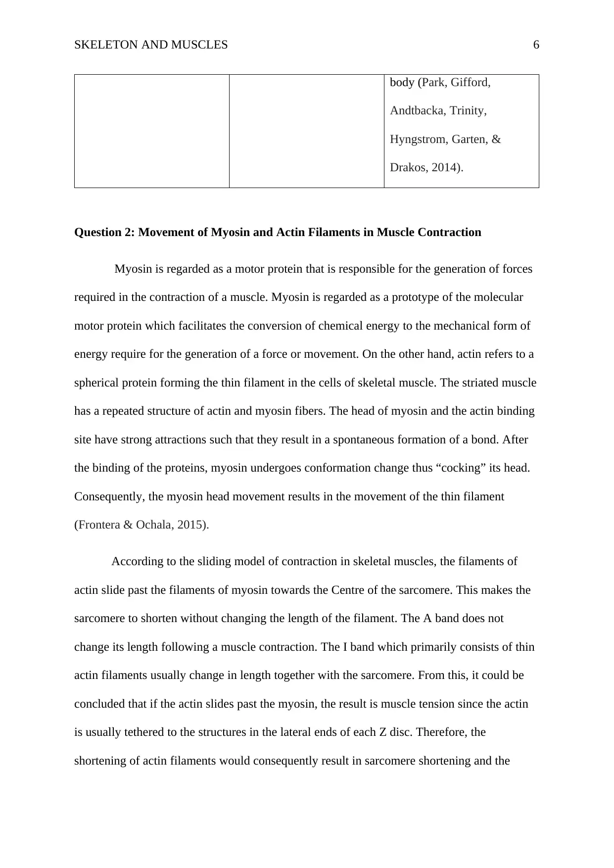
hydrostatic pressure in the
SKELETON AND MUSCLES 6
body (Park, Gifford,
Andtbacka, Trinity,
Hyngstrom, Garten, &
Drakos, 2014).
Question 2: Movement of Myosin and Actin Filaments in Muscle Contraction
Myosin is regarded as a motor protein that is responsible for the generation of forces
required in the contraction of a muscle. Myosin is regarded as a prototype of the molecular
motor protein which facilitates the conversion of chemical energy to the mechanical form of
energy require for the generation of a force or movement. On the other hand, actin refers to a
spherical protein forming the thin filament in the cells of skeletal muscle. The striated muscle
has a repeated structure of actin and myosin fibers. The head of myosin and the actin binding
site have strong attractions such that they result in a spontaneous formation of a bond. After
the binding of the proteins, myosin undergoes conformation change thus “cocking” its head.
Consequently, the myosin head movement results in the movement of the thin filament
(Frontera & Ochala, 2015).
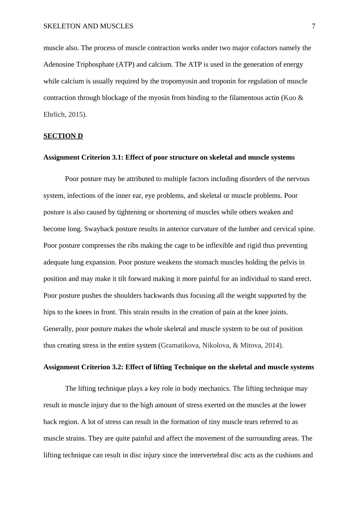
According to the sliding model of contraction in skeletal muscles, the filaments of
actin slide past the filaments of myosin towards the Centre of the sarcomere. This makes the
sarcomere to shorten without changing the length of the filament. The A band does not
change its length following a muscle contraction. The I band which primarily consists of thin
actin filaments usually change in length together with the sarcomere. From this, it could be
concluded that if the actin slides past the myosin, the result is muscle tension since the actin
is usually tethered to the structures in the lateral ends of each Z disc. Therefore, the
shortening of actin filaments would consequently result in sarcomere shortening and the
SKELETON AND MUSCLES 7
muscle also. The process of muscle contraction works under two major cofactors namely the
Adenosine Triphosphate (ATP) and calcium. The ATP is used in the generation of energy
while calcium is usually required by the tropomyosin and troponin for regulation of muscle
contraction through blockage of the myosin from binding to the filamentous actin (Kuo &
Ehrlich, 2015).
SECTION D
Assignment Criterion 3.1: Effect of poor structure on skeletal and muscle systems
Poor posture may be attributed to multiple factors including disorders of the nervous
system, infections of the inner ear, eye problems, and skeletal or muscle problems. Poor
posture is also caused by tightening or shortening of muscles while others weaken and
become long. Swayback posture results in anterior curvature of the lumber and cervical spine.
Poor posture compresses the ribs making the cage to be inflexible and rigid thus preventing
adequate lung expansion. Poor posture weakens the stomach muscles holding the pelvis in
position and may make it tilt forward making it more painful for an individual to stand erect.
Poor posture pushes the shoulders backwards thus focusing all the weight supported by the
hips to the knees in front. This strain results in the creation of pain at the knee joints.
Generally, poor posture makes the whole skeletal and muscle system to be out of position
thus creating stress in the entire system (Gramatikova, Nikolova, & Mitova, 2014).
Assignment Criterion 3.2: Effect of lifting Technique on the skeletal and muscle systems
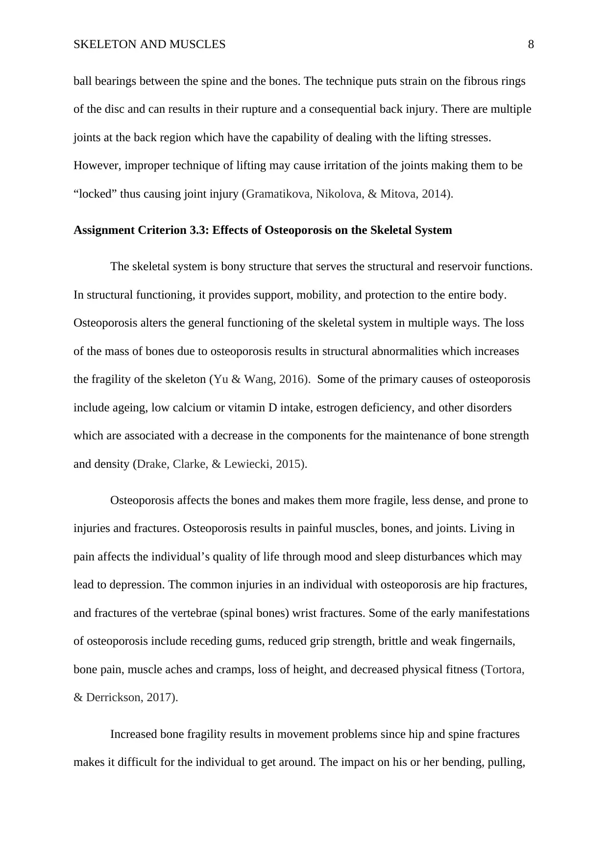
The lifting technique plays a key role in body mechanics. The lifting technique may
result in muscle injury due to the high amount of stress exerted on the muscles at the lower
back region. A lot of stress can result in the formation of tiny muscle tears referred to as
muscle strains. They are quite painful and affect the movement of the surrounding areas. The
lifting technique can result in disc injury since the intervertebral disc acts as the cushions and
Paraphrase This Document
Need a fresh take? Get an instant paraphrase of this document with our AI Paraphraser
SKELETON AND MUSCLES 8
ball bearings between the spine and the bones. The technique puts strain on the fibrous rings
of the disc and can results in their rupture and a consequential back injury. There are multiple
joints at the back region which have the capability of dealing with the lifting stresses.
However, improper technique of lifting may cause irritation of the joints making them to be
“locked” thus causing joint injury (Gramatikova, Nikolova, & Mitova, 2014).
Assignment Criterion 3.3: Effects of Osteoporosis on the Skeletal System
The skeletal system is bony structure that serves the structural and reservoir functions.
In structural functioning, it provides support, mobility, and protection to the entire body.
Osteoporosis alters the general functioning of the skeletal system in multiple ways. The loss
of the mass of bones due to osteoporosis results in structural abnormalities which increases
the fragility of the skeleton (Yu & Wang, 2016). Some of the primary causes of osteoporosis
include ageing, low calcium or vitamin D intake, estrogen deficiency, and other disorders
which are associated with a decrease in the components for the maintenance of bone strength
and density (Drake, Clarke, & Lewiecki, 2015).
Osteoporosis affects the bones and makes them more fragile, less dense, and prone to
injuries and fractures. Osteoporosis results in painful muscles, bones, and joints. Living in
pain affects the individual’s quality of life through mood and sleep disturbances which may
lead to depression. The common injuries in an individual with osteoporosis are hip fractures,
and fractures of the vertebrae (spinal bones) wrist fractures. Some of the early manifestations
of osteoporosis include receding gums, reduced grip strength, brittle and weak fingernails,
bone pain, muscle aches and cramps, loss of height, and decreased physical fitness (Tortora,
& Derrickson, 2017).
Increased bone fragility results in movement problems since hip and spine fractures
makes it difficult for the individual to get around. The impact on his or her bending, pulling,
SKELETON AND MUSCLES 9
pushing, and walking capabilities. Osteoporosis increases the individual’s risk of fractures
due to the weakness of bones which can start crumbling. Hip fractures may affect the way the
person moves and does his or her things since they can result in muscle loss, bedsores,
pneumonia, and formation blood clots in his or her legs (Moreira, Oliveira, Lirani-Galvao,
Marin-Mio, Santos, & Lazaretti-Castro, 2014)
Osteoporosis results in the reduction of bone structure and density especially in the
spongy bone and thinning of cortical bone. This results in a consequential change in the
physical soundness and structure of an individual. Fractures secondary to osteoporosis result
in serious impacts on an individual’s quality of life, happiness and health. Vertebral fractures
result in multiple problems including back pain, height loss, deformities, immobility,
increased hospitalization, and deceased pulmonary functioning (Sozen, Ozısık, & Başaran,
(2017).
Additionally, the vertebral fractures affect the individual’s ability to perform him or
her daily activities of living thus increasing his or her level of dependency. Osteoporosis also
presents with socioeconomic impacts due to high costs of medication and surgical care and
loss of income due to inability to work. Lastly, osteoporosis also affects the home life of the
patient based on its severity. When it is severe, it restricts the person from performing home
chores including mobility, transport, driving, and socialization or interaction with family
members, and friends. To prevent the risk of osteoporosis, some of the appropriate measures
that should be implemented include consumption of varied and healthy diet, avoiding
smoking, and ensuring adequate vitamin D, avoidance of non-prescribed medications such as
corticosteroids, and increased physical activity (Geusens & van den Bergh, 2016).
SKELETON AND MUSCLES 10
References
Bowden, B. S., & Bowden, J. M. (2014). An illustrated atlas of the skeletal Muscles. Morton
Publishing Company.
Drake, M. T., Clarke, B. L., & Lewiecki, E. M. (2015). The pathophysiology and treatment of
osteoporosis. Clinical therapeutics, 37(8), 1837-1850.
Frontera, W. R., & Ochala, J. (2015). Skeletal muscle: a brief review of structure and
function. Calcified tissue international, 96(3), 183-195.
Geusens, P. P., & van den Bergh, J. P. (2016). Osteoporosis and osteoarthritis: shared
mechanisms and epidemiology. Current opinion in rheumatology, 28(2), 97-103.
Gramatikova, M., Nikolova, E., & Mitova, S. (2014). Nature, application and effect of
kinesio-taping. Activities in Physical Education and Sport, 4(2), 115-119.
Kuo, I. Y., & Ehrlich, B. E. (2015). Signaling in muscle contraction. Cold Spring Harbor
perspectives in biology, 7(2), a006023.
Moreira, L. D. F., Oliveira, M. L. D., Lirani-Galvão, A. P., Marin-Mio, R. V., Santos, R. N.
D., & Lazaretti-Castro, M. (2014). Physical exercise and osteoporosis: effects of
different types of exercises on bone and physical function of postmenopausal
women. Arquivos Brasileiros de Endocrinologia & Metabologia, 58(5), 514-522.
Park, S. Y., Gifford, J. R., Andtbacka, R. H., Trinity, J. D., Hyngstrom, J. R., Garten, R. S., ...
& Drakos, S. (2014). Cardiac, skeletal, and smooth muscle mitochondrial respiration:
are all mitochondria created equal?. American Journal of Physiology-Heart and
Circulatory Physiology, 307(3), H346-H352.
Secure Best Marks with AI Grader
Need help grading? Try our AI Grader for instant feedback on your assignments.
SKELETON AND MUSCLES 11
Rahman, M., Cil, A., & Stylianou, A. P. (2016). Prediction of elbow joint contact mechanics
in the multibody framework. Medical engineering & physics, 38(3), 257-266.
Roberts, A. (2014). Human Anatomy: The Definitive Visual Guide. Dorling Kindersley Ltd.
Sozen, T., Ozısık, L., & Başaran, N. C. (2017). An overview and management of
osteoporosis. European journal of rheumatology, 4(1), 46.
Standring, S. (Ed.). (2015). Gray's Anatomy International Edition: The Anatomical Basis of
Clinical Practice. Elsevier Health Sciences. Sokoloff, L. (Ed.). (2014). The joints and
synovial fluid (Vol. 2). Academic Press.
Tortora, G. J., & Derrickson, B. H. (2017). Introduction to the human body. John Wiley &
Sons, Incorporated.
Yu, B., & Wang, C. Y. (2016). Osteoporosis: the result of an ‘aged’ bone
microenvironment. Trends in molecular medicine, 22(8), 641-644.
1 out of 11
Your All-in-One AI-Powered Toolkit for Academic Success.
+13062052269
info@desklib.com
Available 24*7 on WhatsApp / Email
![[object Object]](/_next/static/media/star-bottom.7253800d.svg)
Unlock your academic potential
© 2024 | Zucol Services PVT LTD | All rights reserved.


