Subchorionic Haematoma: Pathophysiology, Diagnosis, and Treatment
VerifiedAdded on 2023/04/21
|6
|1497
|296
Report
AI Summary
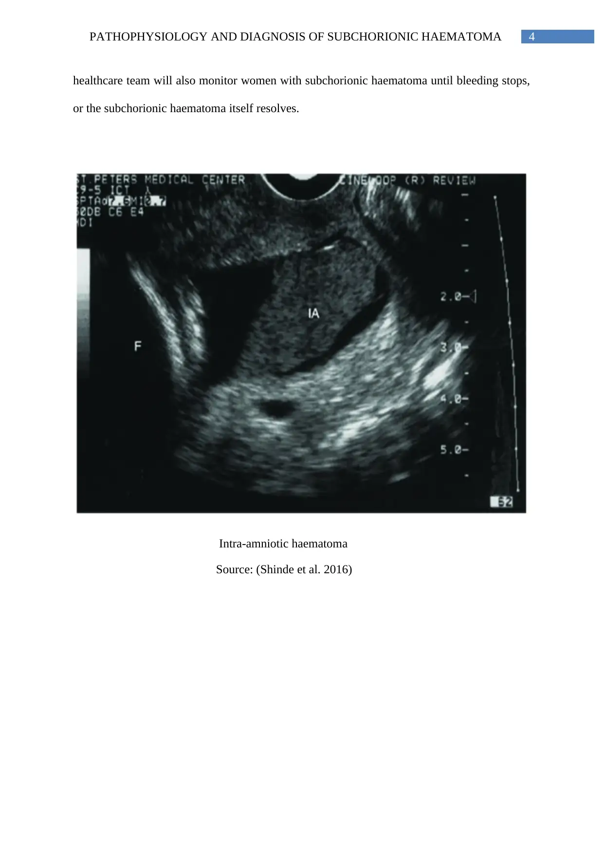
This report delves into the pathophysiology and diagnostic methods for subchorionic haematoma, a common condition characterized by bleeding between the uterine wall and the chorionic membrane during the first trimester of pregnancy. It explores the causes, including poor placentation and disruption of blood vessels, leading to haematoma formation and potential placental detachment. The report details the clinical presentation, including vaginal bleeding and associated symptoms, and emphasizes the role of ultrasound in diagnosis, highlighting sonographic appearances such as hypoechoic lesions. It also discusses potential complications like preterm delivery and placental abruption and touches on the management of subchorionic haematoma, which often involves monitoring and lifestyle adjustments. References to relevant research studies are included.
1 out of 6

Your All-in-One AI-Powered Toolkit for Academic Success.
+13062052269
info@desklib.com
Available 24*7 on WhatsApp / Email
![[object Object]](/_next/static/media/star-bottom.7253800d.svg)
Copyright © 2020–2026 A2Z Services. All Rights Reserved. Developed and managed by ZUCOL.