Synovial Sarcoma in an Athlete: A Detailed Case Study
VerifiedAdded on 2023/06/15
|7
|1303
|476
Case Study
AI Summary
This case study presents the diagnosis, treatment, and nursing care of a 27-year-old female athlete with synovial sarcoma of the left adductor muscle. The patient, C.M., had a history of left leg injuries and presented with a bulge in her upper left thigh. Following a needle biopsy, she was diagnosed with synovial sarcoma and underwent radiation therapy and surgical resection. Post-operatively, nursing care focused on pain management, emotional support, and promoting nutrition. The case highlights the importance of accurate diagnosis and comprehensive treatment for this relatively uncommon tumor, with the athlete ultimately being cancer-free and experiencing only moderate post-operative pain. Desklib provides access to similar case studies and solved assignments to aid students in their studies.

Running head: SYNOVIAL SARCOMA 1
A Case Study on Synovial Sarcoma
Name
Institutional Affiliation
A Case Study on Synovial Sarcoma
Name
Institutional Affiliation
Paraphrase This Document
Need a fresh take? Get an instant paraphrase of this document with our AI Paraphraser
SYNOVIAL SARCOMA 2
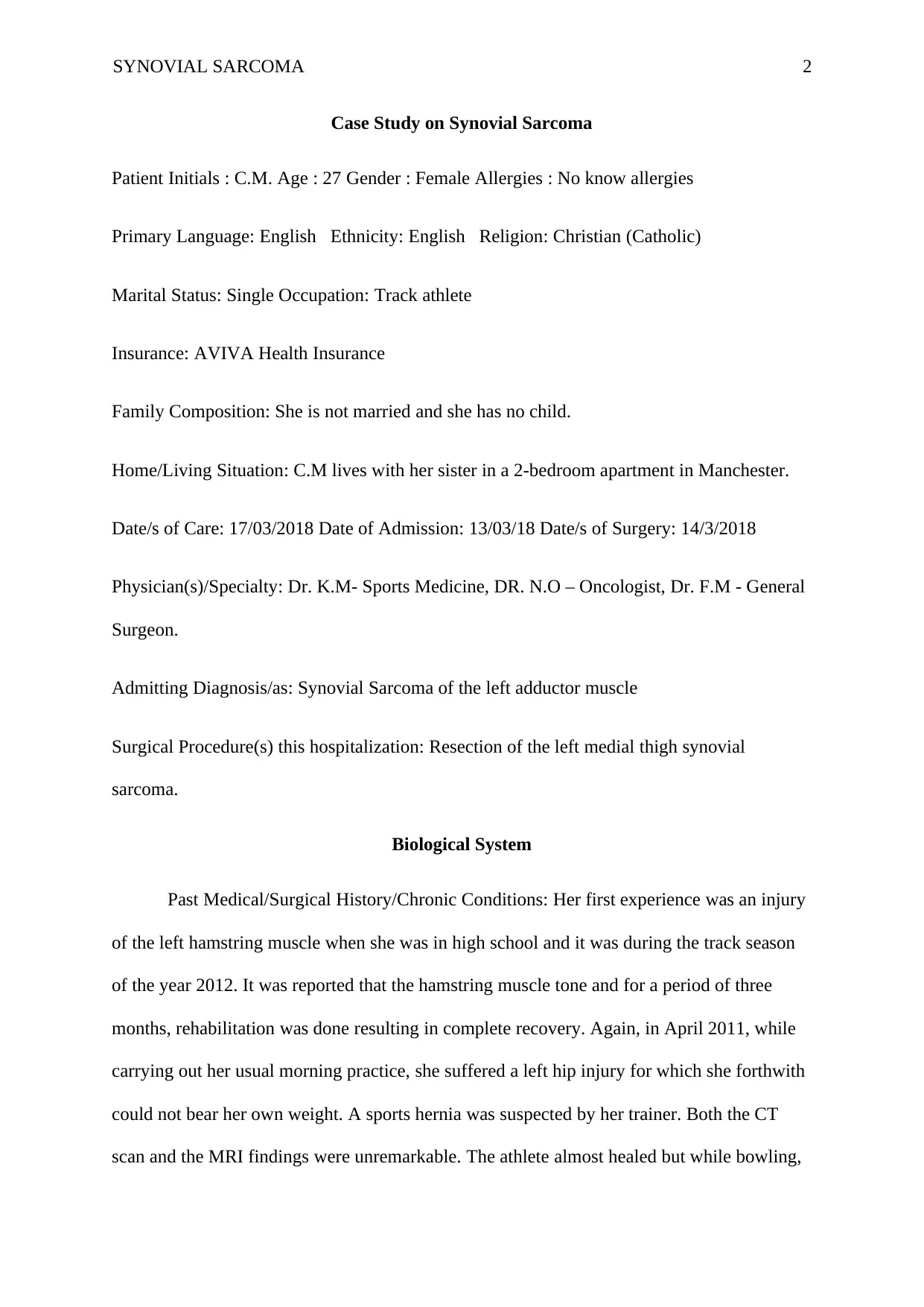
Case Study on Synovial Sarcoma
Patient Initials : C.M. Age : 27 Gender : Female Allergies : No know allergies
Primary Language: English Ethnicity: English Religion: Christian (Catholic)
Marital Status: Single Occupation: Track athlete
Insurance: AVIVA Health Insurance
Family Composition: She is not married and she has no child.
Home/Living Situation: C.M lives with her sister in a 2-bedroom apartment in Manchester.
Date/s of Care: 17/03/2018 Date of Admission: 13/03/18 Date/s of Surgery: 14/3/2018
Physician(s)/Specialty: Dr. K.M- Sports Medicine, DR. N.O – Oncologist, Dr. F.M - General
Surgeon.
Admitting Diagnosis/as: Synovial Sarcoma of the left adductor muscle
Surgical Procedure(s) this hospitalization: Resection of the left medial thigh synovial
sarcoma.
Biological System
Past Medical/Surgical History/Chronic Conditions: Her first experience was an injury
of the left hamstring muscle when she was in high school and it was during the track season
of the year 2012. It was reported that the hamstring muscle tone and for a period of three
months, rehabilitation was done resulting in complete recovery. Again, in April 2011, while
carrying out her usual morning practice, she suffered a left hip injury for which she forthwith
could not bear her own weight. A sports hernia was suspected by her trainer. Both the CT
scan and the MRI findings were unremarkable. The athlete almost healed but while bowling,
SYNOVIAL SARCOMA 3
she suffered a reinjure of the same leg. Two days later, she noted a bulge along the medial
aspect of her upper left thigh. An ultrasound was done on May 21, 2012 and it was concluded
that the bulge was a torn quadriceps muscle.
Reason for Admission: The athlete reported an episodic hip pathology, which did not
show significant improvement with the treatment options provided over a period of one year.
Following a needle biopsy that was performed on 17/10/2017, a diagnosis of synovial
sarcoma of the left medial thigh was made. No distant metastasis to the lungs was indicated
by a chest CT scan. Immediately, the athlete began radiation therapy completing therapy mid-
January 2018 with no other current findings. Following successful radiation therapy, the
athlete was admitted on 13/03/2018 being scheduled for resection of the mass on 14/03/2018.
Procedure: Resection of the left medial thigh synovial sarcoma
I had the opportunity to manage her on her third day post- operative, on 17/03/2018.
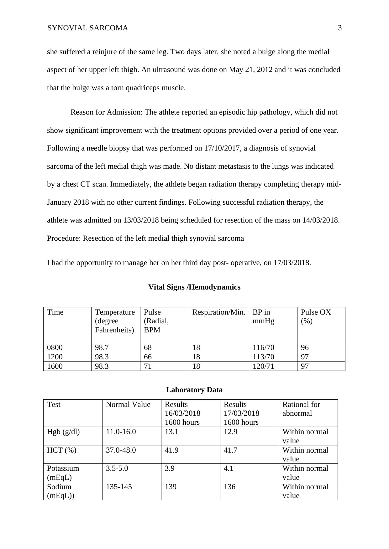
Vital Signs /Hemodynamics
Time Temperature
(degree
Fahrenheits)
Pulse
(Radial,
BPM
Respiration/Min. BP in
mmHg
Pulse OX
(%)
0800 98.7 68 18 116/70 96
1200 98.3 66 18 113/70 97
1600 98.3 71 18 120/71 97
Laboratory Data
Test Normal Value Results
16/03/2018
1600 hours
Results
17/03/2018
1600 hours
Rational for
abnormal
Hgb (g/dl) 11.0-16.0 13.1 12.9 Within normal
value
HCT (%) 37.0-48.0 41.9 41.7 Within normal
value
Potassium
(mEqL)
3.5-5.0 3.9 4.1 Within normal
value
Sodium
(mEqL))
135-145 139 136 Within normal
value
⊘ This is a preview!⊘
Do you want full access?
Subscribe today to unlock all pages.

Trusted by 1+ million students worldwide
SYNOVIAL SARCOMA 4
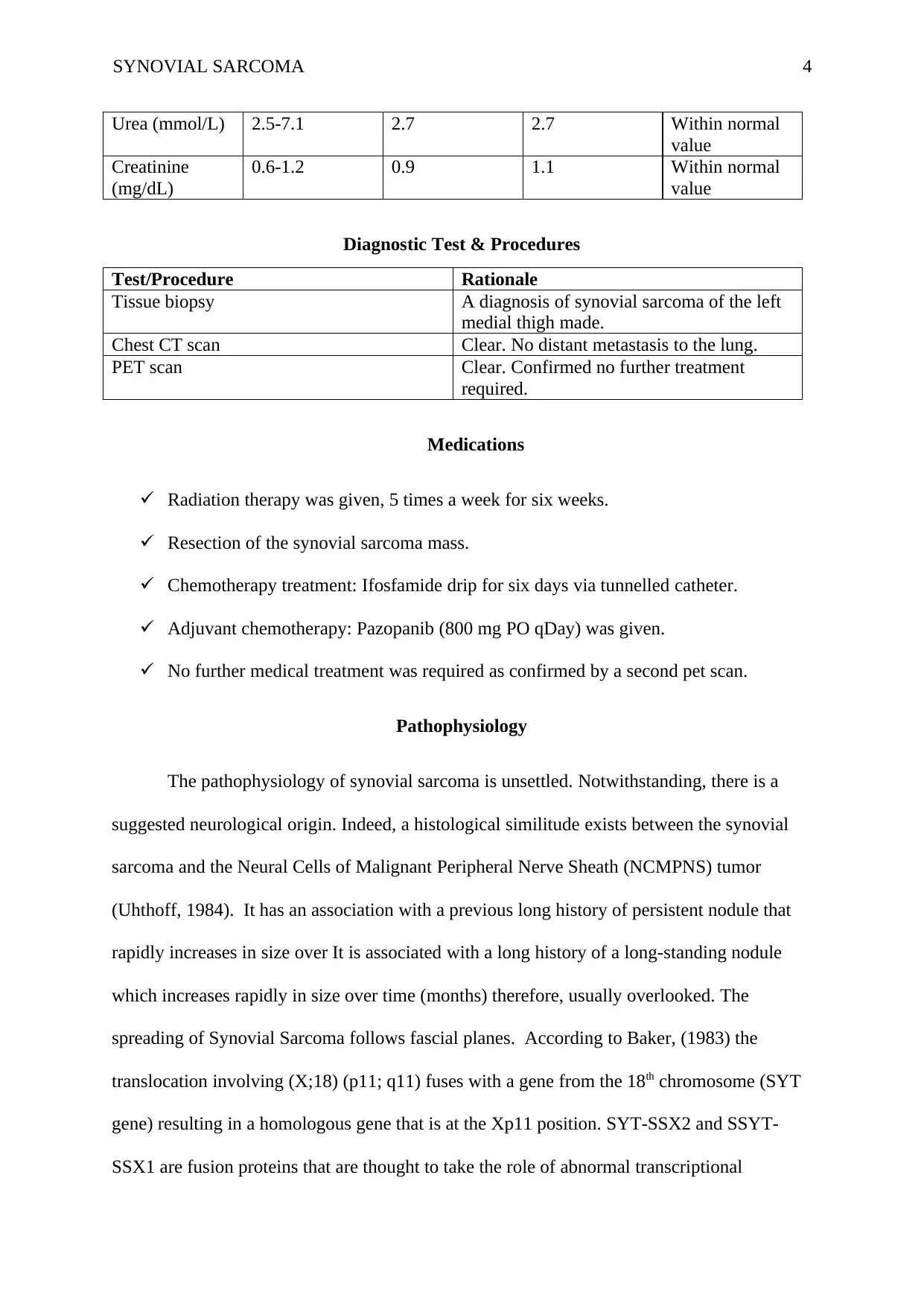
Urea (mmol/L) 2.5-7.1 2.7 2.7 Within normal
value
Creatinine
(mg/dL)
0.6-1.2 0.9 1.1 Within normal
value
Diagnostic Test & Procedures
Test/Procedure Rationale
Tissue biopsy A diagnosis of synovial sarcoma of the left
medial thigh made.
Chest CT scan Clear. No distant metastasis to the lung.
PET scan Clear. Confirmed no further treatment
required.
Medications
Radiation therapy was given, 5 times a week for six weeks.
Resection of the synovial sarcoma mass.
Chemotherapy treatment: Ifosfamide drip for six days via tunnelled catheter.
Adjuvant chemotherapy: Pazopanib (800 mg PO qDay) was given.
No further medical treatment was required as confirmed by a second pet scan.
Pathophysiology
The pathophysiology of synovial sarcoma is unsettled. Notwithstanding, there is a
suggested neurological origin. Indeed, a histological similitude exists between the synovial
sarcoma and the Neural Cells of Malignant Peripheral Nerve Sheath (NCMPNS) tumor
(Uhthoff, 1984). It has an association with a previous long history of persistent nodule that
rapidly increases in size over It is associated with a long history of a long-standing nodule
which increases rapidly in size over time (months) therefore, usually overlooked. The
spreading of Synovial Sarcoma follows fascial planes. According to Baker, (1983) the
translocation involving (X;18) (p11; q11) fuses with a gene from the 18th chromosome (SYT
gene) resulting in a homologous gene that is at the Xp11 position. SYT-SSX2 and SSYT-
SSX1 are fusion proteins that are thought to take the role of abnormal transcriptional
Paraphrase This Document
Need a fresh take? Get an instant paraphrase of this document with our AI Paraphraser
SYNOVIAL SARCOMA 5
regulators. There action leads to either tumour suppressor gene inhibition or
protooncogene(s) activation (Baker, 1983).
Psychological and Spiritual Subsystem
Gulanick and Myers, (2014) poses that normally, in any form of cancer, there is a
usual convoluted process which includes physical, emotional, spiritual, mental and social
responses and by which persons and the people around them embrace an actual, perceived or
an anticipated loss to their own lives. The complex process may be pertinent to a predictable
loss of an individual’s loss of psychological welfare. For instance, the loss of a part of the
body or even change in function. This is usually evidenced by changes in sleep pattern,
libido, activity levels, eating habits and alterations in communication patterns. Chocked
feelings, gross denial and anger are evidence to the grieving process as well.
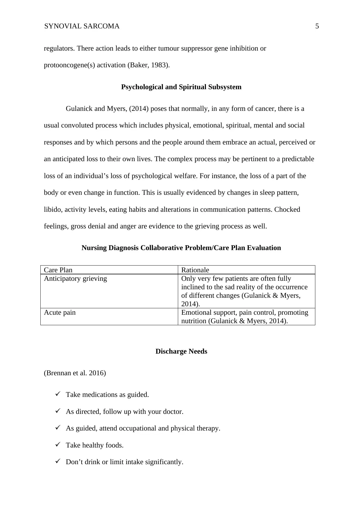
Nursing Diagnosis Collaborative Problem/Care Plan Evaluation
Care Plan Rationale
Anticipatory grieving Only very few patients are often fully
inclined to the sad reality of the occurrence
of different changes (Gulanick & Myers,
2014).
Acute pain Emotional support, pain control, promoting
nutrition (Gulanick & Myers, 2014).
Discharge Needs
(Brennan et al. 2016)
Take medications as guided.
As directed, follow up with your doctor.
As guided, attend occupational and physical therapy.
Take healthy foods.
Don’t drink or limit intake significantly.

SYNOVIAL SARCOMA 6
Conclusion
Generally, synovial sarcomas are comparatively uncommon tumor entities. It is easy
to mistake the different type of sarcomas for a number of reasons, including similarity in
histology. C.M- the athlete is now free of cancer. So far, she has since suffered no
complication save for pain that is moderate in severity along the site where surgery was done.
⊘ This is a preview!⊘
Do you want full access?
Subscribe today to unlock all pages.

Trusted by 1+ million students worldwide
SYNOVIAL SARCOMA 7
References
Baker, L. H. (1983). Soft tissue sarcomas. Boston, MA: Springer US.
Brennan, M. F., Antonescu, C. R., Alektiar, K. M., & Maki, R. G. (2016). Management of
Soft Tissue Sarcoma. Cham: Springer International Publishing.
Gulanick, M., & Myers, J. L. (2014). Nursing care plans: Diagnoses, interventions, and
outcomes. Philadelphia: Elsevier/Mosby.
Uhthoff, H. K. (1984). Current Concepts of Diagnosis and Treatment of Bone and Soft Tissue
Tumors. Berlin, Heidelberg: Springer Berlin Heidelberg.
1 out of 7
Your All-in-One AI-Powered Toolkit for Academic Success.
+13062052269
info@desklib.com
Available 24*7 on WhatsApp / Email
![[object Object]](/_next/static/media/star-bottom.7253800d.svg)
Unlock your academic potential
Copyright © 2020–2026 A2Z Services. All Rights Reserved. Developed and managed by ZUCOL.