Ask a question from expert
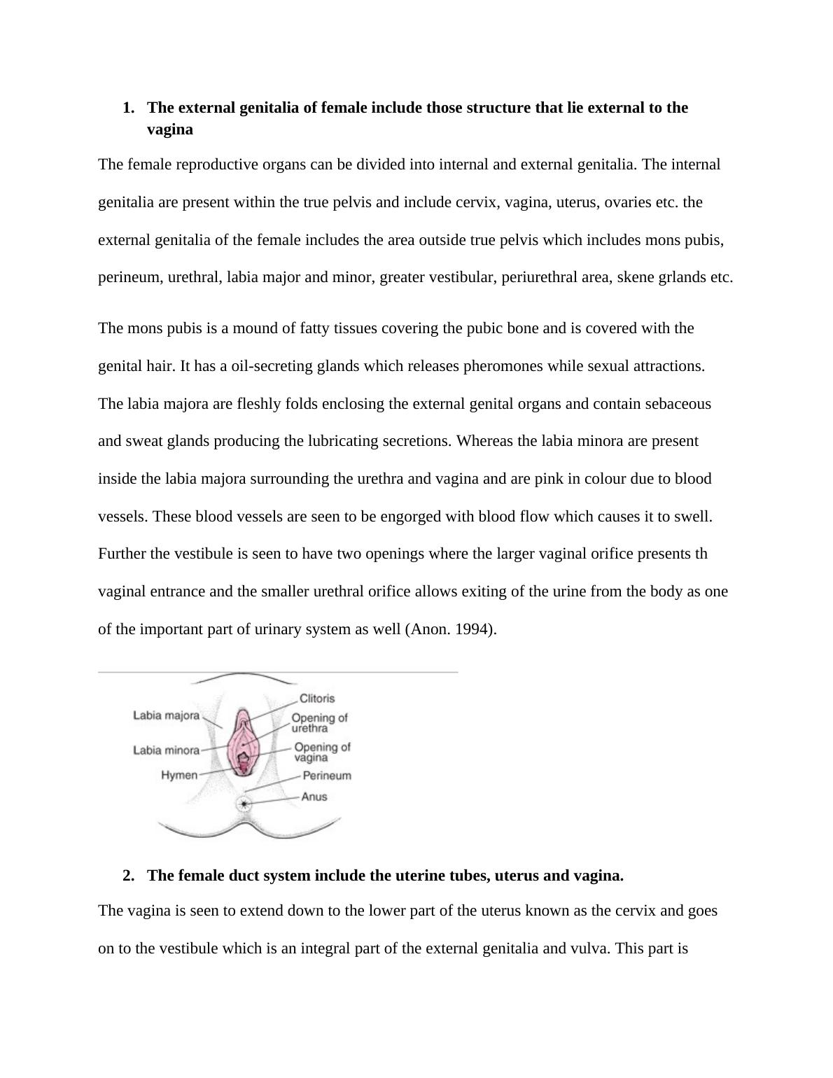
Female Reproductive System: External Genitalia, Duct System, Ovaries, and Mammary Glands
5 Pages928 Words413 Views
Added on 2019-09-20
About This Document
This article discusses the female reproductive system, including the external genitalia, duct system, ovaries, and mammary glands. It explains the functions of each part and their importance in reproduction.
Female Reproductive System: External Genitalia, Duct System, Ovaries, and Mammary Glands
Added on 2019-09-20
BookmarkShareRelated Documents

End of preview
Want to access all the pages? Upload your documents or become a member.
Biology Assignment: Human Reproductive System Assignment
|12
|2089
|625
Structure and Function of Male and Female Reproductive System
|2
|1386
|213
Study On Reproduction - Human Reproduction System
|8
|1454
|49
Human Reproductive System and Gestation in Humans
|10
|2337
|433