Hip Implant Micromotion and Failure Analysis
VerifiedAdded on  2020/07/22
|31
|14473
|325
AI Summary
This assignment delves into the complex interactions between bone, implant, and fibrous tissue in hip replacements, focusing on micromotion's role in fibrous tissue production. It discusses various surgical approaches, patient-related factors influencing implant success, and the analysis of hip replacement failure based on the Swedish hip arthroplasty register.
Contribute Materials
Your contribution can guide someone’s learning journey. Share your
documents today.
Medical Science and bone
Replacement
Replacement
Secure Best Marks with AI Grader
Need help grading? Try our AI Grader for instant feedback on your assignments.
Introduction
Total Hip Replacement (THR) or Arthroplasty is the medical procedure wherein
damaged cartilage and bone in the body is replaced with the prosthetic components. It is one of
the successful surgical procedures in the field of orthopaedic surgery. Patients who are suffering
health issues due to osteoarthritis, congenital deformities, avascular neurosis, rheumatoid
arthritis and post-traumatic disorder can get over rid of pain through hip joints restoration. THR
not only helps to reduce pain but also helps patients in better sleeping which helps in improving
their quality of life.
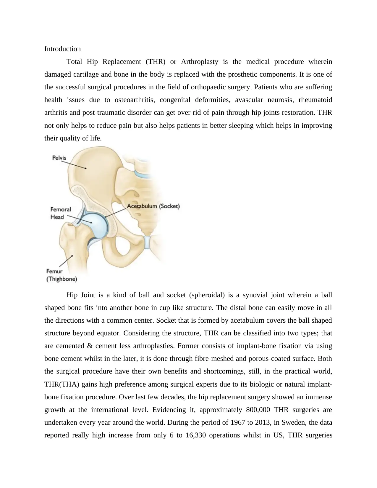
Hip Joint is a kind of ball and socket (spheroidal) is a synovial joint wherein a ball
shaped bone fits into another bone in cup like structure. The distal bone can easily move in all
the directions with a common center. Socket that is formed by acetabulum covers the ball shaped
structure beyond equator. Considering the structure, THR can be classified into two types; that
are cemented & cement less arthroplasties. Former consists of implant-bone fixation via using
bone cement whilst in the later, it is done through fibre-meshed and porous-coated surface. Both
the surgical procedure have their own benefits and shortcomings, still, in the practical world,
THR(THA) gains high preference among surgical experts due to its biologic or natural implant-
bone fixation procedure. Over last few decades, the hip replacement surgery showed an immense
growth at the international level. Evidencing it, approximately 800,000 THR surgeries are
undertaken every year around the world. During the period of 1967 to 2013, in Sweden, the data
reported really high increase from only 6 to 16,330 operations whilst in US, THR surgeries
exceeded 285,000 surgeries per year however, total fractures reported by the end of year 2040 is
forecasted to go beyond 500,000. Australia reported 46.50% in the hip joint surgeries from 2003
and also projected the rate will go up in the forthcoming period. On the other side, in England &
Wales, National Joint Registry stated 620,400 THR between the period of 2003 to 2013, whereas
osteoarthritis reported 93% case. In this, the cement-less THR surgery reported increase from
16.8% to 42.5% with the decline in cemented THR from 60.5% to 33.2%. In India, National
Institute of Arthritis reported 470,000 incidence rate per year. Out of these, 65% of the reported
surgeries have been undertaken through cement-less. The surgical procedure reported a good
progress and success in the past few decades at a failure estimation of 10%. The failed cases is
founded in the first revision surgery which requires revision surgery after 10 years taking into
account patient health conditions & implant used. Although, there are multiple of reasons behind
such failure, still, most of the cases reported bio mechanical reasons for the same. Implant
indulged adverse remodeling & excessive implant interface stress, which in turn, leads to
progressive interface debonding that have been discovered as two most important bio mechanical
reasons for failure that compromised cement fewer prostheses' durability. Besides this, lack of
stability due to excessive micro-motion has a strong influence over the cement-less arthroplasties
which compromises biological attachment that took place between femur and implant.
The mechanism's cumulative or individual effect may result in gross aseptic loosening of the
implant or in cases of extreme scenario, femur fracture.
Keeping aside key aspects, including surgical procedure and patient conditions, the femoral
implant 's design or geometry is also known to be effective on these mechanisms (Huiskes and
Boeklagen, 1988; Viceconti et al., 2001).
Although a wide variety of cement-less femoral implants have a commercial availability in the
market (Fig. 1.1), for maximum of those the outcomes of design remain to be unexplored,
primarily due to a lack in the clinical data. The mechanical designs of hip implant, thus, can be
assessed preclinically and search for the optimal geometry may be conducted for the
minimization of the effects of all the failure in the mechanisms.
The solutions of the design of a prosthesis may be either structural or functional, or both.
However, investigations of the prosthesis have been centralised mostly on the structural aspects.
Considering the various structural aspects of hip prosthesis, its is determined that geometry of
the femoral implant plays a critical role in the outcome of design. Although the gross appearance
of a femoral stem has changed hardly since it was mentioned for the first time (Gluck, 1891), the
overall shape being non-primitive hence offers a lot of scope for a complex study by profile
alteration of stem transverse sections along the stem-length by usage of the state-of-the-art solid
modeling process and finite element (FE) analysis software.
Moreover, with the arrival of the technically advanced high-performance computers, large
number of stem shapes may be assessed in a relatively small period, and one which is most
suited may be selected on basis of its predicted outcomes of design. Therefore, a combination of
suitable design optimization strategy with a solid modeling and FE analysis may result in a more
improved prosthesis design.
Optimization of shape (or geometry) is said to be a particular stage in structural optimization,
which deals with the search of the design domain with optimal configuration of it. (Fraternali et
al., 2011). In optimization of shape, introduction of design variables is done to control the
geometry of the structure and an FE model is required for methodology that changes during
optimization process. The growth in interest of optimization of shapes reflects a realization made
by the effectiveness of changes in shape for improvement in the structural performance. By
employing shape optimization as a design tool, evaluation of stem geometries can be done based
on bio-mechanical cost functions, framed on failure principles taken as a base. Therefore, a
suitable cost function must be formed, representing the effects on global level of these failure
mechanisms. It is necessary in order to search for optimal designs of the cement-less femoral
implant that would enhance prosthesis durability. However, for a priori and understanding the
failures associated with THA practically, a study on the hip joint's bio-mechanics is required.
1.2.1 Anatomical planes and directions
Hip joint bio-mechanics may form a basic understanding of the human anatomy. Typical
orientations of the human body of different anatomical planes are presented in the Fig. 1.2a. The
transverse (axial or horizontal) plane which is noted to be parallel to the ground, apart the
superior (top) from the inferior (bottom) part of the human body. The coronal (frontal) plane,
with perpendicular anatomy towards the ground, separates the anterior (front) from the posterior
(back).
The sagittal (median) plane is noted to be perpendicular to the transverse and coronal plane, and
it separates the left part of the body from the right part. The medial and lateral directions refers to
the direction towards and away from the mid-line of the body respectively (Fig. 1.2b). The
direction towards the front and the back side of the body are termed as The ‘anterior’ and
‘posterior’ directions respectively. The term ‘proximal’ describes the direction which is towards
the limbs origin, while the part which is away from the origin of the limbs is known as the
‘distal’.
This clinical definition done is based on their proximity with their respect to the head. The
‘superior’ and ‘inferior’ directions point towards the top and the bottom parts of the body,
respectively.
Fig. 1.2:
1.2.2 Hip anatomy
The hip joint forms the primary connection between the bones of the lower and upper limbs of
the human skeletal system, which is scientifically referred as the acetabulofemoral joint. The
hip-joint is said to support weight of the body and transfer load from upper limb to the lower
limb. A ball (femoral head) and a hemi-spherical socket (acetabulum) (Fig. 1.3) form the main
parts of this joint.
Secure Best Marks with AI Grader
Need help grading? Try our AI Grader for instant feedback on your assignments.
Fig. 1.3:
The femoral head is located at the top of the thigh bone (femur) and it is made to fit into the
acetabulum in the pelvis by means ligaments (hip capsule), a band of tissue. The function of
ligaments is also to provide stability to the joint.
The movement process is facilitated by a smooth durable cover of articular cartilage (a protein
substance) which cushions the ends of the bones between the two bones ‘surfaces (femoral head
and acetabulum).
The hip joint's remaining surface is covered by a thin, smooth tissue called the synovial
membrane. In a healthy hip, this membrane's function is to generate a small amount of fluid that
lubricates and improves frictional resistance in the hip joint. All of these parts of the hip-joint
work in simultaneously, allowing easy, painless moving of hip.
1.2.3 STRUCTURE OF FEMUR
It is the longest and stronger bone in the human skeletal system. Body weight is
supported by the femurs during many activities like running, jumping, walking, standing etc. It
consists two part, the primarily part is diaphysis or central shaft and two wider and rounded
bulges known as epiphyses (figure.1.4). Each epiphysis attach with diaphysis through conical
regions known as metaphysis. The diaphysis is generally belonged to hard cortical bone with a
small spongy core while the epiphyses and metaphysis contain mostly cancellous or spongy bone
within a thin shell of cortical bone. A head, a neck, a greater trochanter and lesser trochanter are
the nearest part of the femur the transition area between the head and neck is quite rough due the
attachment of muscles and hips joint capsule.(figure.1.4).
The head is conducted upward, medial-ward, and a little ahead and it designs 2/3 of a
sphere and surface of it is smooth and cover with ossein tissue in the fresh condition excluding
depression, clinically known as fovea capitis femoris. The flattened pyramidal part of the bone is
neck which connecting the head with femoral shaft and assemble with the recent wide corner
opening medial ward. The angle of affection of neck to the pole in the front of plane is known as
neck-shaft angle. The neck jutting a little ahead due to change in femur body by projecting
upward and middle side. The angle of inclination of the neck shaft in cross plan is also known as
the angle of interversion. Femur bone of neck has an irregular cross-section and spheroid at the
upper end and approximately ovoid with major and minor axes in the ratio about 1.6 at the lower
end which is close to femoral shaft.
The body of femur is long, slender and almost cylindrical in form. It is little arched, to be
convex in front, and concave behind, where it is strengthened by a prominent longitudinal ridge.
Proximal the lateral ridge of the linear aspera becomes the gluteal tuberosity while the medial
ridge continues as the pectineal line. The shaft has two other bordes one is lateral and other is
medial border. Due to the vast musculature of the thigh the shaft can not be palpated.
The femoral neck suffering regressive changes because of declining. The upper part of neck-
shaft provides attachment sites for number of muscles and build most palpable part of the femur
situated at the junction which torchanter is large, irregular, quadrilateral eminence. It dirtected
the mini part which is about 10 mm lower than the head in adult. The minor trochanter is a
conical eminence on the medial side of the femur (Fig.1.4)
2.2.1 Human gait cycle
It is the way of motion which is achieved by power of body, while maintaining harmony
with the help of human limbs. Nature of gait follows common pattern when it varies from
individual to individual. It is bipedal and biphasic. The two phases of gait cycle which are the
stance phase and swing phase and this both are distinct and interconnected phase and this phases
can further subdivide into 8 different phases as described in terms of percentage of each gait
cycle. 60% of normal walking cycle in stance phase in this the foot remains in contact with the
ground that means it begins from heels and touch the start of foot.
The right foot comes in flat contact with the ground before the heels rises. Lifting the toe
off the ground marks to the end in the stance phase. The remaining 40% is known as swing phase
in this foot moves in the air during the right swing phase and the left legs solely supports the
body. This phase ends with the heel contact and the cycle repeats itself and vice versa. The
duration between feet and ground up to remain in contact is known as double support. A typical
cycle is presented in fig.1.5.
3rd combined musculoskeletal and finite element modelling.
2.REVIEW OF THE HIP JOINT
2.1. Anatomy
The joint of hips made with the ilium, ischium, and pubis bones and the femur which is a
ball and socket comprising Figure 1. The femoral head act as a ball within the socket of the
establishes and formed at the joint between the three pelvis bones. This allows the to all three
rational degrees of freedom ans restricted only by the capsular ligaments and the depth of the cup
Figure 1.
The joint of the hips rotate in all three axes but does not allow translation between the
femur and pelvis. Fort the purpose of this study the y-axis lies parallel to the length of the femur
shaft with the z-axis lies at 90 degree from femoral head to the greater trochanter and the axis is
the product of the two other axe.
2.2 Osteology
The long bones have the unlike properties and the young modulus have an imaginary line
of 17.4GPa approximately, while at an angle of 90° the imaginary line is about 11.7GPa
approximately. The structure of the bone is divided into two types Cortical and the Cancellous
which are not homogeneous. The majority of central part of the long bone is made by compact
bone or by the cortical bone and the outer shell is at the end part of the bone. The lower density
spongy bone i.e. the Cancellous bone makes the major end part of the bone i.e. epiphyses. The
spongy structure of Cancellous bone is made up of bar forming a framework of the bones which
is known as the trabeculae. These trabeculars are in very close contact to the bones and the
internal blood supply.
2.2.1. Bone properties
Cortical bone which is also known as Compact bone or Lamellar bone. The extremely
hard exterior is formed by the cortical bone which has a density of 1700-2100 kg/m-3
approximately. Cancellous bone which also known as spongy or trabecular bone is full of tiny
holes and has two types of bone tissues with highly changeable nature, its density can fall up to
50 kg/m-3 and are found in human body. Through some studies it has been noted that it is
difficult to differentiate between both Cortical bone with a low density and Cancellous bone with
a high density, the density of the Cancellous bones have the similar type of tiny holes as in
Cortical bones which other studies disagrees. With the growing age the density and strength of
the bone increases till the maturity of the bone which is till the age of 35 years and then starts
declining. The bones have the adaptive properties and can change them according to its density
and maturity. The general relationship of density of the bone and modulus density , i.e. the
density of the bone (p, g/cm-3) is proportional to its modulus (E, GPa) is described in the
equation 1.
Equation 1
The cubic relationship between the modules and the density was found by Carter and Hayes in
Paraphrase This Document
Need a fresh take? Get an instant paraphrase of this document with our AI Paraphraser
(1977). Rice et a. (Rice et al. 1988) a slightly better correlation was found with a squared
relationship and also it was found in other studies the power, p, is slightly less than 2
(Hodgskinson and Currey 1992 ; Morgan et al. 2003). According to the equation 2 it was also
found by Carter and Hayes that the modulus – density was marked for a effect by the strain rate.
Equation 2
While it should be noted that this is one of the many relationships to found be between modulus
and density. A large degree of variation between studies was found between variation and studies
when a review of the potential relationship of modulus and density of bone was conducted by
Helgason et al. 2008)
The dependency of the bone density has been shown to depend on the tensile strength and yield
strength of the bone (Figure 4) (carter and Hayes 1977; Kopperdahl and keaveny 1998; cowin
2001). The bone strength is considered to be dependent on its density, whereas the yield strain is
noted to be independent of elastic modulus, yield stress and density (Cowin 2001; Morgan and
keaveny 2001), however a variation has been reported in the vaiues of yield strain. Across
severai anatomicai sites it researches conducted by kopperdahl et al. (1998) the yield
compression for cancellous bone was found to be 8400pe (± 1000 s.d.) and between 7000pe
(±500 s.d.) and 8500pe (±1000 s.d.) by Morgan rt al. (2001). kopperdahl et al. found the yield
strain of cancellous bone to be 7800pe and for Morgan et al. it varied between 6100pe (±500 s.d.)
and 7000pe (±500 s.d.) in tension. In Ebacher et al. (2007) research it was found that cancellous
bone is more ductile than cortical bone with an Ultimate strain of approx 10000-15000pe. The
cortical bone was found to be having a yield strain of approx 4000pe in tension and yield strain
was higher and more variable in compression than in tension, between 6500pe and 10000pe.
Figure 4
Strain gauges were attached to human tibia by Lanyon et a. (1975) to measure the surface strain
during normal walking. A variation was noted in the surface strain through gait cycle from 640pe
(±70) to 2370pe (±180) in tension and it peaked in the strain when the foot left the floor. The
compressive strain measured a hike as the load carried by the subject was increased and strain
hiked to highest when the subject was seen running on a treadmill 8470pe (±590). the bone on
the surface of tibia is cortical bone during the walking process the strain noted was lower than
the yield strain. In an investigation Keyak and Rossi (2000) investigated which failure criteria
could most accurately predict the failure of bone using finite element models. A finite element
model was taken to compare the experimental results of different failure criteria which included
maximum and normal strain, stress and shear strain. The predictions made by all the failure
criteria included the load which caused femoral fracture in the vitro, when using different loading
scenarios the shear strain and hoffmann criterion was found to be robust. Schileo et al. (2008)
also compared failure criteria; von Mises stress, maximum principle stress and principal strain.
The failure of bone was found in all three criteria at the location of fracture. When a compressive
principle strain limit 10400pe nad tensile limit 7300pe was used, maximum principle strain
criteria had a prediction of localised failure, in the femur, which corresponded to experiments.
Wolff's law states that in response to mechanical stimulus the bone density and orientation of
trabeculae changes. Remodelling of bone adapt is based on Wolff's law and is used to predict
where bone will be deposited due to stress stimulus. The density in the bone is increased by
deposition and the bone is resorbed In area of low stress and density lowered (figure 5). between
the threshold values high and low there is lazy or dead zone in which change in stress rate doesnt
change the bone. It was found by Beaupre et al. (1990) that the DSS was approximately
50MP/day which generates a cyclic normal strain of approx 400pe assuming 10000 walking
cycles a day. Based on the adaptive model in fig 5 using 20% stress stimulus as the width of lazy
zone a calculation was on done on change in bone density and predicted a bone density
distribution consistent with that found in vivo.
Figure 5
Myology
2.3.1.Structure
Ligament, a fibrous tissue that connect bones together, and muscle are responsible for movement
and control in the body. The whole of the joint of hip ligament is covered by capsular ligament.
Movement of the skeleton is created by skeletal muscle. Perimsysuim (figure 6) is a collectively
wrapped sheath of muscle belly fibers bundled together. Tendons are made to provide connection
between the muscle belly and the bones on which they act. Spiral cord contain neurons tha fire
electrical impulses down the pathways called axons to muscle fibers. There can be increase on
muscle fibers when additional motor units are activated. (Whiting and zernicke 1998).
Figure 6/ Figure 7
Twenty muscle cross the hip joint approx (Figure 7). All these muscles have different attachment
points in the skeleton which allows different actions and muscles functions to take place Table 1.
Normally a skeleton muscle is attached to bone in the body by tendons minimum two points,
known as origin and insertion points. The origin point end is attached to a relatively stationary
part of the body muscle. The area which is moved by contraction muscle is called insertion point.
It is noted that a muscle may not always have two specific points that they are attached to, many
muscles have large area on the bone on to which the tendon is attached. Such a muscle is gluteus
maximus.
Figure 7
2.3.2 Generation of muscle Force
Figure 8
The muscle strength is directly related to the intellectual cross sectional area of muscle
and alignment of muscle fibres. Further, the muscle fibre direction is vertical to the cross
sectional area of muscle. Moreover, there are two elements of muscle tension which are, the
active tension and the passive tension. Whereas, the active tension is produced by the activity of
muscle fibre while the passive tension is generated by the physical lengthening of tendon and
muscle. Therefore, when the length of tension increases by the force of muscle it leads to
increase the strength of muscle and then continuously decrease the muscle extension till there is
an increase in passive tension. The muscle force is also equivalent to the velocity of the
contraction. Hence, the Hill’s muscle model related to velocity and force was experimentally
figured by Bressler and Clinch (1974) using the sartorii muscle from a toad.
Figure 9:
The result of force vector for a muscle can be analysed as the line of action which is
affected by the orientation of the fibres in the muscle and in the routes of the muscles presented
in the human body. The path of muscles in the body in not the direct route to joints instead it is
interrupted by the soft tissues or by the part of bones. Moreover, it can be said that when the
skeleton of body moves there are chances that the blocked parts of the body change its position
Secure Best Marks with AI Grader
Need help grading? Try our AI Grader for instant feedback on your assignments.
with the relative changes in muscle which will affects the muscle's path. Besides, the Pennate
muscle in the body are made up of fibre which do not run in one line and further fluctuates the
strength of muscle. The angle made between the line of action of muscle and fibre is termed as
Pen nation angle. Therefore, the angle is used to compare the length of muscles fibres which is
used in a Hill type muscle model to calculate the strength of the muscle.
2.3.4. Muscle injury and repair
Muscles injury in minimizes the ability to produce force and during hip arthroplasty
muscles can be divided or dissected. Muscle lacerations heal but can be rebuilt with the use of
dense connective scar tissue instead of muscle tissue which minimizes the functions of muscle.
Lacerations which intersect the muscle fibres are the most harmful type of laceration for
recovery of muscle, specially when they dissect blood supply to areas of the muscle. Further,
there are three stages of muscle recovery; degeneration and inflammation, regeneration and
fibrosis. Hence, in fibrosis stage scar tissue is formed which minimizes the strength of the healed
muscle. Generally, this stage starts in two to three weeks after it is injured and it takes more than
6 weeks for normal functioning.
Besides, the mechanical and biological properties of a muscle can be identified to
investigating how the muscle is affected like by, laceration and healing. The test to is to find the
ways to recover from muscle injury, various methods used by doctors recovery of muscles after
laceration and also it will evaluate the weakest muscle in the skeleton. The elongation of the
healed muscles was measured by applying pressure to injured muscle. Scholars found a
minimizing technique in both type of lengths of muscle, the one which is injured and the non
operated muscle which is also termed as contralateral leg. Further, the failure load is identified
by recovering only to approximately 50% of the contralateral muscle which is attributed to the
atrophy of the muscle instead of damage caused by the laceration. Moreover, the minimization in
elongation is related to the scar tissue which is formed at the laceration site as the scar had a
higher elastic modulus.
In contrast to the fluctuation mechanical properties the biological response of a muscle
can also be affected by injured muscle. According to researcher the force of muscle can be
minimized by some injury so repair methods used by Huard et al., (2002) to measure the
strengths of muscle. Moreover, Garrett and Duncan (1988) used the extensor digitorum muscle
of the New Zealand white rabbit to identify the impact of full and partial laceration on strength
and shortening ability of muscle. It was examined that the partial cut of 50 to 75 percent and the
full dip cut along the width of the widest section of the belly muscle is made. Further, when the
muscles were allowed to heal for 3 months in order to provide muscles the ability stiff and
shorten. Garrett and Duncan stated that full deep cut only recover by 54 percent of strength while
the partial cut regain 62 percent of control. The muscle ability of shortening is also affected by
the cutting method because that minimizes the stiffness. In contrary partially cut muscles attain
80 percent of stiffness and control. Hence, it was examined that the proximal section that is the
origin point and cut point performs the similar functions of healing. Further, due to the limited in
vivo data on shortening ability of the sections of full wide cut in muscle has not been included
into models assuming muscle strength.
Crow et al., (2007) has carried a study over rabbits to examine several repair methods of
dividing the belly muscle in the extensor digitorum longus. Scholars compared various methods
of recovering the dip cut in muscle by testing the force produced by electric stimulus or by
performing tensile test. Further, the electrical stimuli chosen by the researchers needed twice and
ten times the threshold voltage to establish involuntary contraction between muscle. Moreover,
scholars found that using ten times the threshold, 1 0T, after 3 months of healing sutured muscles
had majorly 75% of the muscle strength compared to control. However, when the scholar tested
twice the threshold, 2T, gave only 40% of the strength of the control over healed muscles.
Hence, the study states that the minimization of strength on injured muscle is not constant over
the pressure on muscles and that is the reason why lacerating muscle is unable to recover full
strength. Henceforth, the muscle which were ignored and left unpaired after 3 months at 10 T
attained 61 percent over the control muscle strength and at 2T the unrepaired muscle had greater
strength of approximately 56% of the control.
Therefore, muscles that get injured do not recover their full strength when cured. Various
factors that include the way of improvement and the level of damage determine the strength of
wounded muscle. Partially healed muscle have more strength as compared to completely
compound muscle and it is possible that the part of body that is effected by partial wound might
also affect the whole strength. However, no literature studies have been found that compare
different levels of laceration and therefore the variation in muscle strength caused by different
quantities of laceration cannot yet be determined. During hip arthroplasty surgery many of the
muscles are divided along the line of the muscle fibers rather than lacerated. This will affect the
muscles differently to laceration across the body of the muscle and even the position of
laceration can affect the wounded muscle strength. After getting cured also lacerated muscles can
not reach their original level of strength and therefore they cannot be acknowledged or
considered to be as normal subject.
1. Hip Movement
The hip can rotate in three directions; flexion-extension, abduction-adduction and internal and
external rotation (Figure 10). Flexion of the hip can be decreased by the angle between the leg
and the boot by raising the leg in front of the body. The motion extent is approximately 90° but
this can be increased if the knee is in flexion and even further, to approximately 150°, if the knee
is drawn to the chest (Kingston 1996). Extension of the hip increases the angle between the
anterior surface of the thigh and the boot from the anatomical position. The range of motion is
increased from approximately 40° if the knee is in flexible position, to a maximum extension
angle of approximately 60° if the knee is moved towards the back (Kingston 1996). Abduction of
the hip increases the angle between the mid line of the body and the thigh and range lies
approximately 30° (Kingston 1996). Adduction of the hip is the opposite of abduction and has a
similar range of motion. Medial or internal rotation of the hip brings the frontal thigh and knee
closer to the midline of the body and has an approximate range of motion of 30° whereas lateral
or external rotation is the opposite movement but has a larger range of motion, approximately
60° (Kingston 1 996).Figure 10
1.1.1 Gait
Gait can be referred as any type of movement which is done on foot such as running or
walking. For the analysis of Gait normal walking speed is often taken, however other activities
such as climbing on stairs are also taken for the investigation of the different ranges of motion
which are produced by the Joints in the Body and their angle changing because of the applied
force. Normal walking is comparatively easier to study because it is an activity which is
performed by all ambulating patients on regular basis. The Gait cycle can be defined by a pattern
which starts as heel strike, which is the first point where heel touches the ground. When the heel
strikes the weight of the body is supported by both the supportive legs which we call double leg
stance, and as further the Gait cycle is progresses the weight is transferred on the opposite leg
which is known as single leg stance. After, the original foot lifts off from the ground the toe off
occurs just before the foot leaves the surface of the ground. This area of Gait cycle in which the
Paraphrase This Document
Need a fresh take? Get an instant paraphrase of this document with our AI Paraphraser
foot is in contact with the ground can be referred as the stance phase and then the process of leg
swings from the start of the cycle is referred as swing Phase.
Figure 11
As according to the way in which person walks can be used for determination and
investigation of disease or to come to know about the recovery in treatment process relating to
the lower limbs, For an example in life of individual their gait pattern can change along with the
range of their angle of Flexion-extension which can be measured with the increases in walking
speed and decreases with age. With the help of these Gait-analysis, it can be used in comparing
stride length, moment and angle changing of Joints, and the velocity of walking. This Gait can be
monitered with the help of cameras on the skin of person. By using markers which are retro-
reflective or emits light which can help cameras in the recording movement of subjects which is
moving in predefined area. However, the monitoring of Gait by using this technique may
produce error and also some markers may block during the process. The software which is used
in this comparing process and output marker position, calculates the hidden position of the
marker by using previous frames and their information on the blind spots of the system, instead
of whole this process user can also manually input the position of marker. This process examines
the Gait pattern of individual by analysing the position of lower limbs and pelvis.
11). These different position can further be used in the calculation of torque during the Gait cycle
in leg and in the analysis of coincidence with the data which is regarding to the ground reaction
force and in the prediction of net force in leg.
Figure 12
The relative position of a marker on the skin, as compared to the connected branch which
is a underlying point taken to represent, can act as a major source of error which is associated in
Analysis of Gait. The markers are used in the analysing the movement of limb, however the skin
moves along with the movement which arises the error associated in the use of skin markers. The
magnitude of error directly depends on the position of marker on the limb, amount of muscles
and fat on the limb and on the position of the limb. For to reduce the error from soft tissues and
skin Bone landmark can be used, for example- placing of the marker close to the knee instead of
placing it on calf muscles. Some studies were also conducted in relation to provide more reliable
output and stability by drilling the pins into the bone. These Bone markers can cause pain which
affects the normal movement of Gait process and in the movement of bone and other soft skin
and tissues movement. These may also cause infection to some patients.
However on comparing skin marker to the Pin marker it is come to know that, these skin
markers are placed up to 20mm distance from the underlying bone which we are attempting to
record. Hence from the study it is come to know that these marker errors are not constant and
therefore can't be removed with particular systematic process, whereas form the study it is come
to know about that the fast movement can increase the chances of causing of error. The flexion
angle of the knee is calculated on offsetting at 30° for the skin marker and found increasing with
increased flexion-angle. While, recording the marker trajectories ground reaction forces are also
sometimes measured.
Once, the movement of the Patient is recorded by the use of Gait analysis software
different patterns of patient's Gait can be studied along with more detailed analysis can take
place. The Torque at each joint can be calculated along with the range of different angles which
each joint obtains. However, in this process the force of muscle can't be calculated directly, the
force can only be predicted by the use of equation optimisation additional information
concerning about different strength, position of the muscles and the mass of inertia of patient.
2.5. Measured forces in the body
The investigation have been found in the studies which are patient specific that after
arthroplasty surgery people experience a force on the hip. A joint contact force is measured in
vivo with the help of artificial joints but force of the muscles is difficult to measure in the body
directly. There have been various studies which have measured the force acting across the hip
with the help of an instrument for hip implant. It has been able to give an explanation based on
the examination being done on the patients. (Rydell 1966; Davy et al. 1988; Bergmann et al.
1993; Brand et al. 1994; Taylor et al. 1997; Bergmann et al. 2001; Taylor and Walker 2001).
However, the research was conducted only on few patients due to their undesirable nature.
Moreover, the patients provide forces used in computational models in order to compare designs
of the implant.
1.1.1. Forces of hip contact
A number of strain gauges are attached to the prosthesis of the hip. The measurement
were published for the first time in 1966 with respect to contact force involved in hip
replacement. It happened when Rydell (1966) used wires in order to carry the signal from gauges
to hip prosthesis with the help of skin. There is a limited time in which the experiment has to be
performed by removing the gauges once the trial was completed after 6 months of implantation.
Researchers have experienced various internal batteries (Davy et al. 1988) and external induction
coils (Bergmann et al. 1993; Taylor et al. 1997; Bergmann et al. 2001) so that a power is
transmitted in wireless form which further monitor the forces acting on the hip. The force acting
on the hip can be measured throughout the gait cycle. However, data that are presented in the
research address the peak forces only. Moreover, there is a different impact on every patient
depending upon the immunity and body type. Adequate steps are taken to normalize the data
bsed on body weight or multiple of body weight.
Rydell (1966) have recorded variety of forces through different activities and have
analysed that walking speed approximately 1m/s produced peak forces of 1.59BW and 3BW.
The test was conducted on two patients. Moreover, the force assessed in stance phase and swing phase
as well in both the patients are mentioned in Table 2.
Table 2
There are various studies that have been noticed about hip contact force from instrumented hip
prostheses. However, in Rydell's study different types instrumented prosthesis designs and other
methods of strain gauge data transferring are been used. An implant can also be used which can
transmit signals out from the body to the external Antenna. Different forces were measured on
the Hip on regular intervals for up to 31 days of objectivity. Its first trial was performed just after
3 days where the patient were walked using the help of walker, from the study the force of 1BW
was recorded. The force which is measured on regular interval are increasing which is on day 6th
and later at day 16 they remain constant till the end of the trial on day 31st. The obtained data is
providing peek in the gait which is 2.6-2.8 BW. Even though patients are allowed to use
crutches and walkers helping them in Walking. The obtained results were very much similar of 2
patients from the study by Rydell (1966) unlike from the patients from the study of Davy which
was using walking aids. Taylor et.al., (1997) considered the Shaft force in femur instead of using
Secure Best Marks with AI Grader
Need help grading? Try our AI Grader for instant feedback on your assignments.
the force produced o the process of hip contact, However, from all the studies it is confirmed that
the internal force of the body increases with the time post operations.
Some forces Across the hip were also measured by Bergman et.al., (1993; 2001). In this
the prosthesis which are used was instrumented with the help of Strain gauges which is powered
with the help of induction coils and placed at Femoral neck. In this one is externally and the
other is internally implanted which allows studying patient for a longer period as compared to
before in previous work. The peek force in this study is measured as 2.1 to 3 BW. For the
alteration of hip contact force, walking speed is being found and in general with the increase in
the speed of walking the contact force also increases in proportional to this, some researchers
predetermines the speed of walking which later actually become the normal walking speed of the
patients which affects their contact force with respect to hip.
The patients with Bilateral replacement of hip were also investigated by Bergmann et.al.,
(1993). On comparing it with Gait cycle the magnitude force and the direction were different for
two hips. This difference is due to muscle strength surgery in two hips or due to physiological
difference and different placement of implants at the position between right and lest sides of te
patient. This shows and its is a clear indication that the investigations is very much important at
behind this phenomenon. From some differences in between the right and left side of normal
subjects, from them it can be assumed that they were having both their hip in normal center of
rotation, by using Gait analysis and muscular-skeletal models this have been predicted this is the
reason behind finding inequality in bilateral hip replacement patients posses due to natural
variations.
With the help of some studies it is also used to measure the force which is obtained while
performing other activities. Some activities like stair climbing and descending that is 2-1-2
stance, generates the largest force which includes other such as walking at normal and fast speed.
In all these activities the contact force of hip is approximately around 2.5 BW and 3 BW. In this
the most extreme force which is recorded is which patient stumbled, from this study it was
recorded on stumbling the force producing is around 7.2 BW and 8.7 BW across the hips.
However, patients are unable to generate such forces subsequently by their own.
Commonly the literature illustrates that during the Gait process, the highest force at heel
strike is very much greater than at the force on toe off. It is shown by Brand et.al, (1994) that in
patients the toe off and heel strike ratio is not always remains constant. From the study it is also
confirmed that some patients have greater peak force at their toe off as compared to heel strike.
By the above study, another study is been conducted in which the patient were carrying partial
loads using crutches where only one from the three patients was having toe off force greater than
heel strike force. Other patient in study were having eight normal speed gaits Gait cycles, where
two were showing larger or similar toe off force as compared to heel strike force, but because of
limited number of studies and the different condition are not allowing in the conduction of
definitive conclusion which can be drawn. However, from the range of different studies and
results allowing in to illustrate the range of variability within patients and their inter-patient
variability.
By using instrumented hip replacement the force is measured and found to be average peak
contact hip force at normal walking speed without using any type of aid the recorded speed was
2.69 BW. By using this study, there is a very large spread of data with the use of minimum value
of peak force in the Gait cycle founded in the study form Taylor et.al., at 4BW in other two
separate gait cycles. It is found to be difficult to obtain realistic average values from such limited
data, Yet all the patients are had received replacement of hip even though it is tough to measure
the data and the force can only be expected not surely.
As illustrated by Brand et.al (1994) and Bergmann et.al. (2001) the unevenness between
the forces in the posterior anterior direction within Gait cycle, in this they come to know about
the significance between the different forces in direction of Posterior. From the study the
approximate measured force was 0.75 BW in the direction of Anterior at approximately HS, this
force remain in the direction of Anterior in the whole Gait cycle. Where as, from the study of the
patients at Bergmann et.al (2001) their peak posterior force was between 0.2 BW and 0.6 BW at
HS.
From the report it is found that the force from the Implanted hip are approximately
around 3.5BW which is excluding several extreme events like stumbling. Form this range of data
it covers a variety of instrumental designs and a variety of patients. This peak force increases to
particular constant level of the magnitude force which is commonly around 16 days. Although
the range is around 2.11- 3.5 BW for the peak force in literature for normal Gait. It is also very
much important to understand the force which is being experienced at the hip and allows the
Joint replacement to be tested under reasonable conditions.
2.5.2. Forces proposed by the Muscles
Currently there are techniques for the measurement of muscle force directly, therefore
some indirect methods are developed for the measurement of muscle force ad several activities
are developed. Musculoskeletal models ca be helpful in the prediction of muscle force,
Electromyography (EMG) shows electric signals from the muscles which can be recorded. This
technique uses a pair of electrodes with an Galvanic potential, in which the voltage electronic
potential is recorded in across the muscles. The recorded potential is directly relating to the
electronic impulses which are produced during the movement in the muscles. Mainly there are 2
types of electrodes which are indwelling and surface.
In this the surface electrode measures and investigate only about the muscles on the surface,
whereas for the indwelling electrodes these are used for the studying underlying muscles with the
help of wired electrodes which can be inserted inside the skin to the muscles by using needles.
EMG can be used to find out which muscle is active during the process of Gait cycle and
further can be taken as in the use to compare the activity level from the musculoskeletal models.
2.6 Diseases which affects several movements of the Hip
The various disease and enquiry may have effect on hip joints.Due to this the person may have
problem in movement of hips. The continous sitting and lying in fixed position for longer time may also cause
the spinal problem. If femoral head or femoral neck get injured then it requires surgical intervention either in
the form of surgical pins, plates or even an artificial replacement joint. The cartilage protects the bearing
surfaces in both the femoral head and the acetabula.However damaged cartilage has little reportable capability
to improove (Suh et al. 1995) and therefore there are some diseases which have effect on cartilage such as
arthritis, osteoarthritis and rheumatoid arthritis can be problematic. The failure in function of cartilage can lead
to the bones of the joint rubbing against each other, leading to pain and loss of function. There are drugs used
to treat arthritis, however in cases with severe problems the joint can be replaced with an artificial prosthesis.
Although arthritis can affect all age groups it mainly affects elderly people and osteoarthritis is the main reason
Paraphrase This Document
Need a fresh take? Get an instant paraphrase of this document with our AI Paraphraser
for hip arthroplasty (National Joint Registry 2005).
During this back problem was being found in elderly people. Therefore, hip replacement was only the
way to eliminate or to reduce the pain associated with disease or osteoarthritis in elderly patient. These patients
are typically inactive and the replacement joint usually outlived the patient. However, there is increase in
demand for replacement of joint due to several reasons. First, due to increase in population and life span the
elderly population require a longer life from a hip prosthesis. Second, Due to improper diet and lack of
exercise there in increase number of younger patients, currently approximately 1 2% of patients having hip
replacement in England and Wales are under 55 years old (National Joint Registry 2005) and a replacement hip
is expected to allow them to live an active lifestyle. The success of the standard filling hip replacement
procedure, 95% after 10 years (Malchau et al. 2002), makes total hip arthroplasty a popular alternative to the
pain caused by disease in the joint. However, the age of patients at the time of surgery and current increases in
life expectancy mean hip replacements will be required to last for longer and allow as active and functional a
lifestyle as possible.
2.7.1. Different types of replacement in Joints and their methods of fixation-
During arthroplasty surgery the femoral head is removed and a hole is reamed in
the femur. A metal stem that contains on its top is inserted down to the shaft of femur to replace
its head. The femoral head is usually made of either ceramic or CoCr. The metal cup is then are
being fixed in acetabulum with two or three screws. The cup replaces the ace-tabular socket in
the pelvis and is usually made of Ultra High Molecular Weight Polyethylene (UHMWPE).
The alternate solution to traditional total hip surgery is a resurfacing. This is normally a
metal hemisphere which covers the femoral head may provide the reason to remove it. However,
a resurfacing is suggested by surgeon in case when the femur is viewed to be in sufficiently good
condition. The hip resurfacing is considered to be more beneficial than hip replacement because
it protects bone on the femoral side however the cup revision is same as in case of conventional
arthroplasty. Resurfacing cannot be used in all situations this is done only when femoral is not
very much damaged and it should be in its natural shape to allow the implant to fit into position.
They have a large head size, closer to anatomical size which has the theoretical advantage of a
greater range of motion and lower stress concentrations in the liner of the ace tabular cup
(Siopack and Jergesen 1995; Kludges et al. 2007). However, the Charley prosthesis, the hip
replacement is considered as ‘gold standard’ as compared to other replacement joints
(Wroblewski and Sine y 1993), has a smaller-than-anatomical femoral head and has performed
well clinically for more than 30 years (Charnley 1972). Most resurfacing have metal bearing
surface which brings down the level of wear particles, unfortunately there are concern associated
with it as it may have effect on health of patient. As there is fear in patient mind that
accumulation of metal particles in the body may give birth to other dangerous disease.(Witzleb et
al. 2006).
There are two ways of repairing an implanted joint to the bone, either it can be cemented or
untormented. A third hybrid option can be used where one component is untormented and the
other is fixed with cemented method. Once mixed, bone cement sets quickly with an exothermic
reaction, therefore it is mixed only at the time of surgery and must be used within a limited
amount of time. The cement fills the gap between the bone and the implant and used traditionally
for fixing implants in place. In England and Wales in 2005,in 57% surgery the cement was used
for fixing implants.
Cemented hip stem can be either smooth, porous coated or it can be combination both. Using a
press-fit between the bone and implant smooth stem is fixed to the femur. Few Studies have
shown that the stress in the bone surrounding a press-fit stem are higher than with a porous-
coated stem (Huskies 1990) and therefore many stems have some permeable coating. A stem
with porous coating has rough surface to allow for bone to grow. The surface often had several
layers of cobalt-chromium beads which are separated by 50-400 pm to encourage the growth of
bone onto the implant surface (Bauer and Schils 1999). Some stems only have a very little
porous coating to transfer the hip load through more of the proximal femur. Fully coated stems
may cause Diseases that could have effect on the movement of the hip.
The hip joint can become damaged either through injury or disease and this may prone to pain or
may have effect on function in the hip. Damage to the femoral head or femoral neck due to
injury requires surgical operations that can be in the form of surgical pins, plates or even an
artificial replacement joint. The bearing surfaces in both the femoral head and the ace tabular are
protected by cartilage. However, damaged cartilage has little reported ability to heal (Suh et al.
1995) and therefore diseases which affect the cartilage such as arthritis, osteoarthritis and
rheumatoid arthritis can be problematic. The breakdown of cartilage can lead to the bones of the
joint rubbing against each other, leading to pain and loss of function. There are drugs used to
treat arthritis, however in cases with severe problems the joint can be replaced with an artificial
prosthesis. Although arthritis can affect all age groups it mainly affects elderly people and
osteoarthritis is the main reason for hip arthroplasty (National Joint Registry 2005).
During Earlier hip replacement was only conducted in elderly patients usually to remove or
reduce the pain associated with disease or osteoarthritis. These patients are typically not active
and the replacement joint usually outlived the patient. Now there is increase in demand for hip
implants is growing due to various reasons. First, the life span is increased therefore the elderly
population require a longer life from a hip prosthesis. Secondly, due to lack of exercise and
unhealthy life style among youth there is a growing number of younger patients, currently
approximately 1 2% of patients having hip replacement in England and Wales are under 55 years
old (National Joint Registry 2005) and a replacement hip is expected to allow them to resume an
active lifestyle. The success of the standard cemented hip replacement procedure, 95% after 10
years (Malchau et al. 2002), makes total hip arthroplasty a popular alternative to the pain caused
by disease in the joint.
The age of patients at the time of surgery and current increases in life expectancy mean hip
replacements will be more in demand for hip replacement as it allow patient to live as active and
functional lifestyle as possible.
Types of joint replacement and fixate been adopted to transfer load more distally through the
stem which can lead to bone resorption in the proximal femur (Tensi et al. 1 989) .The surface of
a porous coated stem is often sprayed with a coating of hydroxyapatite (HA) as it was found that
the coating improved the success rate of the implant fixation (Bauer and Schiller 1999). HA has
a similar composition as the mineral component of bone and is said to be osteoclast conductive,
is the element that provide a powerful bond to be created between the bone and the implant. An
HA coating can encourage a shell of bone around the implant within approximately two weeks of
implantation and therefore the initial mechanical stability of a cement less implant regardless of
coating relies on the press- fit provided by the surgeon (Bauer and Schild 1 999).
Motion of the implant surface in relation to the surrounding bone also affects the possibility of
bone to grow(Pillar et al. 1 986; Jasty et al. 1 991; Szmukler-Moncler et al. 1998) (Section 2.7.3)
and only fibrous tissue can only grow in the region around the bone with excessive relative
motion which gives poorer clinical results (Engh et al. 1987). Various procedural studies have
been done to make Concrete stems model to compute the relative motion that is taking place
Secure Best Marks with AI Grader
Need help grading? Try our AI Grader for instant feedback on your assignments.
between the implant and bone (Section 3.2.1). The result of these studies is that they have
founded that the area covered by porous coating and implant geometry (2010) has little effect on
motion and guessed that this would have effect on the growth of bone.
The temperature in an exothermic reaction for the setting of bone cement is much high
enough that it can harm and kill the surrounding bone. After some time cement can also worsen
and may produce small parts of it which at future cause in faction in the body of the patient or
may reduces the effectiveness of the implanted cement to bear the load and transfer it to the
surrounding bones. However, these cemented implants are having great history of successful
procedures.
They allow the patient to put weight on their operated leg just within a day or on the same
day, unlike cement-less techniques. This technique unlike cemented took takes time for the bone
to grow and by the formation of bond between the implant and femur and this resting and lack of
mobility for that part and to the patient can act harmful for their muscles specially for the elders
who losses their muscle power or strength very rapidly and it takes too much of time for them to
regain their muscular-strength back.
2.7.2. Hip Arthroplasty Surgical methods
This method of Surgical procedure involves the cutting of both skin and the soft tissues.
In this the scratch is made and as according to the need of surgery other soft tissues were
removed by the surgeons. There are many of the approaches which are employed, and each of
them have particular advantage and disadvantage. In this the most frequently using techniques of
approaches are anterior or anterolateral approaches, and on comparing the main difference found
is anterolateral or anterior approach which divides the abductor muscles. Whereas in relation to
this context, the posterior approach surgery is the quicker and better way in the postoperative
function and for Gait pattern. But there is a main disadvantage of posterior approach is that it
reduces the risk of post operative dislocation.
In context to this, surgeons have started using technique to reduce the length of cut and in
some cases makes two cuts instead of making bigger cut to reduce harm to the soft tissues. In
promotion to this, Minimally invasive surgeries (MIS) are promoted and developed to reduce the
loss of blood in surgery. This also reduces other effects such as Hospital stay, length of scar and
others. For this an alternative approach is advocated by Charnley, which is a trans trochanteric
approach which involves trochanteric osteotomy, cutting of the greater trochanteric instead of
taking apart the surrounding muscles. This method was suggested i
The temperature in an exothermic reaction for the setting of bone cement is much high enough
that it can harm and kill the surrounding bone. After some time cement can also worsen and may
produce small parts of it which at future cause in faction in the body of the patient or may
reduces the effectiveness of the implanted cement to bear the load and transfer it to the
surrounding bones. However these cemented implants are having great history of successful
procedures.
They allow the patient to put weight on their operated leg just within a day or on the same
day, unlike cement-less techniques. This technique unlike cemented took takes time for the bone
to grow and by the formation of bond between the implant and femur and this resting and lack of
mobility for that part and to the patient can act harmful for their muscles specially for the elders
who losses their muscle power or strength very rapidly and it takes too much of time for them to
regain their muscular-strength back.
2.7.2. Hip Arthroplasty Surgical methods
This method of Surgical procedure involves the cutting of both skin and the soft tissues.
In this the scratch is made and as according to the need of surgery other soft tissues were
removed by the surgeons. There are many of the approaches which are employed, and each of
them have particular advantage and disadvantage. In this the most frequently using techniques of
approaches are anterior or anterolateral approaches, and on comparing the main difference found
is anterolateral or anterior approach which divides the abductor muscles. Whereas in relation to
this context, the posterior approach surgery is the quicker and better way in the postoperative
function and for Gait pattern. But there is a main disadvantage of posterior approach is that it
reduces the risk of post operative dislocation.
In context to this, surgeons have started using technique to reduce the length of cut and in
some cases makes two cuts instead of making bigger cut to reduce harm to the soft tissues. In
promotion to this, Minimally invasive surgeries (MIS) are promoted and developed to reduce the
loss of blood in surgery. This also reduces other effects such as Hospital stay, length of scar and
others. For this an alternative approach is advocated by Charnley, which is a trans trochanteric
approach which involves trochanteric osteotomy, cutting of the greater trochanteric instead of
taking apart the surrounding muscles. This method was suggested in this, the bone can heal
perfectly smooth but it puts scar on the muscle tissue. The technique of Trochanteric osteotomy
has decreased in the direction of popularity, but these can be used in case of additional problems
like the deformity of femoral and others. It is been proved with the help of several studies and
shown the disadvantage to the trochanteric approach in case of the dislocation of Femoral or hip
bone, longer stays at the hospitals and other factors like greater blood loss etc. In this case other
studies does not found any significant approach and advantage of using the approach. In study
the posterior, anterolateral and trans-trochanteric surgical approaches are compared in which it is
come to know about that trochanteric bursitis was twice common and there are multiple increase
in the incidence of Haematoma while using the trans trochanteric approach. The haematoma are
considered as the state of mind which is concerns about the approach since 58% (Vicar and
Coleman 1984).
Vicar and Coleman, 1984 found from the study that it is four time more common that
dislocation take place more likely with the posterior approach than either the anterolateral
approaches. With the help of study the rate of dislocation with the patients as compared with
either anterolateral or posterior trans-trochanteric approach, and found the increased risk of in
posterior approach to the patient during the initial 14 days after the surgery. Robinson et al.
(1980) Masonis and Bourne (2002) from this it is found that the patients having posterior
approach are having greater risk of getting dislocation as compared to the lateral approach in
which it is found the less risk of damaging the soft tissues.
In addition to this, it can be said that posterior approach contains high level of risk in
context to dislocation. It is mainly referred among the factors which are employed under surgical
approaches. The range of motion at the hip also becomes wider during the application of
posterior approach (Whatling et al. 2008). It can also be compared with the anterolateral in order
to have better outcome (Madsen et al. 2004). Along with this, trunk inclination is considered as a
weakness of abductor. Madsen et al. (2004) has argued that anterolateral approach is more
beneficial when it is compared with poster lateral approach. However, Gore et al. (1982) has said
that posterior approach allows to have close monitoring of patients in respect to the issue.
Moreover, the outcomes of diverse studies indicates that surgical approach have impact on the
functional areas which need to be referred properly. Downing et al. (2001) has explained that
there are not much differences in the process of posterior and lateral approach. It means the
significance of both patterns can be referred same. Pospischill et al. (2010) has stated that there is
Paraphrase This Document
Need a fresh take? Get an instant paraphrase of this document with our AI Paraphraser
no such difference in the range of motion or gait kinematics. It means the concern parties can
have application of both anterior and lateral approach patients. In addition to this, it can be said
that surgical approaches are being considered as functionality aspects which are associated with
hip. It means the same time recovery cannot be attained in respect to the issue.
Along with this, it has been noticed that anterior approach patients can have recovery in normal
manner after one year. However, anterolateral patients can have two years’ time period for
recovery from the issue. Normally the hip function affected by the damage of muscles with a
posterior approach is extension. It is necessary to consider the Smith-Peterson approach for
better outcome from the study. In addition to this, it can be said that sparing of the gluteus
minimums is also a critical factor which need to be referred in desired manner. It is also
necessary to have proper analysis of posterolateral approach. It also indicate that
flexion/extension can be referred as a weakness of overall process. Surgical process need to be
considered in appropriate manner by having a use of minimally invasive surgery (MIS). It is
mainly conducted at the time period when impact on the muscles and tendons need to be
reduced. In addition to this, it can be said that by having an effective application of diverse
treatment methods the ratio of recovery can be advanced. Moreover, the speed of recovery
process can also be advanced in significant manner. It impacts the functionality of the joint post
so essential standards need to be set out. With an assistance of MIS the overall outcome can be
advanced in desired way because it decreases the muscles issue. In other words, it can be stated
that MIS lead the process to the success of the surgery with application of standards. Moreover,
the gait analysis does not indicate any such kind of factor in desired manner. n this, the bone can
heal perfectly smooth but it puts scar on the muscle tissue. The technique of Trochanteric
osteotomy has decreased in the direction of popularity, but these can be used in case of
additional problems like the deformity of femoral and others. It is been proved with the help of
several studies and shown the disadvantage to the trochanteric approach in case of the dislocation
of Femoral or hip bone, longer stays at the hospitals and other factors like greater blood loss etc.
In this case other studies does not found any significant approach and advantage of using the
approach. In study the posterior, anterolateral and trans-trochanteric surgical approaches are
compared in which it is come to know about that trochanteric bursitis was twice common and
there are multiple increase in the incidence of Haematoma while using the trans trochanteric
approach. The haematoma are considered as the state of mind which is concerns about the
approach since 58% (Vicar and Coleman 1984).
Vicar and Coleman, 1984 found from the study that it is four time more common that
dislocation take place more likely with the posterior approach than either the anterolateral
approaches. With the help of study the rate of dislocation with the patients as compared with
either anterolateral or posterior trans-trochanteric approach, and found the increased risk of in
posterior approach to the patient during the initial 14 days after the surgery. Robinson et al.
(1980) Masonis and Bourne (2002) from this it is found that the patients having posterior
approach are having greater risk of getting dislocation as compared to the lateral approach in
which it is found the less risk of damaging the soft tissues.
In addition to this, it can be said that posterior approach contains high level of risk in
context to dislocation. It is mainly referred among the factors which are employed under surgical
approaches. The range of motion at the hip also becomes wider during the application of
posterior approach (Whatling et al. 2008). It can also be compared with the anterolateral in order
to have better outcome (Madsen et al. 2004). Along with this, trunk inclination is considered as a
weakness of abductor. Madsen et al. (2004) has argued that anterolateral approach is more
beneficial when it is compared with poster lateral approach. However, Gore et al. (1982) has said
that posterior approach allows to have close monitoring of patients in respect to the issue.
Moreover, the outcomes of diverse studies indicates that surgical approach have impact on the
functional areas which need to be referred properly. Downing et al. (2001) has explained that
there are not much differences in the process of posterior and lateral approach. It means the
significance of both patterns can be referred same. Pospischill et al. (2010) has stated that there is
no such difference in the range of motion or gait kinematics. It means the concern parties can
have application of both anterior and lateral approach patients. In addition to this, it can be said
that surgical approaches are being considered as functionality aspects which are associated with
hip. It means the same time recovery cannot be attained in respect to the issue.
Along with this, it has been noticed that anterior approach patients can have recovery in normal
manner after one year. However, anterolateral patients can have two years’ time period for
recovery from the issue. Normally the hip function affected by the damage of muscles with a
posterior approach is extension. It is necessary to consider the Smith-Peterson approach for
better outcome from the study. In addition to this, it can be said that sparing of the gluteus
minimums is also a critical factor which need to be referred in desired manner. It is also
necessary to have proper analysis of posterolateral approach. It also indicate that
flexion/extension can be referred as a weakness of overall process. Surgical process need to be
considered in appropriate manner by having a use of minimally invasive surgery (MIS). It is
mainly conducted at the time period when impact on the muscles and tendons need to be
reduced. In addition to this, it can be said that by having an effective application of diverse
treatment methods the ratio of recovery can be advanced. Moreover, the speed of recovery
process can also be advanced in significant manner. It impacts the functionality of the joint post
so essential standards need to be set out. With an assistance of MIS the overall outcome can be
advanced in desired way because it decreases the muscles issue. In other words, it can be stated
that MIS lead the process to the success of the surgery with application of standards. Moreover,
the gait analysis does not indicate any such kind of factor in desired manner.
2.7.3. Failure modes of joint replacements
If the arthroplasty of hip is fail than it will be clear through either femoral or acetabular
components which require replacement. The inadequate components replaced with new
components with the help of revision surgery but these operations take more time, costly, and
more difficult than any other operations. In this operation it is difficult to remove old implant
bone because quality of bone is not so good than real bone. To supervise the details of hip
arthroplasty surgery and reasons for its failure , arthroplasty register's have been set up in many
countries and surgery should be taken in revision through pattern of operation. taken to be
revision surgery. Before operation the information related to bone should be taken which are
variety of factor such as implant designs, hospital and reasons for primary hip replacement. The
Swedish hip arthroplasty register is expanded in nation because of its success operation.
( Malchau et al. 2002) England and Wales is not recording hip arthroplasty surgery as compare
with it because Swedish maintains its register since 1979 ( National joint register 2005). and this
register reached at maximum limit which is enough for long term analysis.
There are many reasons of revision for cementless hip replacement which are dislocation(33%),
loosening(23%), deep infection(12%) and fracture of femur(17%) (karrholm et al.2008) and the
reasons for cemented hip replacement is same as cement-less because there is higher rate of
division and infection but a lower % of fracture. Surgical error, pain and implant fracture can be
Secure Best Marks with AI Grader
Need help grading? Try our AI Grader for instant feedback on your assignments.
revised but not more than 13% . The aseptic loosening is main reason for failure which can be
for long period but short term failure caused by dislocation or by infection( Ulrich et al. 2008)
A posterior approach to the hip increase the risk of dislocation because it is affected by surgical
approach, implant design, operation of components , possible to stretch the muscle and pressure
in abductor muscle also increase the risk of dislocation if it reached to limp. ( McCallum and
gray 1990). as described in section 2.7.2. The possibility of retrieve cup is increased with the
help of posterior approach. It is possible when it revolves in opposite direction by the angle of
the patient's pelvis (McCallum and Gray 1990; Archbold et al. 2006).
If movement in muscles is extreme than it is necessary do alteration in surgery because of
loosening. Many factors are responsible for aseptic loosening of an arthroplasty component
reason for this that it can be happen through high stress on bones which is newly implanted is
cement-less as well cemented, those particles which are damaged, the surface where bone is
implant is not able to growth, if weight is decreasing.
If material strength is lower than the interface strength than the interface bones will not work
and this cause movement in the component of bones.(huiskws 1993)
Modules can modify if bones which is flexible and directly and indirectly depends on the
pressure which is applied to it (section 2.2). The area which implanted where possibility of
suction in bones due to normal pressure. Hip prosthesis structure is much higher than of bone
because in natural hip situation is different in this body weight is automatically transferred
through the bones, for example modulus of typical prosthesis is approximately 220GPa which is
made of cobalt chromium but the modulus of bone is only 17.4GPa (callister 2000). it may
renovate the implant bone which is loosening (huiskes et al. 1987). absorption of bone which is
caused by stiffness of implant is called stress-shielding and it can be seen by post operative x-
rays of patient's as radiolucent areas. The force is transfer through femur and reconstruction of
bone has also affected by the stiffness of the stem (1990) and the surface area of fixation (1995)
(section 3.2.1).
To obtain inflammatory response from the bone due to those particles which are damaged and
this cause gross loosening the implant are necessary to remove( Ingham and fisher 2000). the cell
in the body and the healthy bone is used to remove those particles which are damaged and
infectious for bone. The damaged particles and excessive wear particles are distributed and give
direction to gross loosening of the stem or cup due to fluid flow around the implant-bone and
again revision of implant is required. This process is called cement disease and the cement-less
hip arthroplasties is normally damaged from the bearing surface so it covers this phenomenon as
well. This material is used for reduce the damages in joint, ceramic, ceramic on polyethylene,
and metal on damage surface (Dumbleton and Manley 2005 ; Essner et al. 2005 ). This material
have some disadvantages it increases the risk of cancer( Dumleton and Manley 2005). if any
bone is producing low wear that means surface of ceramics is tough. The eak nature of this bone
cause fracture (Anwar et al. 2009). The basic balance is created with energy in cement-less
implants which is perfect for implant and bone. For the growth of area where bone is implant has
motivated so that fixation can become strong ( section 2.7.1). On the surface of stem, the growth
of bone is slow because there is difference between the bone and implant. The gap between
implant and bone is less than 2 mm which can be filled by cancellous bone and if infant surface
do contact with bone than it is possible to developed denser bone ( Bobyn et al. 1981). For the
strong fixation the gap should be less than 1mm for a strong fixation between the implant and
bone with HA coating ( dalton et al. 1995). The growth of the bone is stopped If any
correspondence movement between the bone and implant on the surface where bone is implant.
Micromotion less than 20 pm ( jasty et al.1991) or 28 pm ( piller et al.1986) this allow bonw to
developed and micromotion greater than 40pm( jasty et al.1992),50pm (szmukler-moncler et al..
1998) or 150pm ( pilliar et al. 1986) cause growth in fibrous tissue and decrease the growth of
bone on the surface where bone is implant. The gap between fibrous tissue and implant cause
micromotion and help in the production of fibrous tissue because these tissues are weak and
fixed for temporary (Viceconti et al. 2001). Micromotion between bone and implant is parallelly
increased with the increment in the layer of fibrous tissue because their fixation is weak (2001).
For strong fixation immobilisation is necessary because it reduces the fibrous tissues layer so it
can be generated with a coated implant. With the help of HA coating fixation can become
stronger ( soballe et al. 1993).
Hip replacement is failed on basis of three areas which are implant related, surgical related, and
patient related. The Swedish hip arthroplasty register suggested that the surgery is a last source
of changes in the lifetime because hip replacement is difficult operation and it has positive
chances of success and record the transaction which are related to survivorship and itb depends
on different type of clinic that how patient is treated ( Malchau et al.2002). There is always
difference in plans , the orientation and placement of the prostheses is not same because there is
variety of treatment and there is always difference in surgeon mistakes to actual conditions. We
can say it is different from expected conditions. When process of implant is running various
guidances is given to surgen so that they can get better knowledge of surgery. It is optional to
use CT scans before any implant to get three dimensional view of patient and x- ray guidelines
also helps to get two dimensional view of patient because CT scan is expensive and also risky so
this option is not always use unless it necessary. There are various surgical approach for used to
implant the prosthesis and this all technique require different surgeon for applicability. It divides
according to muscles and ligaments ( section 2.7.2).
Patients themselves plays very crucial role and are extremely important in the whole lifetime
measures of correctively until the failure of implant, the patient's age their activities of Post-
surgery and some original reasons for arthroplasty all put their affect. Hip Surgery is advised to
be postponed as much as possible and not much necessary. Any type of are avoided until it is
very much important on the basis of survivorship livelihood. The rate of implant increases with
the increase in the age of the patient. Widely patient assumes that their hip replacement surgery
will allow them to the same level of activity which they enjoy before and this replacement will
allow them to work normally same as like others, but this is not a primary reason which makes
them to proceed in direction to the replacement of hip and surgery, the high level of activity
reduces the implant lifetime. Leading surgeons and in this field and other postoperative care
workers try to reduce as much they can to for the use of these surgical activities.
1 out of 31
Your All-in-One AI-Powered Toolkit for Academic Success.
 +13062052269
info@desklib.com
Available 24*7 on WhatsApp / Email
Unlock your academic potential
© 2024  |  Zucol Services PVT LTD  |  All rights reserved.