Transport and Respiration Report - Desklib
VerifiedAdded on 2023/04/22
|17
|3788
|192
AI Summary
This report covers the functions of blood, structures of key tissues and components of the heart, pulmonary embolism, stages of cardiac cycle, function of arteries, veins and capillaries, and respiratory system. It also explains the nasal cavity, pharynx, larynx, trachea, pleural membranes and pleural cavity.
Contribute Materials
Your contribution can guide someone’s learning journey. Share your
documents today.
Running Head: Transport and respiration 0
Transport and respiration
Report
Student name:
1/16/2019
Transport and respiration
Report
Student name:
1/16/2019
Secure Best Marks with AI Grader
Need help grading? Try our AI Grader for instant feedback on your assignments.
Transport and respiration 1
Task 1: 1.1 a)
Blood:
Blood is a red colour fluid that circulates in the arteries and veins of the human body and
some other vertebrate animals. It is a connective tissue and performs various functions of the
body. Blood comprises of white blood cells (WBC), red blood cells (RBC), plasma and
platelets (Wignall, 2015).
Functions:
Major function of the blood is transportation of nutrients and oxygen to the tissues of
the body and lungs.
It helps in formation of clots during excessive blood loss.
Blood also carries antibodies, which help to fight against infection in the body.
Blood helps in bringing excretory products to the kidney and liver that helps the blood
to filter and clean.
It also helps in regulation of body temperature.
It helps in removal of carbon dioxide.
b) The four main components of blood are:
Red blood cells: this is also called as erythrocytes. The primary function of the RBCs
is transportation of oxygen from the lungs to the cells of the body. RBC comprises
proteins, which are known as haemoglobin. Haemoglobin is responsible of red colour
of the blood (Bailey, 2018).
White blood cells: this is also called as leukocytes. The function of WBCs is to help
battle against the bacteria, viruses or unknown antigens attacking the body. This is the
important part of the immune system (Birch, 2012).
Transport and respiration 2
Plasma: the light yellow liquid present in the blood is plasma. It contains salts in
plasma and various enzymes. The function of plasma is transportation of hormones,
nutrients, and proteins to various portions of the body.
Platelets: the main function of platelets is that it secretes procoagulants, which clots
blood and stop excessive loss of blood. This also secretes vasoconstrictors that help in
constricting blood vessels further causing vascular shudders in broken blood vessels.
Task 2: 2.1 a)
b) Structures of key tissues and components of the heart:
Components:
Heart chambers
Heart valves
Blood vessels
Heart wall
Conduction system
Tissues:
connective tissues
nervous tissues
muscular tissues
epithelial tissues
Functions of heart:
transportation of nutrients
pump the blood to the lungs
removes the metabolic wastes (carbon dioxide, nitrogenous wastes)
The heart is important organ in the circulatory system (Bottomley, 2015). The structure of
circulatory system have a primarily role of distributing the exchange of blood and carrying all
Transport and respiration 3
the nutrients to each parts of the body. This continuous process of circulation improves the
role of the heart as a most important organ of the body whose normal process is continuously
required in the body (Shen and Chang, 2013). The process of blood-pumping cycle in the
heart is also known as cardiac cycle; this process makes sure that blood is circulated through
the entire part of the body. The circulation of oxygen procedure starts when blood containing
oxygen-free arrives in the heart from the right atrium, and then enter in the right ventricle,
and further goes to the lungs to refill oxygen and carbon dioxide is released, and then the
blood is transferred in the left chambers, prepared to be redistributed again in the body. Blood
is transported (approx. 5.6 litres) to the body and in this way it completes three cardiac cycles
per minute(Miranda, 2019).
Task 3: 3.1 a)
coronary pulmonary systematic
Coronary arteries help
in transportation of
blood to the heart
muscle. Like every
other tissues in the
body, the heart muscle
requires oxygen-rich
blood to function(S,
2018).
The pulmonary circulation is
the part of the circulatory
system that transports
deoxygenated blood from the
right ventricle of the heart to
the lungs, and comes back
with oxygenated blood in
the left atrium and ventricle
of the heart (wiseGEEK,
2019).
In systemic circulation
oxygenated blood is
transported from the left
ventricle, and then entering
through the arteries, to the
capillaries in the tissues of the
body. The deoxygenated blood
is transported from the tissue
capillaries and then comes
back through arrangement of
veins to the right atrium of the
heart (TheFreeDictionary.com,
Secure Best Marks with AI Grader
Need help grading? Try our AI Grader for instant feedback on your assignments.
Transport and respiration 4
2019).
b)
The above example comes under the sign of pulmonary embolism. An embolism is a process
where the artery is blocked due to a foreign particle in the body, such as formation of a blood
clot or an air bubble in the body (Blann, 2015). The most important symptoms of a stroke are
sagging face, weakness or unresponsiveness in the right arm, and indistinct tongue or an
inability to speak at all. Stroke is where the blood source is cut off to reach the brain cells
(Gillard, 2013).
Pulmonary embolism is a process where an alien particle enters the body and blocks the
artery which transports the blood from the heart to the lungs which is also known as the
pulmonary artery (Chan, Woods and Shorr, 2010).
Pulmonary embolism also causes serious health problems such as cancer, obesity, heart
disease, and in women during pregnancy it can cause blood clots inside the veins of a
women’s body without any bleeding conditions. And this kind of blood clot can be
transportable in the bloodstream before it gets trapped and begins to be an obstacle in the
movement of blood transportation from an organ to a limb. Another main cause of pulmonary
embolisms is blood clotting in the veins of a person’s leg which is also known as Deep vein
thrombosis (DVT).
Transport and respiration 5
Task 3.2 a)
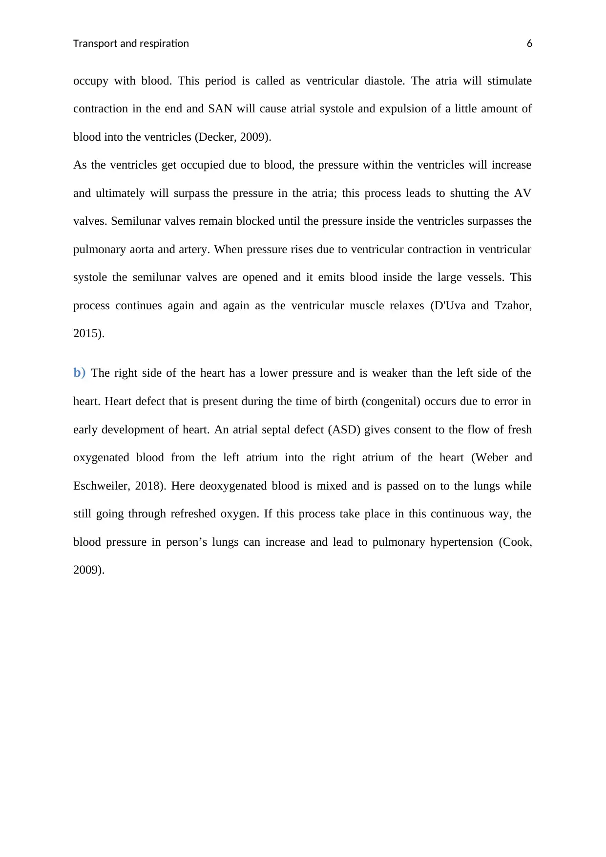
Source: (Opentextbc.ca, 2019)
Stages of cardiac cycle:
The cardiac cycle contains sequence of compartments or cambers which is known as systole
and diastole. Systole is the process where the cardiac muscle contract to form blood
expulsion from the corresponding compartments to another. Diastole is the process where the
compartments get occupied with blood and the muscle gets relaxed during this process. Atrial
diastole is the easy process to begin the cycle. In atrial diastole, the heart where the atrium is
located is occupied with blood from their individual vessels that is for left side it is the
pulmonary veins while for the right side it is the vena cava. This is occurred when the
pressure in the atria decreases in the veins.
Once the pressure in the atria is filled appropriately, it will surpass the pressure in the
ventricles so that the atrio-ventricular valves will start to unlatch and ventricles will begin to
Transport and respiration 6
occupy with blood. This period is called as ventricular diastole. The atria will stimulate
contraction in the end and SAN will cause atrial systole and expulsion of a little amount of
blood into the ventricles (Decker, 2009).
As the ventricles get occupied due to blood, the pressure within the ventricles will increase
and ultimately will surpass the pressure in the atria; this process leads to shutting the AV
valves. Semilunar valves remain blocked until the pressure inside the ventricles surpasses the
pulmonary aorta and artery. When pressure rises due to ventricular contraction in ventricular
systole the semilunar valves are opened and it emits blood inside the large vessels. This
process continues again and again as the ventricular muscle relaxes (D'Uva and Tzahor,
2015).
b) The right side of the heart has a lower pressure and is weaker than the left side of the
heart. Heart defect that is present during the time of birth (congenital) occurs due to error in
early development of heart. An atrial septal defect (ASD) gives consent to the flow of fresh
oxygenated blood from the left atrium into the right atrium of the heart (Weber and
Eschweiler, 2018). Here deoxygenated blood is mixed and is passed on to the lungs while
still going through refreshed oxygen. If this process take place in this continuous way, the
blood pressure in person’s lungs can increase and lead to pulmonary hypertension (Cook,
2009).
Paraphrase This Document
Need a fresh take? Get an instant paraphrase of this document with our AI Paraphraser
Transport and respiration 7
Task 3.3
Cardiac output: The cardiac output is calculated by the heart per minute pumped to
produce quantity of blood. The cardiac output is also calculated through the heart rate
product that is also known as the number of beats per minute, and the stroke volume
that is quantity pumped per beat (Young, 2010).
CO = HR X SV
Blood pressure: this is the pressure that is applied by the blood leading to the
surface of the blood vessels and arteries. This also differs from the muscular
competence of the heart, the volume of blood and viscosity, the individual’s age
and health, and the condition of the vascular wall(Beevers, 2011).
The equation for cardiac output is:
HR x SV = Q. Hence, to evaluate Q we must first begin with HR and SV. An
example at rest is shown below (Gellman and Turner, 2013). The example below
describes the cardiac output in rest.
Transport and respiration 8
HR (70BPM) x SV (70ml) = 4900ml/min or 4.9 litres per minute
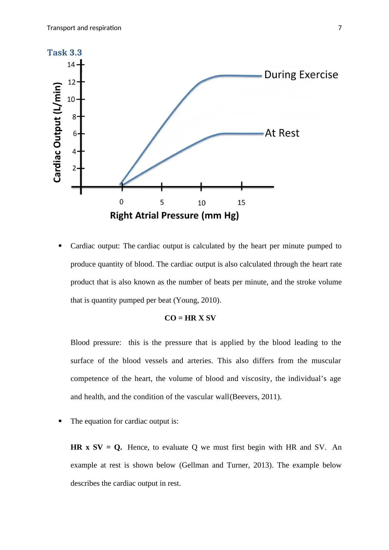
From the above graph, we can see that in rest the cardiac output is low as the volume of blood
is decreased because in a rest body the pressure on the vessel walls is reduced (Gulgun,
2016). While during exercise the cardiac out is increased due to increase in blood pressure in
the body as it affects the heart rate (HR) or stroke volume (SV) which may stimulate in
pumping the blood pressure on the vessel walls.
Task4.1.a)
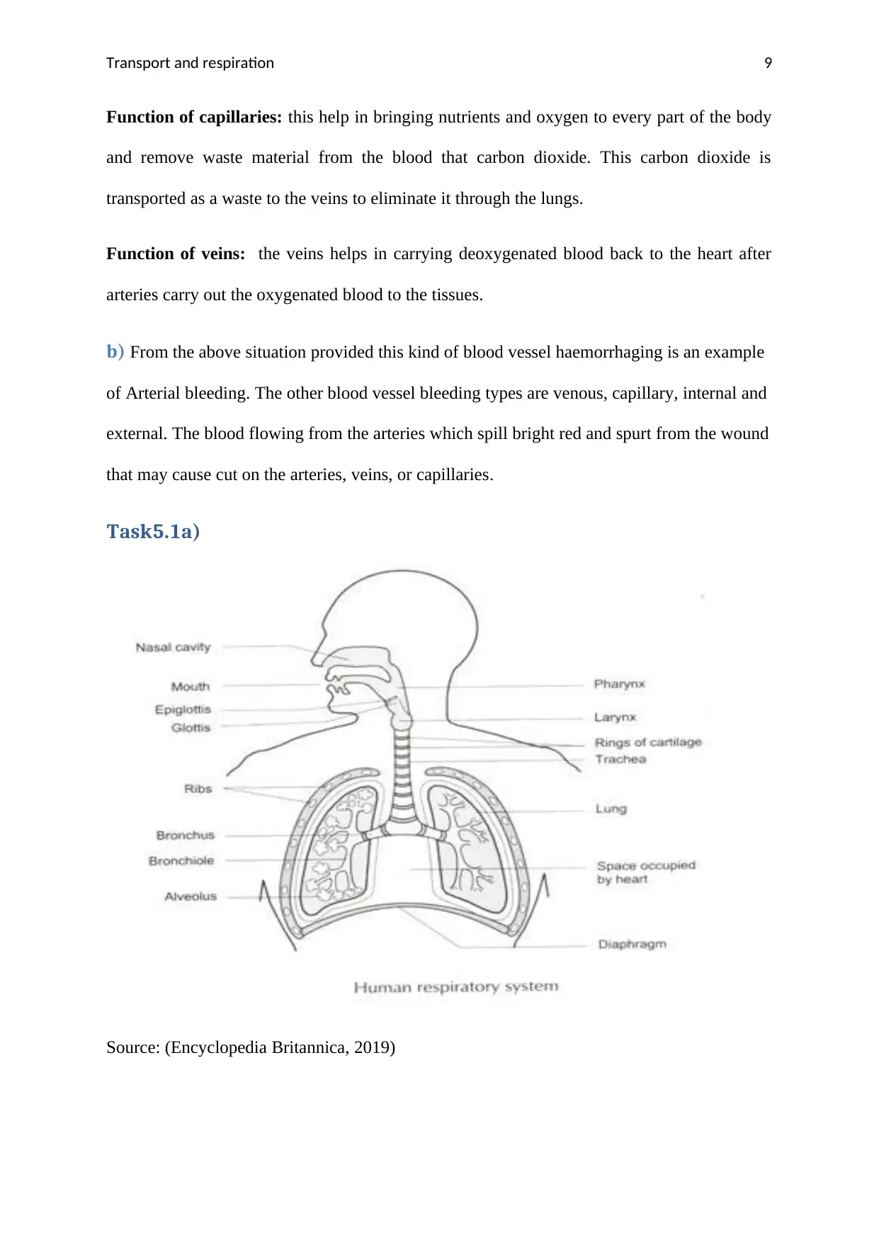
Source: (ScienceDaily, 2019)
Function of arteries: arteries are the important part of circulatory system that helps in
transportation of oxygen and nutrients to every part of the body. Its main function is to carry
blood which contain low amount of oxygen to the lungs in the right side of the ventricle and
carry back the blood containing oxygen from the left ventricle and towards other tissues and
organs.
Transport and respiration 9
Function of capillaries: this help in bringing nutrients and oxygen to every part of the body
and remove waste material from the blood that carbon dioxide. This carbon dioxide is
transported as a waste to the veins to eliminate it through the lungs.
Function of veins: the veins helps in carrying deoxygenated blood back to the heart after
arteries carry out the oxygenated blood to the tissues.
b) From the above situation provided this kind of blood vessel haemorrhaging is an example
of Arterial bleeding. The other blood vessel bleeding types are venous, capillary, internal and
external. The blood flowing from the arteries which spill bright red and spurt from the wound
that may cause cut on the arteries, veins, or capillaries.
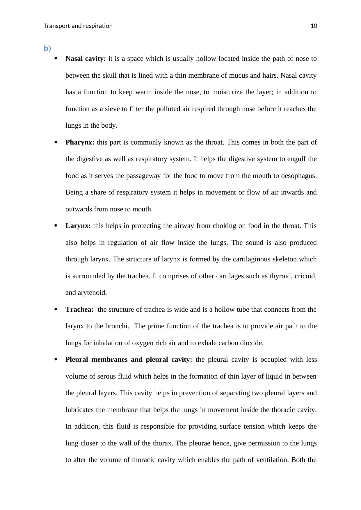
Task5.1a)
Source: (Encyclopedia Britannica, 2019)
Secure Best Marks with AI Grader
Need help grading? Try our AI Grader for instant feedback on your assignments.
Transport and respiration 10
b)
Nasal cavity: it is a space which is usually hollow located inside the path of nose to
between the skull that is lined with a thin membrane of mucus and hairs. Nasal cavity
has a function to keep warm inside the nose, to moisturize the layer; in addition to
function as a sieve to filter the polluted air respired through nose before it reaches the
lungs in the body.
Pharynx: this part is commonly known as the throat. This comes in both the part of
the digestive as well as respiratory system. It helps the digestive system to engulf the
food as it serves the passageway for the food to move from the mouth to oesophagus.
Being a share of respiratory system it helps in movement or flow of air inwards and
outwards from nose to mouth.
Larynx: this helps in protecting the airway from choking on food in the throat. This
also helps in regulation of air flow inside the lungs. The sound is also produced
through larynx. The structure of larynx is formed by the cartilaginous skeleton which
is surrounded by the trachea. It comprises of other cartilages such as thyroid, cricoid,
and arytenoid.
Trachea: the structure of trachea is wide and is a hollow tube that connects from the
larynx to the bronchi. The prime function of the trachea is to provide air path to the
lungs for inhalation of oxygen rich air and to exhale carbon dioxide.
Pleural membranes and pleural cavity: the pleural cavity is occupied with less
volume of serous fluid which helps in the formation of thin layer of liquid in between
the pleural layers. This cavity helps in prevention of separating two pleural layers and
lubricates the membrane that helps the lungs in movement inside the thoracic cavity.
In addition, this fluid is responsible for providing surface tension which keeps the
lung closer to the wall of the thorax. The pleurae hence, give permission to the lungs
to alter the volume of thoracic cavity which enables the path of ventilation. Both the
Transport and respiration 11
pleural sacs are separated from each other so that if any sac gets punctured the other
one continues the process effectively.
Lungs: the central function of the lungs is the process of exchanging gases through
respiration. Lungs fill oxygen and release out carbon dioxide. The lung consists of
trachea (windpipe), bronchi, diaphragm, bronchiole, pleura and alveoli.
o Bronchi: this is an extension of trachea that helps in respiration which
functions of exchanging the gases, oxygen and carbon dioxide.
o Bronchiole: the function of bronchiole is to transport the incoming air to
every alveolus. It makes a pathway through which the air is passed from
the nose or mouth to the alveoli or air sac of lungs.
o Alveoli: the function of alveoli is to exchange the gases, that are oxygen
and carbon dioxide molecules from the blood flow. The structure of
alveoli is in balloon like shaped air sacs which is arranged in cluster inside
the lungs.
Intercostal Muscles: the intercostal muscles are located internally as well as
internally. The external one is responsible for involuntary and silent inhalation. The
external intercostal muscle also helps in raising the ribcage and expands the chest
cavity. The internal intercostal muscles help in involuntary exhalation.
Diaphragm: its main function is it helps in respiration. Diaphragm is the organ which
separates the thoracic cavity which carries lungs and heart, from the abdominal cavity
and then the process of respiration takes place. During the process of respiration the
contraction of diaphragm takes place, and the volume of the thoracic cavity is
increased which helps the air to flow into the lungs.
Transport and respiration 12
Task 6: a)
Source: (Quizlet, 2019)
Pulmonary ventilation is also known as breathing. The function of pulmonary ventilation is
to flow the air into the lungs by inhalation and exhalation. The mechanisms that initiate
pulmonary ventilation are atmospheric pressure (Patm); the pressure of air inside the alveoli
which is also called as alveolar pressure (Palv); and the pressure inside the pleural cavity is
also called as intrapleural pressure (Pip)(Goldsmith and Karotkin, 2011).
In pulmonary ventilation while inspiration the diaphragm contracts downwards while during
expiration the diaphragm relaxes and it is pushed upwards. During inhalation the lungs
expands which leads in decrease of pressure inside the chest while during exhalation the
Paraphrase This Document
Need a fresh take? Get an instant paraphrase of this document with our AI Paraphraser
Transport and respiration 13
lungs is compressed due to increase in pressure which leads in relaxation of diaphragm. In
inspiration the air is drawn inside through the nose or the mouth while in expiration the air is
passed outside through nose and mouth.
The pressure is dependent on the relaxation and contraction of muscles of the diaphragm and
thorax. The lungs always create a passive movement that helps in inhalation and expiration.
The process of respiration takes place due to adhesive nature of the pleural fluid that
pressurise the lungs to pull outward during inspiration when the movement in thoracic wall
takes place. The shrinking of the thoracic wall takes place during expiration.
b) From the above situation, the patient has been admitted due to a penetrating injury to the
chest that has gone through the pleural membranes. In penetrating injury, between the lung
and the chest wall abnormal amount of air is collected in the pleural space. In this condition,
the patient is going through problem in pulmonary ventilation. Here, the quantity of air is
increased in the chest as one path of valve is moulded by a part of damage cells which further
leads to tension pneumothorax. In this cause, the patient may face oxygen shortage and low
blood pressure in his/her body. In any case if the patient continues to face pneumothorax
he/she may cause serious health issues which include infection or problem in the lungs also
known as collapsing of lungs.
References:
Bailey, R. (2018) Red Blood Cells (Erythrocytes) [Online]. Avaiable from
https://www.thoughtco.com/red-blood-cells-373487 [Accessed: 02/ 2/19]
Beevers, D. (2011). Understanding blood pressure. Poole: Family Doctor in association with
the British Medical Association.
Transport and respiration 14
Birch, T. (2012). Blood. St Lucia, Qld.: University of Queensland Press.
Blann, A. (2015). Deep Vein Thrombosis and Pulmonary Embolism. M & K Update Ltd.
Bottomley, P. (2015). Sodium MRI in human heart: a review. NMR in Biomedicine, 29(2),
pp.187-196.
Chan, C., Woods, C. and Shorr, A. (2010). Prognostic Scoring Tools for Acute Pulmonary
Embolism: The Pulmonary Embolism Severity Index vs Prognosis in Pulmonary
Embolism. Chest, 138(4), p.399A.
Cook, E. (2009). Gastrointestinal symptoms in ASD, brain structure of identical twins with
ASD. Autism Research, 2(5), pp.285-286.
Decker, L. (2009). Cardiac wellness. Hauppauge, N.Y.: Nova Science.
D'Uva, G. and Tzahor, E. (2015). The key roles of ERBB2 in cardiac regeneration. Cell
Cycle, 14(15), pp.2383-2384.
Encyclopedia Britannica. (2019). human respiratory system | Description, Parts, Function, &
Facts. [online] Available at: https://www.britannica.com/science/human-respiratory-system
[Accessed 2 Feb. 2019].
Gillard, A. (2013). Stroke. Detroit: Greenhaven Press.
Goldsmith, J. and Karotkin, E. (2011). Assisted ventilation of the neonate. St. Louis, Mo.:
Elsevier/Saunders.
Gulgun, M. (2016). Heart Rate Variability, Heart Rate Recovery, and Heart Rate Turbulence
Represent Different Components of the Cardiac Autonomic Nervous System. Medical
Principles and Practice, 26(1), pp.98-99.
Transport and respiration 15
Miranda, G. M. (2019) Structure and Function of the Heart[Online].Available from
https://www.news-medical.net/health/Structure-and-Function-of-the-Heart.aspx [Accessed:
02/ 2/19]
ScienceDaily. (2019). Blood vessel. [online] Available at:
https://www.sciencedaily.com/terms/blood_vessel.htm [Accessed 2 Feb. 2019].
Shen, F. and Chang, A. (2013). ESTABLISHING A COORDINATION SYSTEM FOR
CONSTRUCTION PROJECT ORGANIZATIONS BY SIMULATING HUMAN BODY
CIRCULATORY SYSTEM. Journal of Civil Engineering and Management, 19(4), pp.492-
504.
S, S. (2018). Histology of Coronary Arteries in Relation to the Coronary Sclerosis. Journal of
Human Anatomy, 2(2).
TheFreeDictionary.com. (2019). systemic circulation. [online] Available at: https://medical-
dictionary.thefreedictionary.com/systemic+circulation [Accessed 2 Feb. 2019].
Opentextbc.ca. (2019). 19.3 Cardiac Cycle – Anatomy and Physiology. [online] Available at:
https://opentextbc.ca/anatomyandphysiology/chapter/19-3-cardiac-cycle/ [Accessed 2 Feb.
2019].
Quizlet.(2019). Pulmonary ventilation Diagram | Quizlet. [online] Available at:
https://quizlet.com/233714637/pulmonary-ventilation-diagram/ [Accessed 2 Feb. 2019].
Weber, M. and Eschweiler, S. (2018).Professionelles Management in ASD.ASD-
LeitungskräftebrauchenspezifischeKompetenzen. unserejugend, 70(10), p.419.
wiseGEEK. (2019). What is Pulmonary Circulation? (with pictures). [online] Available at:
https://www.wisegeek.com/what-is-pulmonary-circulation.htm [Accessed 2 Feb. 2019].
Secure Best Marks with AI Grader
Need help grading? Try our AI Grader for instant feedback on your assignments.
Transport and respiration 16
Wignall, K. (2015). Blood. Minneapolis: Carolrhoda Lab.
Young, D. (2010). Control of cardiac output. San Rafael, CA: Morgan & Claypool Life
Sciences.
Wignall, K. (2015). Blood. Minneapolis: Carolrhoda Lab.
Young, D. (2010). Control of cardiac output. San Rafael, CA: Morgan & Claypool Life
Sciences.
1 out of 17
Related Documents
Your All-in-One AI-Powered Toolkit for Academic Success.
+13062052269
info@desklib.com
Available 24*7 on WhatsApp / Email
![[object Object]](/_next/static/media/star-bottom.7253800d.svg)
Unlock your academic potential
© 2024 | Zucol Services PVT LTD | All rights reserved.


