Urine Analysis to Identify Disease: Biology Lab Report, FFSC031H4
VerifiedAdded on 2022/09/01
|8
|1338
|19
Report
AI Summary
This biology lab report details the analysis of urine samples to identify potential diseases. The report begins with an introduction to the excretory system, kidney structure, and the components of urine. The methodology section describes the procedures for testing urine samples for glucose, protein, and bile using Benedict's, Biuret, and sulfur tests, respectively. The results are presented in a table format, including observations, test outcomes, and diagnostic inferences. The discussion section interprets the results, explaining the significance of positive and negative test outcomes. Additionally, the report includes an evaluation of the experiment, acknowledging limitations and suggesting improvements. The report also addresses application questions, including calculations related to protein concentration using the formula V1S1= V2S2 and the Lambert-Beer law, and discusses the limitations of the Benedict's test in detecting sucrose.
Running head: URINE ANALYSIS TO IDENTFY DISEASE
URINE ANALYSIS TO IDENTFY DISEASE
Name of the student
Name of the university
Author note
URINE ANALYSIS TO IDENTFY DISEASE
Name of the student
Name of the university
Author note
Paraphrase This Document
Need a fresh take? Get an instant paraphrase of this document with our AI Paraphraser
1
URINE ANALYSIS TO IDENTFY DISEASE
Introduction
The kidney is a bean-shaped filter organ found in pairs in the side of the spine in
every human being. The primary role of the kidneys is to filter the blood from substances that
are toxic for the cell and oxidative end products and to extract and maintain water and
electrolytes by producing urine that is discharged into the ureter and urinary bladder.
Nephrons in the kidney play the chief part in producing urine (Sherwood 2015). The standard
components of urine are urea, chloride, potassium, sodium, creatinine, dissolved organics,
and inorganic ions. Urinalysis (UA), also referred to like routine and microscopy (R&M), is a
series of urine components test measures and mostly used in medical diagnostic techniques
to identify the components present in urine (Fox 2015).
Method:
The urinary glucose concentration detection test requires to perform Benedict's test.
The Benedicts test is done in a few steps. 5ml benedicts reagent are taken in a pipette, and
few drops of sample urine were added to it. The mixture was then heated for 5 to 10 minutes.
After that, the test tube is cooled by keeping them under running water. The change of the
solution color will indicate the concentration of glucose in the blood (Tawfik et al, 2019).
The urine proteins can be detected by performing the biuret test. The urine sample
was acidified, and precipitants were added to the sample. The sample was then centrifuged
and put in the analyzer for the estimation of protein—the system mix the NaOH and biuret
reagent. The test result is estimated in a colorimetric way (Kumar and Gill, 2018).
The bile salt of urine can be detected by the sulfur test. Some dry sulfur powder is
sprinkled on the surface of the fresh urine. If the bile salt is present in the urine, the sulfur
powder will be sink (Mohapatra et al, 2016).
2
URINE ANALYSIS TO IDENTFY DISEASE
Result:
URINE ANALYSIS TO IDENTFY DISEASE
Result:
⊘ This is a preview!⊘
Do you want full access?
Subscribe today to unlock all pages.

Trusted by 1+ million students worldwide

3
URINE ANALYSIS TO IDENTFY DISEASE
SampleTest Observation Result
(+ve or
–ve)
Inference Diagnosis
A
Benedict's
Test
The solution
remains pale blue
after heating
-ve No glucose
in the urine
No glycosuria
Biuret Test The solution turns
pale violet
+ve Presence
of protein in
the urine
Kidney
diseases
Sulfur Test The sulfur powder
remains on the
surface of the
sample
-ve No bile in
the urine
Liver
function is
normal
B Benedict's
Test
Solution turns
orange
+++
ve
1500-
2000mg/ dl
Presence of
glucose in
urine
Biuret Test No change in color -ve No protein
in
sample
Kidney
function is
normal
Paraphrase This Document
Need a fresh take? Get an instant paraphrase of this document with our AI Paraphraser

4
URINE ANALYSIS TO IDENTFY DISEASE
Sulfur Test Sulfur power
remains on surface
of sample
-ve No bile in
urine
Normal liver
function
C
Benedict's
Test
The solution
remains pale blue
after heating
-ve No glucose
in urine
Blood sugar
is normal
Biuret Test No change in colour -ve No protein
in urine
Kidney
function is
normal
Sulfur Test Sulfur power
remains on surface
of sample
-ve No bile salt
in urine
No jaundice
or liver
disorder
Biuret Test No change in colour -ve No protein
in urine
Kidney
function
normally
Sulfur Test Sulfur power sinks
to bottom of test
tube
+ve Bile present
in the urine
Indicates
jaundice
5
URINE ANALYSIS TO IDENTFY DISEASE
Discussion:
Both monosaccharides like glucose, fructose, galactose, and other disaccharides,
including lactose and maltose, give positive benedict test results.
Glucose observed to be present in the urine is Diabetes mellitus sign. The color
changes from blue to gradually red as the glucose concentration increases.
The biuret reagent reacts with the peptide bonds of the protein present in the urine, changing
the solution color from blue to violet, indicating kidney diseases (Asthana et al, 2019).
The bile salt present in the urine decreases the surface tension, allowing the sprinkled
sulfur to sink, indicating a positive bile test.
Evaluation:
The experiment was done as per protocol to eliminate any error. However, there are
some limitations.
The Benedict test is not recommended to testing diabetes sololy. The test can give
false-positive or false-negative results.
The errors can be eliminated by strict restrictions of any contamination in the solution.
Task:
1)
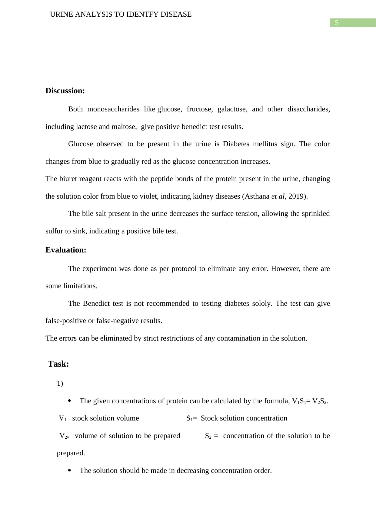
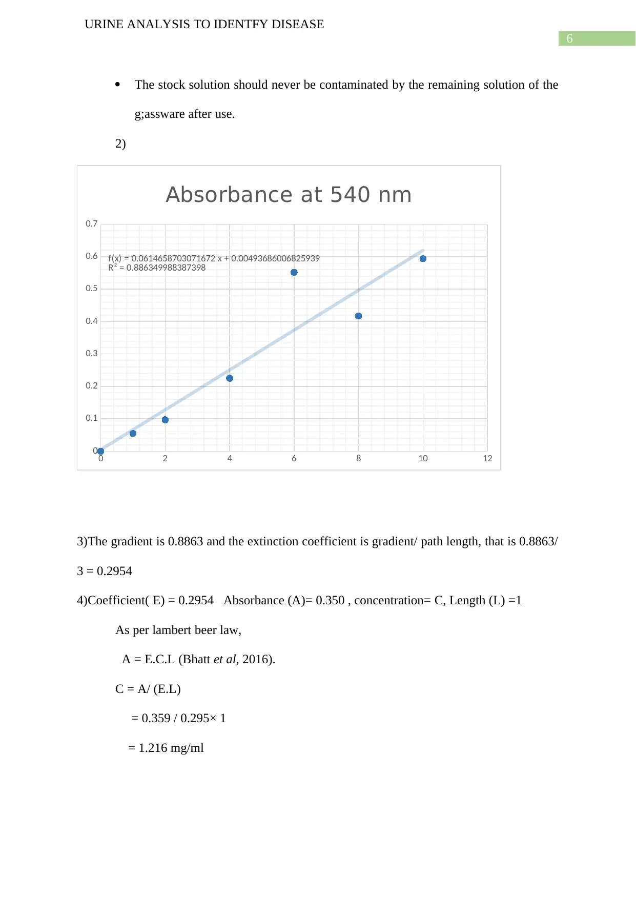
The given concentrations of protein can be calculated by the formula, V1S1= V2S2.
V1 = stock solution volume S1= Stock solution concentration
V2= volume of solution to be prepared S2 = concentration of the solution to be
prepared.
The solution should be made in decreasing concentration order.
⊘ This is a preview!⊘
Do you want full access?
Subscribe today to unlock all pages.

Trusted by 1+ million students worldwide
6
URINE ANALYSIS TO IDENTFY DISEASE
The stock solution should never be contaminated by the remaining solution of the
g;assware after use.
2)
3)The gradient is 0.8863 and the extinction coefficient is gradient/ path length, that is 0.8863/
3 = 0.2954
4)Coefficient( E) = 0.2954 Absorbance (A)= 0.350 , concentration= C, Length (L) =1
As per lambert beer law,
A = E.C.L (Bhatt et al, 2016).
C = A/ (E.L)
= 0.359 / 0.295× 1
= 1.216 mg/ml
0 2 4 6 8 10 12
0
0.1
0.2
0.3
0.4
0.5
0.6
0.7
f(x) = 0.0614658703071672 x + 0.00493686006825939
R² = 0.886349988387398
Absorbance at 540 nm
Paraphrase This Document
Need a fresh take? Get an instant paraphrase of this document with our AI Paraphraser
7
URINE ANALYSIS TO IDENTFY DISEASE
Part 3:
The Benedict test cannot detect the presence of sucrose in the urine, as the sucrose is
non-reducible by the benedict reagent. Thus, it gives a negative result even if there is the
presence of sucrose in the urine. Sucrose is a disaccharide joined by glycosidic bond, which
inhibits the glucose and fructose from isomerizing, following the prevention of the reduction.
The test is, however, can show positive results if the urine sample is boiled with dilute
hydrochloric acid, which can break the glycosidic bond by hydrolysis, allowing the benedict
reagent to react with the reduced sugar (Kumar and Gill 2018).
1 out of 8
Your All-in-One AI-Powered Toolkit for Academic Success.
+13062052269
info@desklib.com
Available 24*7 on WhatsApp / Email
![[object Object]](/_next/static/media/star-bottom.7253800d.svg)
Unlock your academic potential
Copyright © 2020–2026 A2Z Services. All Rights Reserved. Developed and managed by ZUCOL.
