Understanding Agarose Gel Electrophoresis, PCR, and DNA Sequencing
VerifiedAdded on 2019/09/23
|10
|5273
|229
Report
AI Summary
In this assignment content, we learn about Agarose Gel Electrophoresis and Polymerase Chain Reaction (PCR). Agarose gel electrophoresis involves preparing an agarose gel by dissolving agarose in a buffer solution, adding ethidium bromide to bind DNA, pouring the gel into a tray, and allowing it to solidify. The gel is then loaded with samples and run at 100V for about 1 hour. PCR is a laboratory technique used to selectively amplify specific DNA sequences. It involves denaturing, annealing, and synthesizing DNA primers to produce thousands of copies of the target DNA sequence. The materials needed for PCR include an earlier prepared DNA sample, PCR buffer, deoxynucleotide triphosphates, primer #1 and #2, and Amplitaq Gold DNA polymerase.
Contribute Materials
Your contribution can guide someone’s learning journey. Share your
documents today.

Running Head: Virology
Virology
Virology
Secure Best Marks with AI Grader
Need help grading? Try our AI Grader for instant feedback on your assignments.
Virology 1
The Prohibitin
Protein pair of prohibitin 1 and prohibitin 2 is universally expressed as the prohibitin proteins where prohibitin 2 or
PHB2 is also referred as REA (receptor estrogen activity) repressor or BAP-37 (B-cell receptor associate protein).
Both these proteins belong to flotilin, stomatin, HflK/C, and prohibitin superfamily (Mishra, Murphy & Murphy,
2017). It is designated as “prohibitin” because originally PHB1 cDNA transfection led to arrest of cell cycle (Mishra,
Murphy & Murphy, 2006), ("Isolation of a cDNA that hybrid selects antiproliferative mRNA from rat liver -
ScienceDirect", 2006). It has been showed that PHB1 and PHB2 are present in cytosol, mitochondria, nucleus, and in
certain receptors of cell membrane ("Prohibitin Ligands in Cell Death and Survival: Mode of Action and Therapeutic
Potential - ScienceDirect", 1989). At present, PHBs are considered as the best examples that illustrate distinctive and
clear functions which depend on intercellular localization. PHB1 and PHB2 inside mitochondria make an alternating
ring-like heterodimeric complex helpful in mitochondrial stability. On the contrary, both the independent PHBs
inside the nucleus helps in transcriptionally suppressing the target genes (Mishra, Murphy & Murphy, 2006),
("Prohibitin Ligands in Cell Death and Survival: Mode of Action and Therapeutic Potential - ScienceDirect", 1989).
Although PHBs in the nucleus influence cell cycle and many transcription factors, the prohibitin loss of any type and
most of the observed cellular effects are attributed to their mitochondrial function (Merkwirth et al., 2008). Level of
ROS (reactive oxygen species), inflammation, and PHBs’ expressions is correlated; hence, inflammatory component
diseases such as diabetes, cancer, and degenerative disorders of neuromuscles are more likely to be present with
localization and/or expression of altered PHB1/2. In fact, experiments on altered expression of PHB in dissimilar
model systems copies numerous pathologies of inflammation (Merkwirth et al., 2012), (Supale et al., 2013), (Theiss
et al., 2007), (Kasashima, Ohta, Kagawa & Endo, 2006). While majority of researches till date on the PHBs has been
collected from PHB1 studies, this study focuses on recent advancements made in the field of PHB2 which implicates
intercellular communication nature of PHBs between mitochondria and the nucleus (Bavelloni, Piazzi, Raffini,
Faenza & Blalock, 2015).
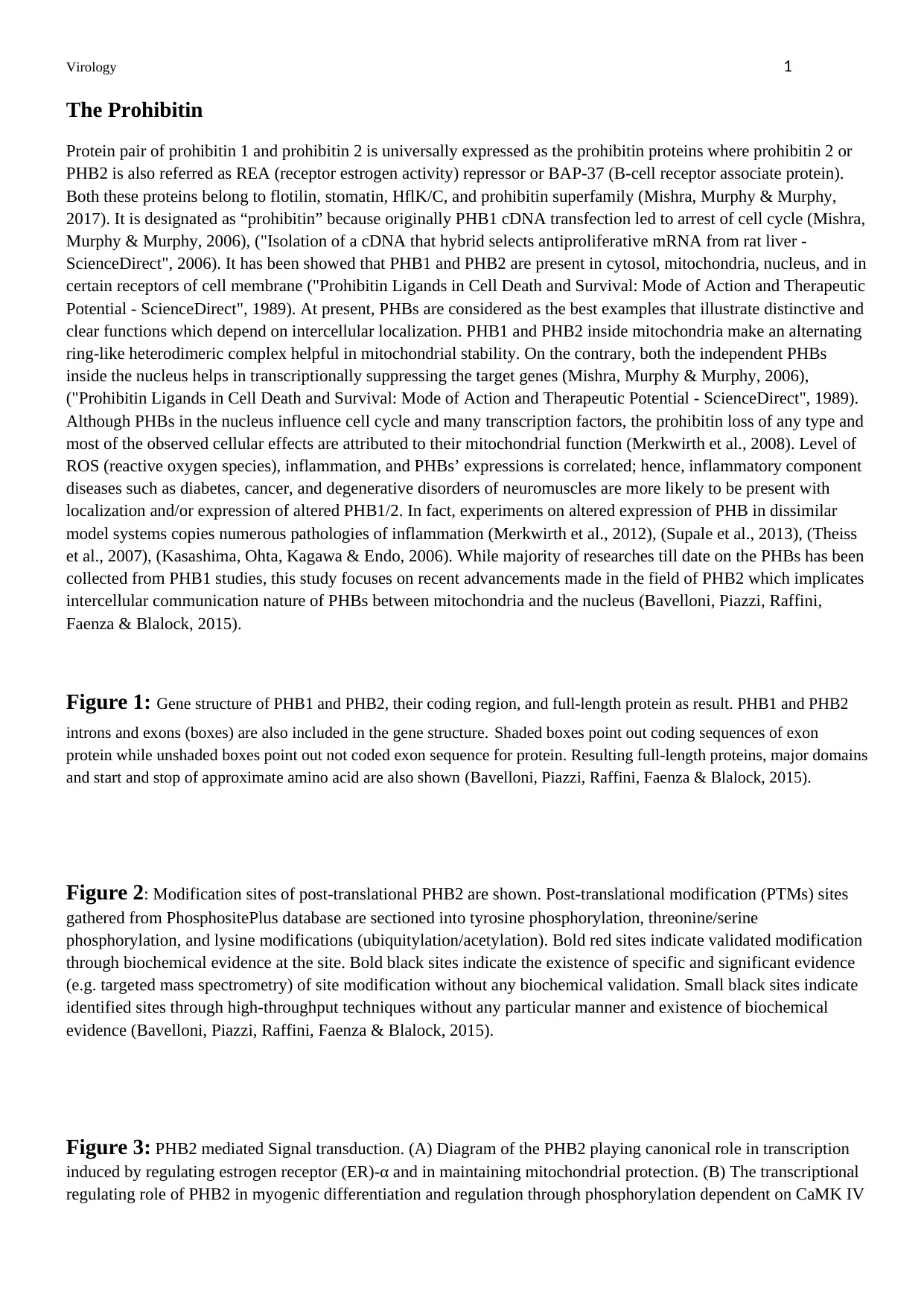
Figure 1: Gene structure of PHB1 and PHB2, their coding region, and full-length protein as result. PHB1 and PHB2
introns and exons (boxes) are also included in the gene structure. Shaded boxes point out coding sequences of exon
protein while unshaded boxes point out not coded exon sequence for protein. Resulting full-length proteins, major domains
and start and stop of approximate amino acid are also shown (Bavelloni, Piazzi, Raffini, Faenza & Blalock, 2015).
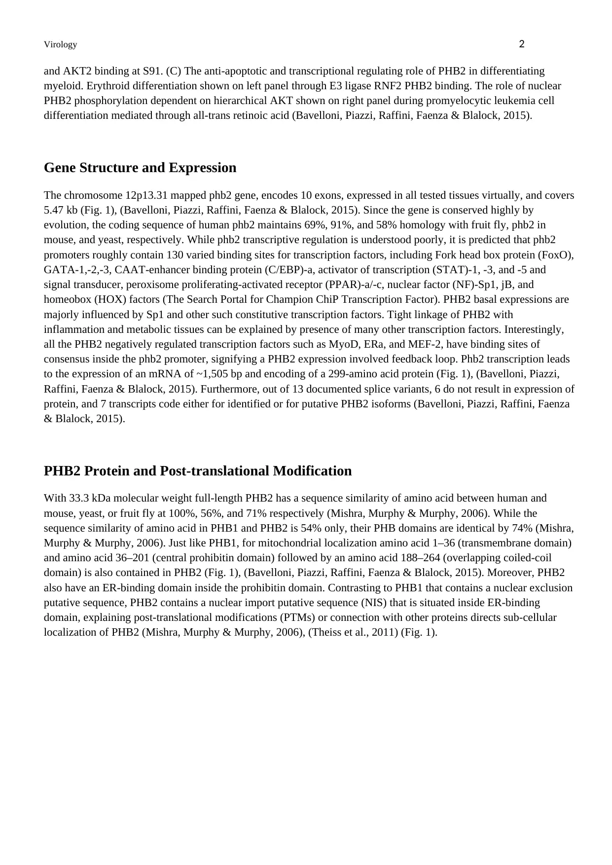
Figure 2: Modification sites of post-translational PHB2 are shown. Post-translational modification (PTMs) sites
gathered from PhosphositePlus database are sectioned into tyrosine phosphorylation, threonine/serine
phosphorylation, and lysine modifications (ubiquitylation/acetylation). Bold red sites indicate validated modification
through biochemical evidence at the site. Bold black sites indicate the existence of specific and significant evidence
(e.g. targeted mass spectrometry) of site modification without any biochemical validation. Small black sites indicate
identified sites through high-throughput techniques without any particular manner and existence of biochemical
evidence (Bavelloni, Piazzi, Raffini, Faenza & Blalock, 2015).
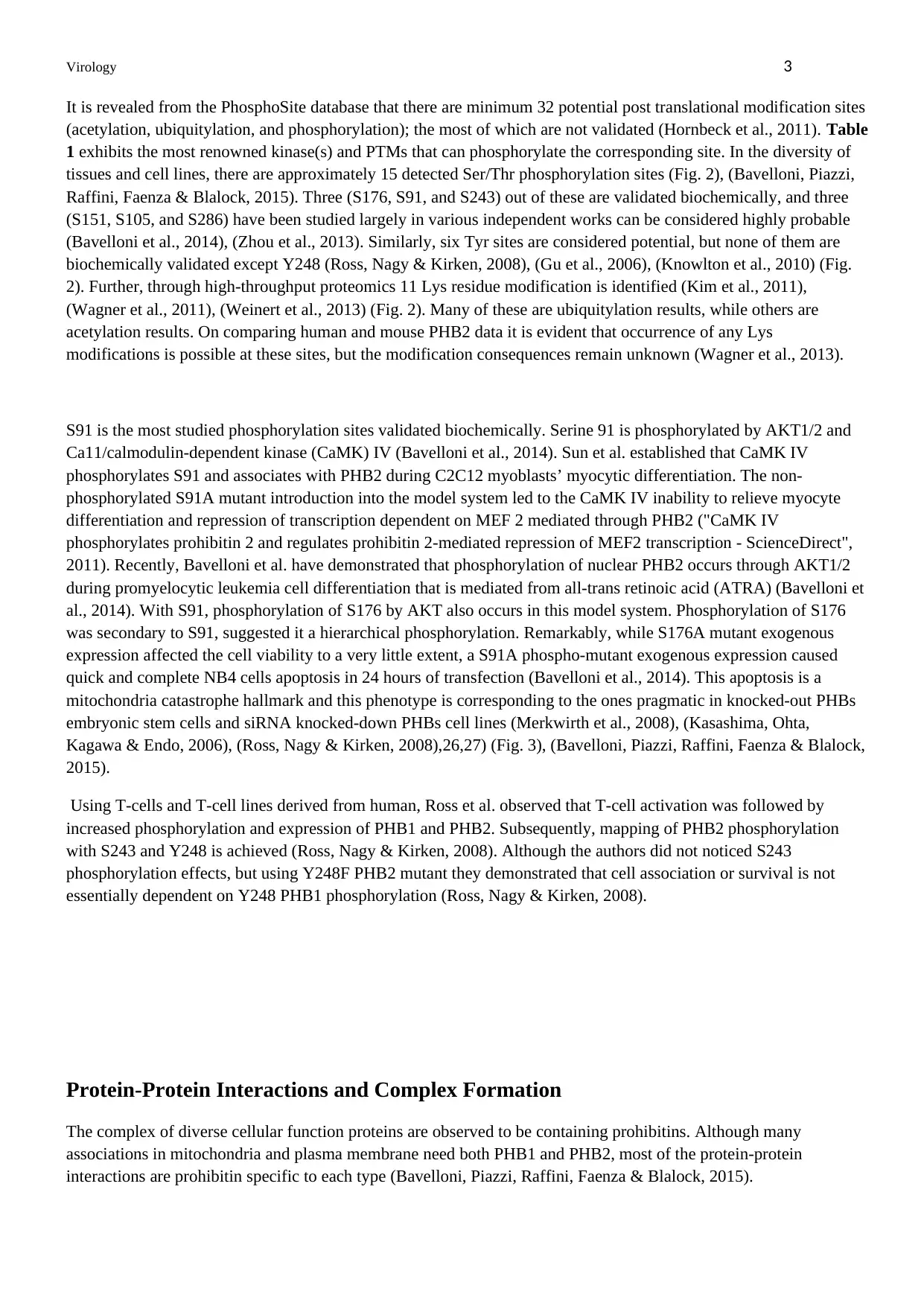
Figure 3: PHB2 mediated Signal transduction. (A) Diagram of the PHB2 playing canonical role in transcription
induced by regulating estrogen receptor (ER)-α and in maintaining mitochondrial protection. (B) The transcriptional
regulating role of PHB2 in myogenic differentiation and regulation through phosphorylation dependent on CaMK IV
The Prohibitin
Protein pair of prohibitin 1 and prohibitin 2 is universally expressed as the prohibitin proteins where prohibitin 2 or
PHB2 is also referred as REA (receptor estrogen activity) repressor or BAP-37 (B-cell receptor associate protein).
Both these proteins belong to flotilin, stomatin, HflK/C, and prohibitin superfamily (Mishra, Murphy & Murphy,
2017). It is designated as “prohibitin” because originally PHB1 cDNA transfection led to arrest of cell cycle (Mishra,
Murphy & Murphy, 2006), ("Isolation of a cDNA that hybrid selects antiproliferative mRNA from rat liver -
ScienceDirect", 2006). It has been showed that PHB1 and PHB2 are present in cytosol, mitochondria, nucleus, and in
certain receptors of cell membrane ("Prohibitin Ligands in Cell Death and Survival: Mode of Action and Therapeutic
Potential - ScienceDirect", 1989). At present, PHBs are considered as the best examples that illustrate distinctive and
clear functions which depend on intercellular localization. PHB1 and PHB2 inside mitochondria make an alternating
ring-like heterodimeric complex helpful in mitochondrial stability. On the contrary, both the independent PHBs
inside the nucleus helps in transcriptionally suppressing the target genes (Mishra, Murphy & Murphy, 2006),
("Prohibitin Ligands in Cell Death and Survival: Mode of Action and Therapeutic Potential - ScienceDirect", 1989).
Although PHBs in the nucleus influence cell cycle and many transcription factors, the prohibitin loss of any type and
most of the observed cellular effects are attributed to their mitochondrial function (Merkwirth et al., 2008). Level of
ROS (reactive oxygen species), inflammation, and PHBs’ expressions is correlated; hence, inflammatory component
diseases such as diabetes, cancer, and degenerative disorders of neuromuscles are more likely to be present with
localization and/or expression of altered PHB1/2. In fact, experiments on altered expression of PHB in dissimilar
model systems copies numerous pathologies of inflammation (Merkwirth et al., 2012), (Supale et al., 2013), (Theiss
et al., 2007), (Kasashima, Ohta, Kagawa & Endo, 2006). While majority of researches till date on the PHBs has been
collected from PHB1 studies, this study focuses on recent advancements made in the field of PHB2 which implicates
intercellular communication nature of PHBs between mitochondria and the nucleus (Bavelloni, Piazzi, Raffini,
Faenza & Blalock, 2015).
Figure 1: Gene structure of PHB1 and PHB2, their coding region, and full-length protein as result. PHB1 and PHB2
introns and exons (boxes) are also included in the gene structure. Shaded boxes point out coding sequences of exon
protein while unshaded boxes point out not coded exon sequence for protein. Resulting full-length proteins, major domains
and start and stop of approximate amino acid are also shown (Bavelloni, Piazzi, Raffini, Faenza & Blalock, 2015).
Figure 2: Modification sites of post-translational PHB2 are shown. Post-translational modification (PTMs) sites
gathered from PhosphositePlus database are sectioned into tyrosine phosphorylation, threonine/serine
phosphorylation, and lysine modifications (ubiquitylation/acetylation). Bold red sites indicate validated modification
through biochemical evidence at the site. Bold black sites indicate the existence of specific and significant evidence
(e.g. targeted mass spectrometry) of site modification without any biochemical validation. Small black sites indicate
identified sites through high-throughput techniques without any particular manner and existence of biochemical
evidence (Bavelloni, Piazzi, Raffini, Faenza & Blalock, 2015).
Figure 3: PHB2 mediated Signal transduction. (A) Diagram of the PHB2 playing canonical role in transcription
induced by regulating estrogen receptor (ER)-α and in maintaining mitochondrial protection. (B) The transcriptional
regulating role of PHB2 in myogenic differentiation and regulation through phosphorylation dependent on CaMK IV
Virology 2
and AKT2 binding at S91. (C) The anti-apoptotic and transcriptional regulating role of PHB2 in differentiating
myeloid. Erythroid differentiation shown on left panel through E3 ligase RNF2 PHB2 binding. The role of nuclear
PHB2 phosphorylation dependent on hierarchical AKT shown on right panel during promyelocytic leukemia cell
differentiation mediated through all-trans retinoic acid (Bavelloni, Piazzi, Raffini, Faenza & Blalock, 2015).
Gene Structure and Expression
The chromosome 12p13.31 mapped phb2 gene, encodes 10 exons, expressed in all tested tissues virtually, and covers
5.47 kb (Fig. 1), (Bavelloni, Piazzi, Raffini, Faenza & Blalock, 2015). Since the gene is conserved highly by
evolution, the coding sequence of human phb2 maintains 69%, 91%, and 58% homology with fruit fly, phb2 in
mouse, and yeast, respectively. While phb2 transcriptive regulation is understood poorly, it is predicted that phb2
promoters roughly contain 130 varied binding sites for transcription factors, including Fork head box protein (FoxO),
GATA-1,-2,-3, CAAT-enhancer binding protein (C/EBP)-a, activator of transcription (STAT)-1, -3, and -5 and
signal transducer, peroxisome proliferating-activated receptor (PPAR)-a/-c, nuclear factor (NF)-Sp1, jB, and
homeobox (HOX) factors (The Search Portal for Champion ChiP Transcription Factor). PHB2 basal expressions are
majorly influenced by Sp1 and other such constitutive transcription factors. Tight linkage of PHB2 with
inflammation and metabolic tissues can be explained by presence of many other transcription factors. Interestingly,
all the PHB2 negatively regulated transcription factors such as MyoD, ERa, and MEF-2, have binding sites of
consensus inside the phb2 promoter, signifying a PHB2 expression involved feedback loop. Phb2 transcription leads
to the expression of an mRNA of ~1,505 bp and encoding of a 299-amino acid protein (Fig. 1), (Bavelloni, Piazzi,
Raffini, Faenza & Blalock, 2015). Furthermore, out of 13 documented splice variants, 6 do not result in expression of
protein, and 7 transcripts code either for identified or for putative PHB2 isoforms (Bavelloni, Piazzi, Raffini, Faenza
& Blalock, 2015).
PHB2 Protein and Post-translational Modification
With 33.3 kDa molecular weight full-length PHB2 has a sequence similarity of amino acid between human and
mouse, yeast, or fruit fly at 100%, 56%, and 71% respectively (Mishra, Murphy & Murphy, 2006). While the
sequence similarity of amino acid in PHB1 and PHB2 is 54% only, their PHB domains are identical by 74% (Mishra,
Murphy & Murphy, 2006). Just like PHB1, for mitochondrial localization amino acid 1–36 (transmembrane domain)
and amino acid 36–201 (central prohibitin domain) followed by an amino acid 188–264 (overlapping coiled-coil
domain) is also contained in PHB2 (Fig. 1), (Bavelloni, Piazzi, Raffini, Faenza & Blalock, 2015). Moreover, PHB2
also have an ER-binding domain inside the prohibitin domain. Contrasting to PHB1 that contains a nuclear exclusion
putative sequence, PHB2 contains a nuclear import putative sequence (NIS) that is situated inside ER-binding
domain, explaining post-translational modifications (PTMs) or connection with other proteins directs sub-cellular
localization of PHB2 (Mishra, Murphy & Murphy, 2006), (Theiss et al., 2011) (Fig. 1).
Virology 3
It is revealed from the PhosphoSite database that there are minimum 32 potential post translational modification sites
(acetylation, ubiquitylation, and phosphorylation); the most of which are not validated (Hornbeck et al., 2011). Table
1 exhibits the most renowned kinase(s) and PTMs that can phosphorylate the corresponding site. In the diversity of
tissues and cell lines, there are approximately 15 detected Ser/Thr phosphorylation sites (Fig. 2), (Bavelloni, Piazzi,
Raffini, Faenza & Blalock, 2015). Three (S176, S91, and S243) out of these are validated biochemically, and three
(S151, S105, and S286) have been studied largely in various independent works can be considered highly probable
(Bavelloni et al., 2014), (Zhou et al., 2013). Similarly, six Tyr sites are considered potential, but none of them are
biochemically validated except Y248 (Ross, Nagy & Kirken, 2008), (Gu et al., 2006), (Knowlton et al., 2010) (Fig.
2). Further, through high-throughput proteomics 11 Lys residue modification is identified (Kim et al., 2011),
(Wagner et al., 2011), (Weinert et al., 2013) (Fig. 2). Many of these are ubiquitylation results, while others are
acetylation results. On comparing human and mouse PHB2 data it is evident that occurrence of any Lys
modifications is possible at these sites, but the modification consequences remain unknown (Wagner et al., 2013).
S91 is the most studied phosphorylation sites validated biochemically. Serine 91 is phosphorylated by AKT1/2 and
Ca11/calmodulin-dependent kinase (CaMK) IV (Bavelloni et al., 2014). Sun et al. established that CaMK IV
phosphorylates S91 and associates with PHB2 during C2C12 myoblasts’ myocytic differentiation. The non-
phosphorylated S91A mutant introduction into the model system led to the CaMK IV inability to relieve myocyte
differentiation and repression of transcription dependent on MEF 2 mediated through PHB2 ("CaMK IV
phosphorylates prohibitin 2 and regulates prohibitin 2-mediated repression of MEF2 transcription - ScienceDirect",
2011). Recently, Bavelloni et al. have demonstrated that phosphorylation of nuclear PHB2 occurs through AKT1/2
during promyelocytic leukemia cell differentiation that is mediated from all-trans retinoic acid (ATRA) (Bavelloni et
al., 2014). With S91, phosphorylation of S176 by AKT also occurs in this model system. Phosphorylation of S176
was secondary to S91, suggested it a hierarchical phosphorylation. Remarkably, while S176A mutant exogenous
expression affected the cell viability to a very little extent, a S91A phospho-mutant exogenous expression caused
quick and complete NB4 cells apoptosis in 24 hours of transfection (Bavelloni et al., 2014). This apoptosis is a
mitochondria catastrophe hallmark and this phenotype is corresponding to the ones pragmatic in knocked-out PHBs
embryonic stem cells and siRNA knocked-down PHBs cell lines (Merkwirth et al., 2008), (Kasashima, Ohta,
Kagawa & Endo, 2006), (Ross, Nagy & Kirken, 2008),26,27) (Fig. 3), (Bavelloni, Piazzi, Raffini, Faenza & Blalock,
2015).
Using T-cells and T-cell lines derived from human, Ross et al. observed that T-cell activation was followed by
increased phosphorylation and expression of PHB1 and PHB2. Subsequently, mapping of PHB2 phosphorylation
with S243 and Y248 is achieved (Ross, Nagy & Kirken, 2008). Although the authors did not noticed S243
phosphorylation effects, but using Y248F PHB2 mutant they demonstrated that cell association or survival is not
essentially dependent on Y248 PHB1 phosphorylation (Ross, Nagy & Kirken, 2008).
Protein-Protein Interactions and Complex Formation
The complex of diverse cellular function proteins are observed to be containing prohibitins. Although many
associations in mitochondria and plasma membrane need both PHB1 and PHB2, most of the protein-protein
interactions are prohibitin specific to each type (Bavelloni, Piazzi, Raffini, Faenza & Blalock, 2015).
Paraphrase This Document
Need a fresh take? Get an instant paraphrase of this document with our AI Paraphraser
Virology 4
Some PHB2 interacting proteins in the nucleus are both specific and global transcription factors, including: b-
catenin, the cAMP-dependent transcription factor (ATF)-2, COUP-TF1, the estrogen receptor (ER)-a, COUP-TF2,
inter-leukin enhancer binding factor (ILF)-3, MYOD1, MEF2A, transcription factor (TF)-E3, and Runt-related
transcription factor (RUNX3) (Lau et al., 2012), (Ewing et al., 2007), (Sun, 2004). Others include enzymes
modifying DNA such as breast-related carcinoma antigen (BRCA)-1, the histone deacetylases (HDAC1, HDAC3,
HDAC2, and HDAC5), cyclin-dependent kinase (CDK)-2, enzymes associated with DNA repair, proteins of the
complex of PRC2/EEDEZH2 related polycomb, and proteins associated with cell cycle (Ewing et al., 2007), (Sun,
2004), (Neganova et al., 2011),(Kim et al., 2009). Several proteins that bind RNA are needed for RNA processing
(DDX20 ATP-dependent helicase RNA), stability [basophile leukemia expressed protein (Bles03) and Epiplakin
(EPPK1)] and transport (Staufen) associated to nuclear PHB2 (Ewing et al., 2007), (Milev, Ravichandran, Khan,
Schriemer & Mouland, 2012), (Bavelloni, Piazzi, Raffini, Faenza & Blalock, 2015).
Most of the PHB2 interacting proteins in cytosol are connected with the cytoskeletal and cytoskeleton transport
[coatomer subunit gamma 1 (COPG1)], ubiquitylation, cellular signaling [receptor interacting S/T kinase (RIPK)-2
and MDM2] (Kim et al., 2011), (Wagner et al., 2011), (Ewing et al., 2007), (Xu, Cai, Yang, Huang & Ye, 2012).
Other PHB2 interacting proteins in the cytoplasm are linked with proteins integral to cell membrane and cellular
receptors such as integrins like VCAM1 and IGFR1 (Humphries et al., 2009).
As PHBs are also functional inside mitochondria, many important mitochondrial proteins gets associated with PHB2.
Such proteins are comprised of residing proteins that belong to mitochondrial respiratory chain and membrane
translocases and mitochondrial transporters (Richter-Dennerlein et al., 2014). Formation of cristae and structure
maintenance of mitochondria involves many linked proteins, while translation mediated through mitochondria
involves other proteins. Prominent proteins regulating autophagy and mitochondrial apoptosis have been observed to
be linked with PHB2; among these, there are proteins related to apoptosis growth hormone inducible transmembrane
protein (GHITM), SCaMC-1 (SLC25A24), and the E3-ligase RNF185 promoting autophagy (Richter-Dennerlein et
al., 2014). Although many PHB2 protein interactions have been studied, the practical result of PHB2 linkage with
these proteins is still not clear (Bavelloni, Piazzi, Raffini, Faenza & Blalock, 2015).
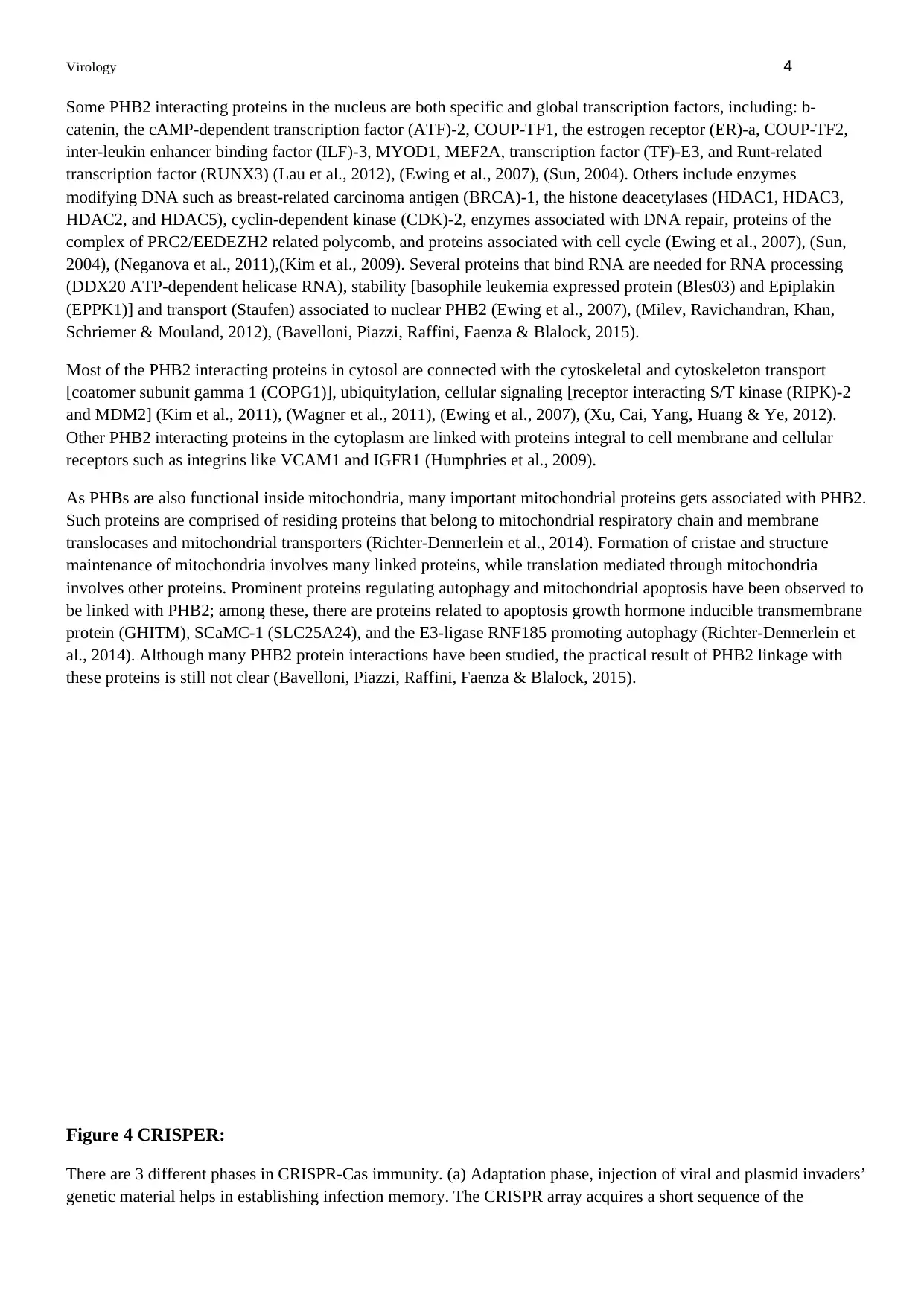
Figure 4 CRISPER:
There are 3 different phases in CRISPR-Cas immunity. (a) Adaptation phase, injection of viral and plasmid invaders’
genetic material helps in establishing infection memory. The CRISPR array acquires a short sequence of the
Some PHB2 interacting proteins in the nucleus are both specific and global transcription factors, including: b-
catenin, the cAMP-dependent transcription factor (ATF)-2, COUP-TF1, the estrogen receptor (ER)-a, COUP-TF2,
inter-leukin enhancer binding factor (ILF)-3, MYOD1, MEF2A, transcription factor (TF)-E3, and Runt-related
transcription factor (RUNX3) (Lau et al., 2012), (Ewing et al., 2007), (Sun, 2004). Others include enzymes
modifying DNA such as breast-related carcinoma antigen (BRCA)-1, the histone deacetylases (HDAC1, HDAC3,
HDAC2, and HDAC5), cyclin-dependent kinase (CDK)-2, enzymes associated with DNA repair, proteins of the
complex of PRC2/EEDEZH2 related polycomb, and proteins associated with cell cycle (Ewing et al., 2007), (Sun,
2004), (Neganova et al., 2011),(Kim et al., 2009). Several proteins that bind RNA are needed for RNA processing
(DDX20 ATP-dependent helicase RNA), stability [basophile leukemia expressed protein (Bles03) and Epiplakin
(EPPK1)] and transport (Staufen) associated to nuclear PHB2 (Ewing et al., 2007), (Milev, Ravichandran, Khan,
Schriemer & Mouland, 2012), (Bavelloni, Piazzi, Raffini, Faenza & Blalock, 2015).
Most of the PHB2 interacting proteins in cytosol are connected with the cytoskeletal and cytoskeleton transport
[coatomer subunit gamma 1 (COPG1)], ubiquitylation, cellular signaling [receptor interacting S/T kinase (RIPK)-2
and MDM2] (Kim et al., 2011), (Wagner et al., 2011), (Ewing et al., 2007), (Xu, Cai, Yang, Huang & Ye, 2012).
Other PHB2 interacting proteins in the cytoplasm are linked with proteins integral to cell membrane and cellular
receptors such as integrins like VCAM1 and IGFR1 (Humphries et al., 2009).
As PHBs are also functional inside mitochondria, many important mitochondrial proteins gets associated with PHB2.
Such proteins are comprised of residing proteins that belong to mitochondrial respiratory chain and membrane
translocases and mitochondrial transporters (Richter-Dennerlein et al., 2014). Formation of cristae and structure
maintenance of mitochondria involves many linked proteins, while translation mediated through mitochondria
involves other proteins. Prominent proteins regulating autophagy and mitochondrial apoptosis have been observed to
be linked with PHB2; among these, there are proteins related to apoptosis growth hormone inducible transmembrane
protein (GHITM), SCaMC-1 (SLC25A24), and the E3-ligase RNF185 promoting autophagy (Richter-Dennerlein et
al., 2014). Although many PHB2 protein interactions have been studied, the practical result of PHB2 linkage with
these proteins is still not clear (Bavelloni, Piazzi, Raffini, Faenza & Blalock, 2015).
Figure 4 CRISPER:
There are 3 different phases in CRISPR-Cas immunity. (a) Adaptation phase, injection of viral and plasmid invaders’
genetic material helps in establishing infection memory. The CRISPR array acquires a short sequence of the
Virology 5
infecting virus or plasmid (Jiang & Marraffini, 2015). The integration of this spacer sequence into the first recurrence
of the array through Cas1 and Cas2 happens with the copying of recurring sequence. The spacer recorded memory is
utilized for protecting the bacterial host against infection (Jiang & Marraffini, 2015). Transcription of spacer
sequence from promoter/leader region is followed by the processing of resulting transcript (pre crRNA or precursor
crRNA) into a short crRNA (biogenesis phase). This crRNA is utilized as a guide for specifying the cleavage’s target
through Cas nuclei (targeting phase) (Jiang & Marraffini, 2015). (b–d) Cas gene content is helpful in differentiating
three CRISPR-Cas system types. They have differences in crRNA biogenesis and targeting mechanisms, and can
possibly differ in adaptation mechanisms also. Closed and open types of arrowheads show cleavage of RNA and
DNA, respectively. Abbreviations: tracrRNA, trans-encoded crRNA; PAM, protospacer-adjacent motif; crRNA,
CRISPR RNA; RNAP, RNA polymerase (Jiang & Marraffini, 2015).
Figure 5 receptors:
There is an interaction between DENV E protein and lectin or other molecules that are expressed on the surface of
the host cell. DC-SIGN recognizes the N-Glycan at 67 position, and N-glycans at 67 and/or 153 positions may get
associated with lectin proteins of host such as proteins binding mannose. E protein is comprises three practical
domains. Domain I (red) is a hinge region that is flexible and helps in bringing protein conformational change under
endosomes’ conditions with low pH. Domain II (yellow) contains amino acid residues that are hydrophobic and
make a fusion loop at the domain tip. Moreover, this domain can also contribute to protein dimerization on the virus
membrane that is mature. Finally, domain III (blue) is considered to be responsible in characterizing direct
interaction with receptor molecules of host. The structure shows DENV3 E protein that was made using PyMol and
PDB accession number 1UZG (Hidari & Suzuki, 2011).
Virology 6
Cell Passage:
Cell passaging is the process of enabling an individual for keeping the cells alive and developing under cultured
conditions for prolonged time periods. When cells are 90%-100% confluent, they must be passed.
Materials:
Ethanol squirt bottle, cultured multi-well plates containing confluent cells, 5 mL, 10 mL sterile pipets, 1.5 ml
centrifuge tube, media, scraper, and automatic pipetters.
Procedure for Passaging Cells
Media is warmed in water-bath at 28C. Cells in cultured multi-well plates are checked under microscope in order to
confirm that cells are 90%-100% confluent. Hood is cleaned using ethanol. All the materials, bottles, etc. loaded in
the hood is sterilized. Gloves are sprayed with ethanol. Liquid jars are sprayed using ethanol. Sterile pipets are to be
placed directly in the hood. Entry of automatic pipetters in the hood is done without sterilization. Gloves are sprayed
again with ethanol. 2 ml media is transferred to 6-well plates using the automatic pipetters. 500 μl media in the
cultured wells (previous one) is discarded. Cells are detached from the wells’ bottom using the scraper. Cells are
checked under the microscope for confirming that cells have been detached from the surface. 100 μl detached cells
are pipetted to new wells with medium. Remaining quantity of the detached cells are pipetted to 1.5 ml centrifuge
tube and labelled for to lysate and extracting DNA (purification).
Cell Freezing:
Information is written on cryovial. Medium is removed through pipetting. Cells are suspended in 900 μl Fetal calf
serum (FCS). Adherent cells are scraped using scraper. 100 μl Dimethyl sulfoxide (freezing medium) is added, and
pipetted up and down for ensuring evenness of mixture and about 1ml is aliquoted into cryovial. Cryovial is kept in
cryovial container according to the freezer. Cells are immediately transferred to the freezer.
Preparing Lysates–Mini Kit
Mammalian Cells Lysate
Heat block or water bath is set at 55°C. 500 μl growth medium is removed from the culture plate and cells are
harvested through scraping and 100 μl is transferred to a new well with 2ml of fresh media for passaging. The rest is
transferred to an Eppendorf tube. It is spun down using minifuge for 1 minute, and supernatant is removed. Cells are
resuspended from previous step into 200 μl PBS. 20 μL of Proteinase K is added to the sample. 20 μL of RNase A is
added to the sample. It is then mixed well by brief vortexing, and incubated for 2 minutes at room temperature. 200
μL of PureLinkR Genomic Lysis/Binding Buffer is added and mixed well by vortexing for obtaining a homogenous
solution. For promoting digestion of protein, it is incubated for 10 minutes at 55°C. 200 μL 96–100% ethanol is
added to lysate. Through vortexing it is mixed for 5 seconds for yielding a homogenous solution. The next step is to
proceed immediately for the process Binding DNA.
Purification Procedure Using Spin Columns:
Binding DNA
PureLinkR Spin column in the Collection Tube is removed from the package. The lysate (~640 μL) that is prepared
with PureLinkR Genomic Lysis/Binding Buffer and ethanol is added to the PureLinkR Spin Column. Column is
centrifuged at 10,000 × g at room temperature for 1 minute. Flow through is discarded and the spin column is
placed into the Collection Tube of PureLinkR that was supplied with the kit. The next step is to proceed for the
process of Washing DNA.
Washing DNA
500 μL of Wash Buffer 1 which is prepared by ethanol (page 23) is added to the column. Column is centrifuged at
10,000 × g at room temperature for 1 minute. Flow through is discarded and the spin column is then placed into the
same collection tube of PureLinkR that was supplied with the kit. 500 μL of Wash Buffer 2 that is prepared by
Secure Best Marks with AI Grader
Need help grading? Try our AI Grader for instant feedback on your assignments.
Virology 7
ethanol (page 23) is added to the column. The column is centrifuged at maximum speed at room temperature for 3
minutes and the flow through is discarded. It is then spun again for 1 minute at maximum speed, and the collection
tube is discarded. The next step is to proceed for Eluting DNA.
Eluting DNA
Spin column is placed in a new collection tube. 25μL sterile MQW water is added to the column. It is incubated for 1
minute at room temperature. The column is centrifuged for 1 minute at maximum speed and at room temperature.
The tube now contains purified genomic DNA. Second elution step is performed using the elution buffer volume as
first elution into the sterile collection tube. The column is centrifuged at maximum speed at room temperature for 2
minutes. Purified genomic DNA is transferred to new Eppendorf tube.
DNA Quantification:
DNA quantification and inspection of protein level and RNA contamination can be done using NanoDrops, which
are referred to by:
- 260/230 ratio: RNA contamination indicator (for value lesser than 1.8).
- 260/280 ratio: Protein contamination indicator (for value lesser than 1.8).
Method:
First step is click on the button by the name of “Nucleic Acid” in the software of Nano Drop, and then compute pure
water of 2 μl to the curtailed podium then curtailed the upper arm next step is to click “Okay” button on the desktop
and wait till the sound of click will arise after the clock sound, drive the upper arm and arid the podium with a swab.
Next step is vacant by computing antiseptic water of 2 μl (MQW), that should be similar which is used in halt of
DNA then reduce the Nano Drop upper arm and click the button by the name of “Blank” on the computer program
and then wait for the measurement which is blank to be created after this step drive the upper arm and arid the
podium with the swab. Next step is to measure the specimen through computing the sample of 2μl in lower podium
then reduce the upper arm after that accommodate all the data and last step is to compute pure water of 2μl to the
curtailed podium after that lower the arm them drive the upper arm and with the help of swab to mop both lower and
upper podium.
Agarose Gel Electrophoresis
Background Information
The basic process of lab for disconnecting DNA by size (such as length in basic combination) is Gel electrophoresis
in order to visualize and purified. Electrophoresis make use of electrical path for shifting the DNA which is of
negatively charged with the help of agarose gel matrix to electrode which is positive. Smaller portion of DNA drift
by the agarose gel quicker than lengthy ones. Therefore, you can identify the rough length of the DNA portion
through working it on an agarose gel along the side of a system of DNA (accommodation of portion of DNA of
familiar lengths).
Protocol: Gel Electrophoresis
PREPARING a 2% AGAROSE GEL:
2 g of agarose is measured. Agarose powder is mixed in a microwavable flask with 100 mL 1xTAE.
Note: It should be ensured that the same buffer is used as the one placed in the gel box (different buffers
should not be mixed and water should not be used).
It is then microwaved for about 1-3 min till the agarose is not dissolved completely (it is suggested to
microwave for only one minute, stop it and swirl the solution, and then it should be boiled). Agarose
solution is left for cooling down for about 6 mins. Ethidium bromide (EtBr) is added to the final
concentration of 5μl for 50 mL (or 10 μg for 100 mL) approximately. EtBr helps in binding to the DNA
and allows visualization of the DNA UV (under ultraviolet) light. (EtBr is a previously known mutagen).
Agarose is poured into the gel tray having in place well comb.
Virology 8
Note: it should be poured slowly for preventing the bubble formation that will disrupt the gel. Bubbles
formed can be pulled away using the pipette tip.
Newly poured gel is placed at room temperature till it gets solidified completely.
Loading Samples and Running an Agarose Gel:
Once the agarose gel is solidified, it is placed in the electrophoresis unit (gel box). Gel box is filled with
1xTAE till the gel gets covered. With the help of parafilm, 2 μl loading dye + 5 μl Water + 5 μl Sample
DNA is added where each sample should be equal to 12 μl. 5 μl of the ladder is carefully loaded in the first
gel lane. 12 μl samples are carefully loaded in the additional gel wells. The gel is ran at 100 V till the line
of dye becomes approximately 75-80% (approx.1 hour) of the gel way down.
Note: Black is considered as negative, red is considered as positive. (The DNA is considered to be
negatively charged and it runs toward the positive electrode.) Always Run toward Red.
Note: Usually run time goes for about 1-1.5 hours, according to the gel voltage and concentration.
The gel is carefully removed from gel box. The DNA fragments are visualized using any UV device and
their bands are analysed by comparing them to the DNA ladder as it will behave as a size guide for each
band.
POLYMERASE CHAIN REACTION
An in-vitro mechanism which is used for the selective amplification of a fragment of double standard
nucleic acid is Polymerase chain reaction. For instance (RNA-Cdna or DNA-DNA). This amplification
system employs two short lengths of single-stranded DNA, oligonucleotide primers which are basically
chosen from the sequence of genome put under investigation to be called as target DNA. The size of the
amplified product called amplicon is determined by measuring the distance between the two primers. As
soon as the so called target DNA is made to heat at a temperature of 90oC-95oC, it separates the two strands
and this can be termed as denature. On reducing the temperature the oligonucleotide primers anneal i.e.
stick to a complementary sequence on DNA so targeted and on each strand. The step ahead to this is the
inducing the synthesis of new strands of DNA by a DNA polymerase enzyme and furthermore adding free
deoxynucleotide triphosphates for extending the annealed primers. The continuation of this cycle for 25
times whereby the three mechanisms play role denaturing, annealing and synthesizing fabricate around one
lakh prints of a single target DNA molecule thus duplicating the number of prints per round.
The materials provided for PCR protocol includes within its ambit an earlier prepared DNA, 10x PCR
buffer, 10mM deoxynucleotide triphosphate mixture, 20μM primer #1, 20μM primer #2, components of
PCR reaction mix, Amplitaq Gold® DNA polymerase and Ultrapure sterile distilled water
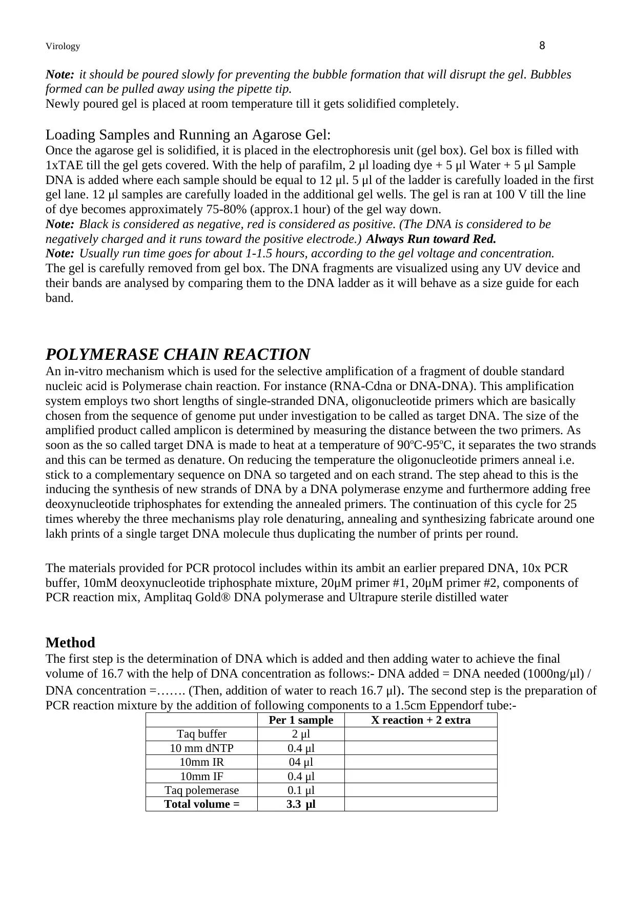
Method
The first step is the determination of DNA which is added and then adding water to achieve the final
volume of 16.7 with the help of DNA concentration as follows:- DNA added = DNA needed (1000ng/μl) /
DNA concentration =……. (Then, addition of water to reach 16.7 μl). The second step is the preparation of
PCR reaction mixture by the addition of following components to a 1.5cm Eppendorf tube:-
Per 1 sample X reaction + 2 extra
Taq buffer 2 μl
10 mm dNTP 0.4 μl
10mm IR 04 μl
10mm IF 0.4 μl
Taq polemerase 0.1 μl
Total volume = 3.3 μl
Note: it should be poured slowly for preventing the bubble formation that will disrupt the gel. Bubbles
formed can be pulled away using the pipette tip.
Newly poured gel is placed at room temperature till it gets solidified completely.
Loading Samples and Running an Agarose Gel:
Once the agarose gel is solidified, it is placed in the electrophoresis unit (gel box). Gel box is filled with
1xTAE till the gel gets covered. With the help of parafilm, 2 μl loading dye + 5 μl Water + 5 μl Sample
DNA is added where each sample should be equal to 12 μl. 5 μl of the ladder is carefully loaded in the first
gel lane. 12 μl samples are carefully loaded in the additional gel wells. The gel is ran at 100 V till the line
of dye becomes approximately 75-80% (approx.1 hour) of the gel way down.
Note: Black is considered as negative, red is considered as positive. (The DNA is considered to be
negatively charged and it runs toward the positive electrode.) Always Run toward Red.
Note: Usually run time goes for about 1-1.5 hours, according to the gel voltage and concentration.
The gel is carefully removed from gel box. The DNA fragments are visualized using any UV device and
their bands are analysed by comparing them to the DNA ladder as it will behave as a size guide for each
band.
POLYMERASE CHAIN REACTION
An in-vitro mechanism which is used for the selective amplification of a fragment of double standard
nucleic acid is Polymerase chain reaction. For instance (RNA-Cdna or DNA-DNA). This amplification
system employs two short lengths of single-stranded DNA, oligonucleotide primers which are basically
chosen from the sequence of genome put under investigation to be called as target DNA. The size of the
amplified product called amplicon is determined by measuring the distance between the two primers. As
soon as the so called target DNA is made to heat at a temperature of 90oC-95oC, it separates the two strands
and this can be termed as denature. On reducing the temperature the oligonucleotide primers anneal i.e.
stick to a complementary sequence on DNA so targeted and on each strand. The step ahead to this is the
inducing the synthesis of new strands of DNA by a DNA polymerase enzyme and furthermore adding free
deoxynucleotide triphosphates for extending the annealed primers. The continuation of this cycle for 25
times whereby the three mechanisms play role denaturing, annealing and synthesizing fabricate around one
lakh prints of a single target DNA molecule thus duplicating the number of prints per round.
The materials provided for PCR protocol includes within its ambit an earlier prepared DNA, 10x PCR
buffer, 10mM deoxynucleotide triphosphate mixture, 20μM primer #1, 20μM primer #2, components of
PCR reaction mix, Amplitaq Gold® DNA polymerase and Ultrapure sterile distilled water
Method
The first step is the determination of DNA which is added and then adding water to achieve the final
volume of 16.7 with the help of DNA concentration as follows:- DNA added = DNA needed (1000ng/μl) /
DNA concentration =……. (Then, addition of water to reach 16.7 μl). The second step is the preparation of
PCR reaction mixture by the addition of following components to a 1.5cm Eppendorf tube:-
Per 1 sample X reaction + 2 extra
Taq buffer 2 μl
10 mm dNTP 0.4 μl
10mm IR 04 μl
10mm IF 0.4 μl
Taq polemerase 0.1 μl
Total volume = 3.3 μl
Virology 9
The third step is placing of each tube in PCR heating block and further setting it to appropriate cycling
program as 94C / 2 minutes, 1 cycle ; 94C / 25 minutes; 54 C / 35 seconds; 68 C / 1 minute 30 cycles and
68 C / 7 minutes; 4 C / 1 hour respectively.
Clean-up mechanism of PCR- the cleaning of PCR product is carried out by using the illustra
exoprostar 1-step method and is carried out firstly by taking 5μl of each PCR product in order to
separate 0.2ml PCR tubes, secondly adding 2μl of illustra exoprostar 1-step to each PCR product and
grinding it well, next step is incubation at 37oC for 15 minutes and lastly for inactivation of enzymes,
incubating at 80oC for 15 minutes.
The concept of DNA sequencing is also known as Sanger sequencing, dideoxy sequencing or chain
termination sequencing and is based on the sole detection of chain-terminating nucleotides which have
been labelled and are thus incorporated by a DNA polymerase during its replication of template.
1 out of 10
Related Documents

Your All-in-One AI-Powered Toolkit for Academic Success.
+13062052269
info@desklib.com
Available 24*7 on WhatsApp / Email
![[object Object]](/_next/static/media/star-bottom.7253800d.svg)
Unlock your academic potential
© 2024 | Zucol Services PVT LTD | All rights reserved.