Advanced Biochemistry Practical: Baculovirus DNA and Plasmid Analysis
VerifiedAdded on 2021/04/16
|14
|2689
|63
Practical Assignment
AI Summary
This practical assignment in advanced biochemistry details the process of plasmid extraction from E. coli cells, followed by digestion with a restriction enzyme. The experiment aims to identify a specific DNA fragment from baculovirus inserted into the pTZ18U plasmid. The methods include plasmid extraction using a Wizard Plus SV Minipreps DNA purification kit, digestion with HindIII restriction enzyme, and gel electrophoresis to analyze the resulting DNA fragments. In silico analysis is also employed to determine which of the 52 baculovirus fragments was inserted into the parent plasmid, and to estimate the size of the insert. The report discusses the use of plasmids as cloning vectors, the properties of baculovirus as biological control agents, and the overall experimental procedures. The conclusion emphasizes the importance of plasmids in genetic research and the application of baculoviruses in pest control.
ADVANCED BIOCHEMISTRY 1
ADVANCED BIOCHEMISTRY
By Name
Course
Instructor
Institution
Location
Date
ADVANCED BIOCHEMISTRY
By Name
Course
Instructor
Institution
Location
Date
Paraphrase This Document
Need a fresh take? Get an instant paraphrase of this document with our AI Paraphraser
ADVANCED BIOCHEMISTRY 2
Table of Contents
1.0 Introduction..........................................................................................................................3
2.0 Aim/objectives.....................................................................................................................6
3.0 Materials and apparatus........................................................................................................6
4.0 Methods................................................................................................................................6
4.1 Plasmid extraction.....................................................................................................6
4.2 Plasmid digestion......................................................................................................8
4.3 Gel Electrophoresis...................................................................................................9
4.4 In silico analysis......................................................................................................10
5.0 Results................................................................................................................................10
6.0 Discussion..........................................................................................................................10
7.0 Conclusion..........................................................................................................................10
8.0 References..........................................................................................................................12
ADVANCED BIOCHEMISTRY 3
1.0 Introduction
Plasmids are very small, circular, double-stranded DNA molecules which are fully defined by
a cell's chromosomal DNA. In the bacteria cells, the plasmids exist naturally, and they also
exist in some types of eukaryotes (Akiyama, 2016, p. 423). Regularly the genes which are
carried in the plasmids supply the bacteria with the genetic advantages which include the
antibiotic resistance. Plasmids have a wide range of the lengths from approximately one
thousand DNA base pairs to the hundreds of thousands of the base pair.
All the plasmids which are contained in the cell are copied when the bacterium divides in a
way that the daughter cells are presented with a copy of each of the plasmid. The bacteria can
also move the plasmids to one another through a process known as conjugation (Ausubel,
2011, p. 34).
Plasmids are used as the cloning vectors because the plasmids are essentially tiny, self-
contained genomes. A plasmid can be moved from one cell to another cell easily. The
plasmid also has the genes which control their replication. Hence they can be applied in the in
the movement and copying of any gene that can be inserted into the plasmid. The plasmids
are also known not to be having any detrimental effects on their host cells.
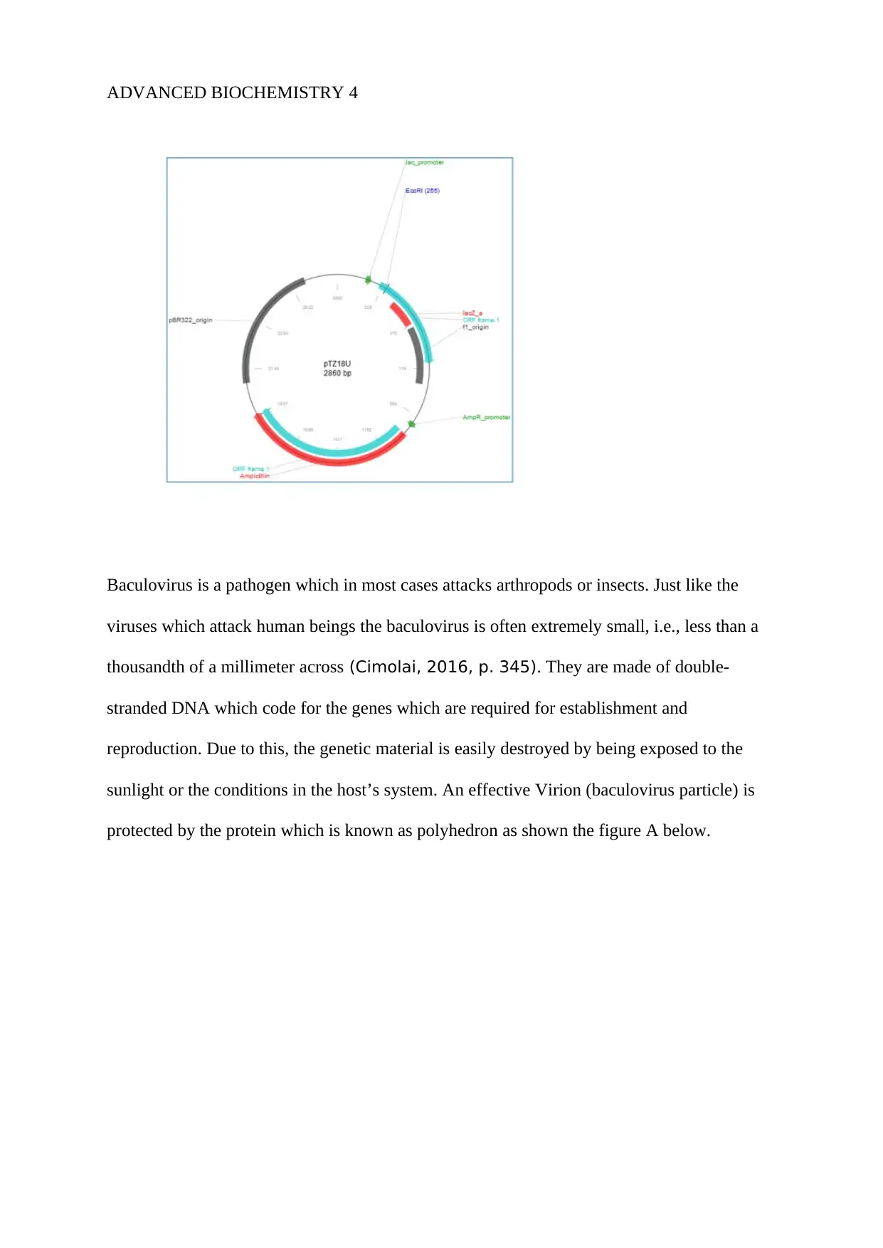
PTZ18U plasmid is a 2860 Pb in size, and it has multiple cloning sites which makes it easier
for cleavage by different restrictions endonucleases which include the single site restriction
which is for the restriction endonuclease. The figure below shows the generated pTZ18U
plasmid map (Biotechnology, 2015, p. 357).
1.0 Introduction
Plasmids are very small, circular, double-stranded DNA molecules which are fully defined by
a cell's chromosomal DNA. In the bacteria cells, the plasmids exist naturally, and they also
exist in some types of eukaryotes (Akiyama, 2016, p. 423). Regularly the genes which are
carried in the plasmids supply the bacteria with the genetic advantages which include the
antibiotic resistance. Plasmids have a wide range of the lengths from approximately one
thousand DNA base pairs to the hundreds of thousands of the base pair.
All the plasmids which are contained in the cell are copied when the bacterium divides in a
way that the daughter cells are presented with a copy of each of the plasmid. The bacteria can
also move the plasmids to one another through a process known as conjugation (Ausubel,
2011, p. 34).
Plasmids are used as the cloning vectors because the plasmids are essentially tiny, self-
contained genomes. A plasmid can be moved from one cell to another cell easily. The
plasmid also has the genes which control their replication. Hence they can be applied in the in
the movement and copying of any gene that can be inserted into the plasmid. The plasmids
are also known not to be having any detrimental effects on their host cells.
PTZ18U plasmid is a 2860 Pb in size, and it has multiple cloning sites which makes it easier
for cleavage by different restrictions endonucleases which include the single site restriction
which is for the restriction endonuclease. The figure below shows the generated pTZ18U
plasmid map (Biotechnology, 2015, p. 357).
⊘ This is a preview!⊘
Do you want full access?
Subscribe today to unlock all pages.
Trusted by 1+ million students worldwide
ADVANCED BIOCHEMISTRY 4
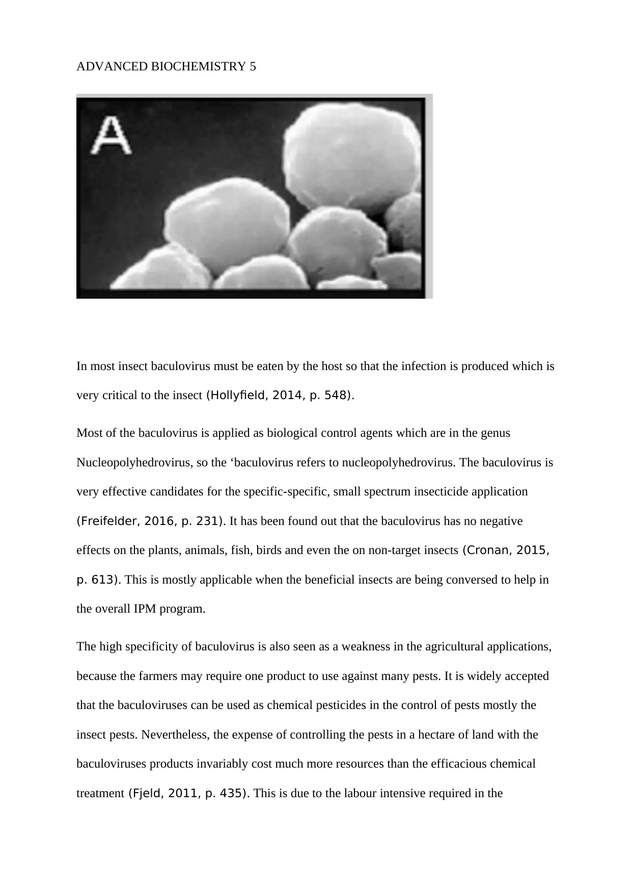
Baculovirus is a pathogen which in most cases attacks arthropods or insects. Just like the
viruses which attack human beings the baculovirus is often extremely small, i.e., less than a
thousandth of a millimeter across (Cimolai, 2016, p. 345). They are made of double-
stranded DNA which code for the genes which are required for establishment and
reproduction. Due to this, the genetic material is easily destroyed by being exposed to the
sunlight or the conditions in the host’s system. An effective Virion (baculovirus particle) is
protected by the protein which is known as polyhedron as shown the figure A below.
Paraphrase This Document
Need a fresh take? Get an instant paraphrase of this document with our AI Paraphraser
ADVANCED BIOCHEMISTRY 5
In most insect baculovirus must be eaten by the host so that the infection is produced which is
very critical to the insect (Hollyfield, 2014, p. 548).
Most of the baculovirus is applied as biological control agents which are in the genus
Nucleopolyhedrovirus, so the ‘baculovirus refers to nucleopolyhedrovirus. The baculovirus is
very effective candidates for the specific-specific, small spectrum insecticide application
(Freifelder, 2016, p. 231). It has been found out that the baculovirus has no negative
effects on the plants, animals, fish, birds and even the on non-target insects (Cronan, 2015,
p. 613). This is mostly applicable when the beneficial insects are being conversed to help in
the overall IPM program.
The high specificity of baculovirus is also seen as a weakness in the agricultural applications,
because the farmers may require one product to use against many pests. It is widely accepted
that the baculoviruses can be used as chemical pesticides in the control of pests mostly the
insect pests. Nevertheless, the expense of controlling the pests in a hectare of land with the
baculoviruses products invariably cost much more resources than the efficacious chemical
treatment (Fjeld, 2011, p. 435). This is due to the labour intensive required in the

ADVANCED BIOCHEMISTRY 6
production of baculoviruses. The pAB2 was constructed by cloning a fragment of the
baculovirus DNA into an EcoRI-digested.
2.0 Aim/objectives.
The aims and objectives of conducting the practical session include:
To separate recombinant plasmid DNA from the E.coli cells and digest the separated plasmid
DNA with the restriction enzyme.
To find out which DNA fragment out of the 52 fragments was inserted into the pTZ18U to
achieve Pab2.
To estimate the size of the insert.
To determine which of the 52 fragments was inserted into the parent plasmid.
3.0 Materials and apparatus
The following materials and apparatus were used during the practical session:
Frozen pellets of E.coli cells which are transformed with the recombinant plasmid AB2
which contains an insert of the baculovirus DNA (Howard, 2017, p. 43).
Wizard Plus SV Minipreps DNA purification kit containing the following: cell lysis solution,
cell resuspension solution, alkaline protease solution, wash solution, neutralization solution,
and spin columns, nuclease-free water, and the collection tubes.
Restriction enzyme HindIII: 10 units/μL (10U/ μL)
Restriction enzyme buffer, i.e., HindIII buffer.
4.0 Methods.
During the practical session, the following procedures were followed.
⊘ This is a preview!⊘
Do you want full access?
Subscribe today to unlock all pages.
Trusted by 1+ million students worldwide
ADVANCED BIOCHEMISTRY 7
4.1 Plasmid extraction
The wizard SV mini Minipreps used for the extraction of the plasmid DNA.
The following procedures were used for Production of the cells Iysate.
The frozen E.coli pallet which was provided was resuspended in the 250μL of cell
resuspension solution and then stirred well.
250μL of the cell Iysis was added and then mixed well by inverting the tubes for a few
seconds.
10μL of the alkaline protease solution was added, and the mixture was mixed well.
The tube was incubated for 5 minutes at room temperature (Whitworth, 2013, p. 56).
350μL of the neutralization solution was added and then mixed thoroughly.
The tube was centrifuged at 14500 rpm for 10mins.
For binding of the plasmid DNA.
The spin column was inserted into the collection tube.
The supernatant was transferred into the spin Column to avoid the pallets.
The tube was centrifuged for 1 minute at 14500 rpm, and the flow through was discarded.
Re-insert the spin column in the same collection tube (Sambrook, 2015, p. 73).
For Washing
750μL of the washing solution was added to the spin column.
The tube was centrifuged for 1 minute, and the flow-through was discarded.
The spin column was re-inserted into the same collection tube.
Paraphrase This Document
Need a fresh take? Get an instant paraphrase of this document with our AI Paraphraser
ADVANCED BIOCHEMISTRY 8
250μL of the washing solution was added to the spin column.
The tube was centrifuged for two mins at 14500rmp, and the collection tube was discarded.
The spin column was transferred into a sterile 1.5 mL microfuge tube (Rhen, 2013, p.
760).
Elution
50μL of the Nuclease-Free water was added to the spin column.
Then centrifuged at 14,000 rpm for 1 minute.
The spin column was discarded, and the 50μL plasmid DNA was stored on the ice for the
next procedures.
4.2 Plasmid digestion
The restrictive enzyme was held on the ice all the time ensuring that it was not left without
ice at any given length of time because the enzymes are highly thermo-sensitive and also they
are very expensive as the other students will require using the same tubes (Preston, 2015,
p. 391).
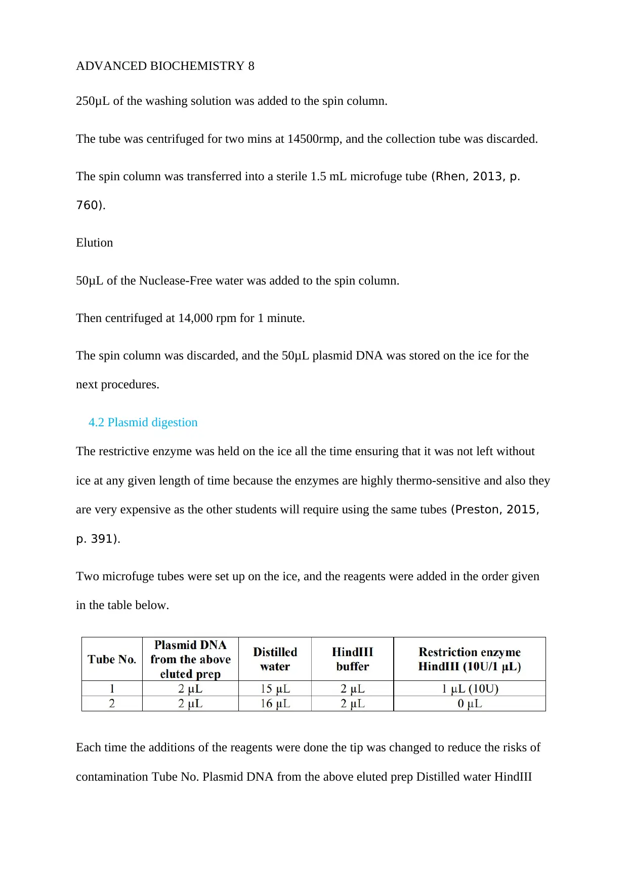
Two microfuge tubes were set up on the ice, and the reagents were added in the order given
in the table below.
Each time the additions of the reagents were done the tip was changed to reduce the risks of
contamination Tube No. Plasmid DNA from the above eluted prep Distilled water HindIII
250μL of the washing solution was added to the spin column.
The tube was centrifuged for two mins at 14500rmp, and the collection tube was discarded.
The spin column was transferred into a sterile 1.5 mL microfuge tube (Rhen, 2013, p.
760).
Elution
50μL of the Nuclease-Free water was added to the spin column.
Then centrifuged at 14,000 rpm for 1 minute.
The spin column was discarded, and the 50μL plasmid DNA was stored on the ice for the
next procedures.
4.2 Plasmid digestion
The restrictive enzyme was held on the ice all the time ensuring that it was not left without
ice at any given length of time because the enzymes are highly thermo-sensitive and also they
are very expensive as the other students will require using the same tubes (Preston, 2015,
p. 391).
Two microfuge tubes were set up on the ice, and the reagents were added in the order given
in the table below.
Each time the additions of the reagents were done the tip was changed to reduce the risks of
contamination Tube No. Plasmid DNA from the above eluted prep Distilled water HindIII
ADVANCED BIOCHEMISTRY 9
buffer Restriction enzyme HindIII (10U/1 μL) 1 2 μL 15 μL two μL one μL (10U) 2 2 μL 16
μL two μL 0 μL (Kendrick, 2017, p. 78).
The above tubes were incubated in the water bath for 45 mins which were set at 37ºC.
After that, the preparation of the gel for electrophoresis started during the incubation time.
Add 5μL of the provided gel loading dye was added after incubation to both of the tubes and
then mixed thoroughly then stored on the ice until loading these samples onto the gels
(Mulhardt, 2012, p. 453).
4.3 Gel Electrophoresis
Prepare 300ml of the 1x TAE electrophoresis buffer by diluting the 10x TAE buffer with
270mL 0f distilled water.
The end of the gel-casting tray was sealed by use of the masking tape and the 8-well slot
former (comb) onto the gel-casting tray about 1 cm from the end.
0.3 g of agarose was weighed out directly into a 100ML conical flask and 30mL of the 1x
TAE buffer added to it and then heated in a microwave for 1 minute.
The flask was carefully taken out of the microwave, then held against light to see if all the
agarose melted to form a clear solution. In the cases where there were particles, the flask was
heated again for 10-20 seconds until all the particles had melted. Then the gel was cooled 50-
55 ºC (Mulhardt, 2012, p. 364).
3μL of the Gel Red Staining agent was added to the gel solution once it cooled to the
required temperature.
The gel solution was poured gently along a side of the flack into the tray, ensuring that there
was no air bubble which was forming. In the cases where the air bubbles formed they were
⊘ This is a preview!⊘
Do you want full access?
Subscribe today to unlock all pages.
Trusted by 1+ million students worldwide
ADVANCED BIOCHEMISTRY 10
quickly broken down by use of the clean pipette tip and then the gel was left to solidify for
15-20 minutes.
Once the gel had cooled, the masking tape was removed and then the comb was removed
gently. Then the gel t6ray was transferred into the gel tank. Then the remaining 1x TAE
buffer into the gel tank.
The four separate wells were load into the gel as follows: (i) tube 1 above: your 10 μL
restriction digest (ii) tube 2 above: your 10 μL digest no-enzyme control; (iii) 4 μL of DNA
molecular weight marker (Gene Ruler), and (iv) 5 μL of EcoRI-digested pAB2 (100ng/μL)
(provided to you).
After all the wells were load into the gel, the electrophoresis equipment was turned on. The
gel was electrophoresed for 35-40 mins at 120V.
When the electrophoresis process was through the demonstrator turned off the equipment,
and the gel tray was carefully removed, and the gel was slid into a press‐seal plastic bag.
The gel was inspected on the UV-light box with a lot of caution of the UV radiations, and the
image of the gel was captured by use of the gel documentation camera.
The electrode was disposed carefully down the sink, and the apparatus was thoroughly
cleaned. The image of the gel was saved on the email or the memory stick from the hard
drive (Marbois, 2015, p. 211).
The image was shown to the demonstrator and ensured that it was recorded.
4.4 In silico analysis
During the process of In silico analysis, the following procedures were followed.
Determination of baculovirus fragment sequence – FASTA format
Paraphrase This Document
Need a fresh take? Get an instant paraphrase of this document with our AI Paraphraser
ADVANCED BIOCHEMISTRY 11
Determination of baculovirus fragment sequence – FASTA format
Determination of parent plasmid pTZ18U sequence - FASTA format.
Construction of recombinant plasmid to identify the correct insert
Identification of the correct insert (Maloy, 2014, p. 115).
5.0 Results.
6.0 Discussion.
7.0 Conclusion.
In conclusion, Plasmids are very small, circular, double-stranded DNA molecules which are
fully defined by a cell's chromosomal DNA. In the bacteria cells, the plasmids exist naturally,
and they also exist in some types of eukaryotes.
Plasmids are used as the cloning vectors because the plasmids are essentially tiny, self-
contained genomes. A plasmid can be moved from one cell to another cell easily. The
plasmid also has the genes which control their replication. It is widely accepted that the
baculoviruses can be used as chemical pesticides in the control of pests mostly the insect
pests (Hurwitz, 2013, p. 56).
The practical session was conducted in two parts. In the first phase, the recombinant plasmid
DNA was isolated from the E.coli cells and the separated plasmid DNA digested with the
restriction enzyme. In the second phase of the practice session the in silico restriction
digestion of pAB2 and baculovirus to estimate which one of the 52 fragments were inserted
into the parent plasmid was performed (Lipps, 2014, p. 42).
ADVANCED BIOCHEMISTRY 12
8.0 References.
Akiyama, H., 2016. Bioelectrics. 4th ed. London: Springer.
Anon., n.d. Microbial Genetics. s.l.:s.n.
Ausubel, F. M., 2011. Current Protocols in Molecular Biology, Volume 1. 4th ed. Texas:
John Wiley & Sons,
Biotechnology, I. D. o., 2015. Annual Report. 5th ed. Paris: Department of Biotechnology,
Ministry of Science & Technology.
Cimolai, N., 2016. Laboratory Diagnosis of Bacterial Infections. 1st ed. London: Taylor &
Francis,
Cronan, J. E., 2015. Can J Microbiol, 1st ed. Texas: National Research Council of Canada.
⊘ This is a preview!⊘
Do you want full access?
Subscribe today to unlock all pages.
Trusted by 1+ million students worldwide
1 out of 14
Related Documents


Your All-in-One AI-Powered Toolkit for Academic Success.
+13062052269
info@desklib.com
Available 24*7 on WhatsApp / Email
![[object Object]](/_next/static/media/star-bottom.7253800d.svg)
Unlock your academic potential
Copyright © 2020–2026 A2Z Services. All Rights Reserved. Developed and managed by ZUCOL.