Analysis of Alveolar Ventilation-Perfusion Relationships in Lungs
VerifiedAdded on 2022/08/14
|5
|1002
|23
Report
AI Summary
This report examines the alveolar ventilation-perfusion (V/Q) relationships within the lungs, focusing on the critical process of gas exchange between the alveoli and pulmonary capillaries. It discusses how diffusion, alveolar ventilation, and pulmonary capillary perfusion maintain gas concentration differences, highlighting the impact of V/Q ratios on oxygen and carbon dioxide levels. The report covers the effects of alveolar hypoventilation, pulmonary shunts, and the calculation of the Bohr-Enghoff dead space and pulmonary shunt fraction. It also addresses the causes of hypoxemia and shock, emphasizing the importance of monitoring and healthcare interventions to prevent respiratory complications. References to key research papers are provided to support the analysis.

Running Head: ALVEOLAR VENTILATION-PERFUSION RELATIONSHIPS
ALVEOLAR VENTILATION-PERFUSION RELATIONSHIPS
Name of the Student:
Name of the University:
Author Note:
ALVEOLAR VENTILATION-PERFUSION RELATIONSHIPS
Name of the Student:
Name of the University:
Author Note:
Paraphrase This Document
Need a fresh take? Get an instant paraphrase of this document with our AI Paraphraser

1ALVEOLAR VENTILATION-PERFUSION RELATIONSHIPS
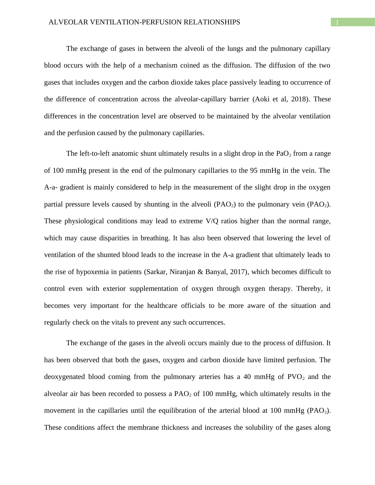
The exchange of gases in between the alveoli of the lungs and the pulmonary capillary
blood occurs with the help of a mechanism coined as the diffusion. The diffusion of the two
gases that includes oxygen and the carbon dioxide takes place passively leading to occurrence of
the difference of concentration across the alveolar-capillary barrier (Aoki et al, 2018). These
differences in the concentration level are observed to be maintained by the alveolar ventilation
and the perfusion caused by the pulmonary capillaries.
The left-to-left anatomic shunt ultimately results in a slight drop in the PaO2 from a range
of 100 mmHg present in the end of the pulmonary capillaries to the 95 mmHg in the vein. The
A-a- gradient is mainly considered to help in the measurement of the slight drop in the oxygen
partial pressure levels caused by shunting in the alveoli (PAO2) to the pulmonary vein (PAO2).
These physiological conditions may lead to extreme V/Q ratios higher than the normal range,
which may cause disparities in breathing. It has also been observed that lowering the level of
ventilation of the shunted blood leads to the increase in the A-a gradient that ultimately leads to
the rise of hypoxemia in patients (Sarkar, Niranjan & Banyal, 2017), which becomes difficult to
control even with exterior supplementation of oxygen through oxygen therapy. Thereby, it
becomes very important for the healthcare officials to be more aware of the situation and
regularly check on the vitals to prevent any such occurrences.
The exchange of the gases in the alveoli occurs mainly due to the process of diffusion. It
has been observed that both the gases, oxygen and carbon dioxide have limited perfusion. The
deoxygenated blood coming from the pulmonary arteries has a 40 mmHg of PVO2 and the
alveolar air has been recorded to possess a PAO2 of 100 mmHg, which ultimately results in the
movement in the capillaries until the equilibration of the arterial blood at 100 mmHg (PAO2).
These conditions affect the membrane thickness and increases the solubility of the gases along

2ALVEOLAR VENTILATION-PERFUSION RELATIONSHIPS
with the increase in the surface are of the membranes. The barriers involving the intrinsic
diffusion present between the alveoli and the capillaries results in the hypoxemia, that poses as
huge threat to the patient.
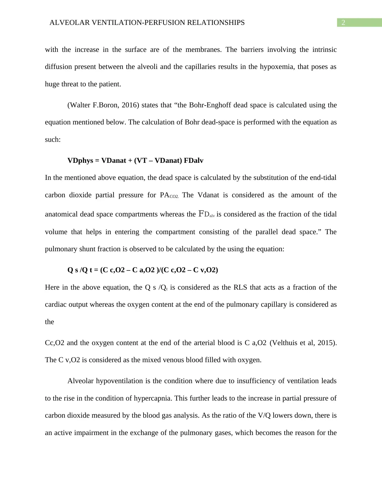
(Walter F.Boron, 2016) states that “the Bohr-Enghoff dead space is calculated using the
equation mentioned below. The calculation of Bohr dead-space is performed with the equation as
such:
VDphys = VDanat + (VT – VDanat) FDalv
In the mentioned above equation, the dead space is calculated by the substitution of the end-tidal
carbon dioxide partial pressure for PACO2. The Vdanat is considered as the amount of the
anatomical dead space compartments whereas the FDalv is considered as the fraction of the tidal
volume that helps in entering the compartment consisting of the parallel dead space.” The
pulmonary shunt fraction is observed to be calculated by the using the equation:
Q s /Q t = (C c,O2 – C a,O2 )/(C c,O2 – C v,O2)
Here in the above equation, the Q s /Qt is considered as the RLS that acts as a fraction of the
cardiac output whereas the oxygen content at the end of the pulmonary capillary is considered as
the
Cc,O2 and the oxygen content at the end of the arterial blood is C a,O2 (Velthuis et al, 2015).
The C v,O2 is considered as the mixed venous blood filled with oxygen.
Alveolar hypoventilation is the condition where due to insufficiency of ventilation leads
to the rise in the condition of hypercapnia. This further leads to the increase in partial pressure of
carbon dioxide measured by the blood gas analysis. As the ratio of the V/Q lowers down, there is
an active impairment in the exchange of the pulmonary gases, which becomes the reason for the
⊘ This is a preview!⊘
Do you want full access?
Subscribe today to unlock all pages.

Trusted by 1+ million students worldwide

3ALVEOLAR VENTILATION-PERFUSION RELATIONSHIPS
cause of low arterial partial pressure of Oxygen (PAO2). Shock can be however, be caused in the
body whenever there is a very poor blood flow or there is an inadequate mitochondrial
oxygenation caused to active lactic acid build up in the tissues or the blood.
cause of low arterial partial pressure of Oxygen (PAO2). Shock can be however, be caused in the
body whenever there is a very poor blood flow or there is an inadequate mitochondrial
oxygenation caused to active lactic acid build up in the tissues or the blood.
Paraphrase This Document
Need a fresh take? Get an instant paraphrase of this document with our AI Paraphraser

4ALVEOLAR VENTILATION-PERFUSION RELATIONSHIPS
References:
Aoki, K., Izumi, Y., Watanabe, W., Shimizu, Y., Osada, H., Honda, N., ... & Nakayama, M.
(2018). Generation of ventilation/perfusion ratio map in surgical patients by dual-energy
CT after xenon inhalation and intravenous contrast media. Journal of cardiothoracic
surgery, 13(1), 43.
Boron, W. F., & Boulpaep, E. L. (2016). Medical physiology E-book. Elsevier Health Sciences.
Petersson, J., & Glenny, R. W. (2014). Gas exchange and ventilation–perfusion relationships in
the lung..
Sarkar, M., Niranjan, N., & Banyal, P. K. (2017). Mechanisms of hypoxemia. Lung India:
official organ of Indian Chest Society, 34(1), 47.
Velthuis, S., Vorselaars, V. M., Westermann, C. J., Snijder, R. J., Mager, J. J., & Post, M. C.
(2015). Pulmonary shunt fraction measurement compared to contrast echocardiography in
hereditary haemorrhagic telangiectasia patients: time to abandon the 100% oxygen
method?. Respiration, 89(2), 112-118.
1 out of 5
Your All-in-One AI-Powered Toolkit for Academic Success.
+13062052269
info@desklib.com
Available 24*7 on WhatsApp / Email
![[object Object]](/_next/static/media/star-bottom.7253800d.svg)
Unlock your academic potential
Copyright © 2020–2026 A2Z Services. All Rights Reserved. Developed and managed by ZUCOL.


