Anatomy & Physiology Chapter 1
VerifiedAdded on 2019/09/16
|110
|57241
|88
Homework Assignment
AI Summary
This document provides solutions to a homework assignment covering Chapter 1 of an Anatomy and Physiology course. The content comprehensively explains the definitions of anatomy and physiology, their subspecialties, and the six levels of structural organization in the human body. It details the eleven body systems, their components, and functions. The document further elucidates the concept of homeostasis, feedback systems (negative and positive), and homeostatic imbalances. Basic anatomical terminology, including directional terms, planes, sections, and body cavities, is thoroughly explained. Finally, it describes various medical imaging techniques and the principles of protein synthesis and cell division (mitosis and meiosis).
ANATOMY AND PHYSIOLOGY DEFINED
OBJECTIVE
• Define anatomy and physiology, and name several subspecialties of these sciences.
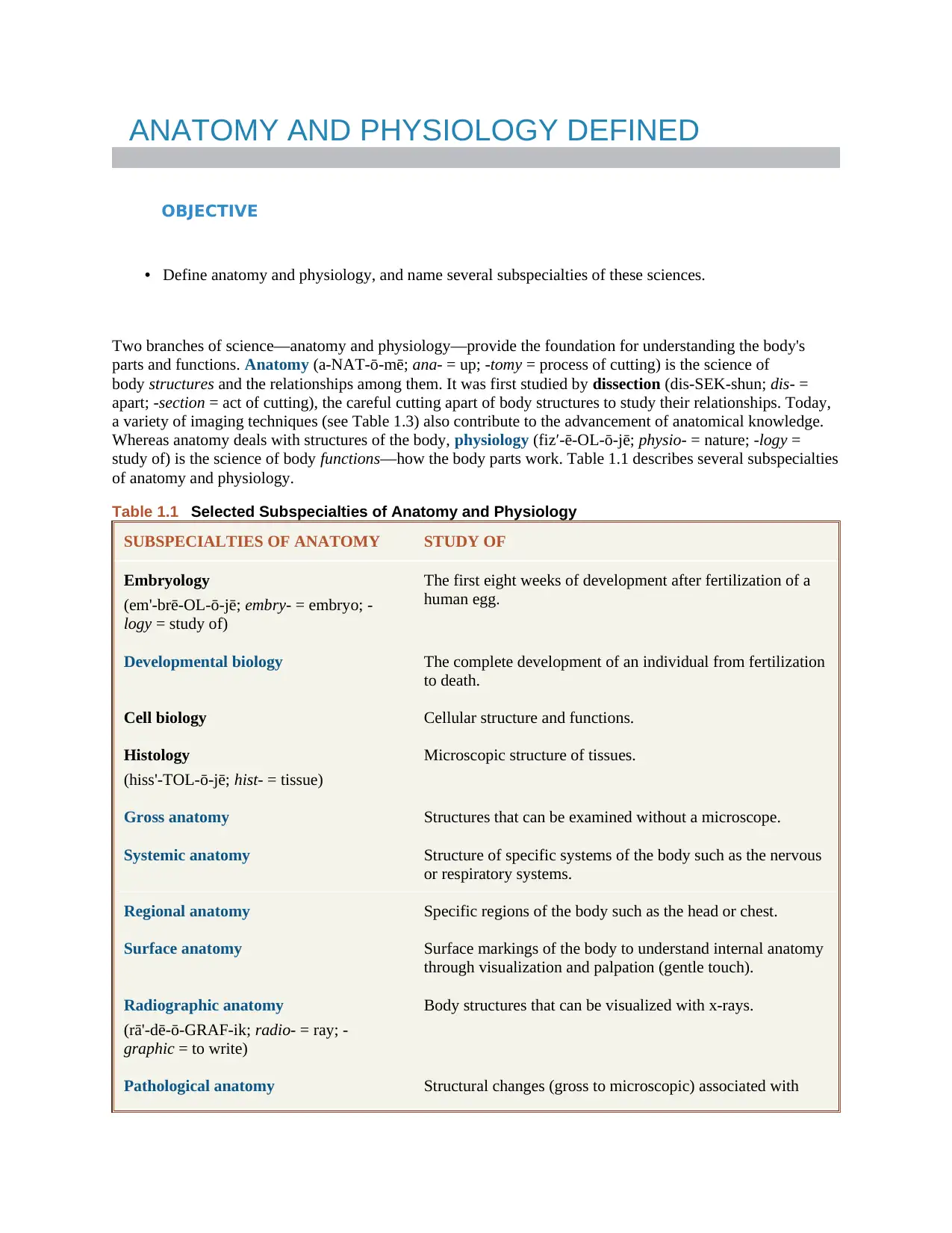
Two branches of science—anatomy and physiology—provide the foundation for understanding the body's
parts and functions. Anatomy (a‐NAT‐ō‐mē; ana‐ = up; ‐tomy = process of cutting) is the science of
body structures and the relationships among them. It was first studied by dissection (dis‐SEK‐shun; dis‐ =
apart; ‐section = act of cutting), the careful cutting apart of body structures to study their relationships. Today,
a variety of imaging techniques (see Table 1.3) also contribute to the advancement of anatomical knowledge.
Whereas anatomy deals with structures of the body, physiology (fiz′‐ē‐OL‐ō‐jē; physio‐ = nature; ‐logy =
study of) is the science of body functions—how the body parts work. Table 1.1 describes several subspecialties
of anatomy and physiology.
Table 1.1 Selected Subspecialties of Anatomy and Physiology
SUBSPECIALTIES OF ANATOMY STUDY OF
Embryology
(em'‐brē‐OL‐ō‐jē; embry‐ = embryo; ‐
logy = study of)
The first eight weeks of development after fertilization of a
human egg.
Developmental biology The complete development of an individual from fertilization
to death.
Cell biology Cellular structure and functions.
Histology
(hiss'‐TOL‐ō‐jē; hist‐ = tissue)
Microscopic structure of tissues.
Gross anatomy Structures that can be examined without a microscope.
Systemic anatomy Structure of specific systems of the body such as the nervous
or respiratory systems.
Regional anatomy Specific regions of the body such as the head or chest.
Surface anatomy Surface markings of the body to understand internal anatomy
through visualization and palpation (gentle touch).
Radiographic anatomy
(rā'‐dē‐ō‐GRAF‐ik; radio‐ = ray; ‐
graphic = to write)
Body structures that can be visualized with x‐rays.
Pathological anatomy Structural changes (gross to microscopic) associated with
OBJECTIVE
• Define anatomy and physiology, and name several subspecialties of these sciences.
Two branches of science—anatomy and physiology—provide the foundation for understanding the body's
parts and functions. Anatomy (a‐NAT‐ō‐mē; ana‐ = up; ‐tomy = process of cutting) is the science of
body structures and the relationships among them. It was first studied by dissection (dis‐SEK‐shun; dis‐ =
apart; ‐section = act of cutting), the careful cutting apart of body structures to study their relationships. Today,
a variety of imaging techniques (see Table 1.3) also contribute to the advancement of anatomical knowledge.
Whereas anatomy deals with structures of the body, physiology (fiz′‐ē‐OL‐ō‐jē; physio‐ = nature; ‐logy =
study of) is the science of body functions—how the body parts work. Table 1.1 describes several subspecialties
of anatomy and physiology.
Table 1.1 Selected Subspecialties of Anatomy and Physiology
SUBSPECIALTIES OF ANATOMY STUDY OF
Embryology
(em'‐brē‐OL‐ō‐jē; embry‐ = embryo; ‐
logy = study of)
The first eight weeks of development after fertilization of a
human egg.
Developmental biology The complete development of an individual from fertilization
to death.
Cell biology Cellular structure and functions.
Histology
(hiss'‐TOL‐ō‐jē; hist‐ = tissue)
Microscopic structure of tissues.
Gross anatomy Structures that can be examined without a microscope.
Systemic anatomy Structure of specific systems of the body such as the nervous
or respiratory systems.
Regional anatomy Specific regions of the body such as the head or chest.
Surface anatomy Surface markings of the body to understand internal anatomy
through visualization and palpation (gentle touch).
Radiographic anatomy
(rā'‐dē‐ō‐GRAF‐ik; radio‐ = ray; ‐
graphic = to write)
Body structures that can be visualized with x‐rays.
Pathological anatomy Structural changes (gross to microscopic) associated with
Paraphrase This Document
Need a fresh take? Get an instant paraphrase of this document with our AI Paraphraser
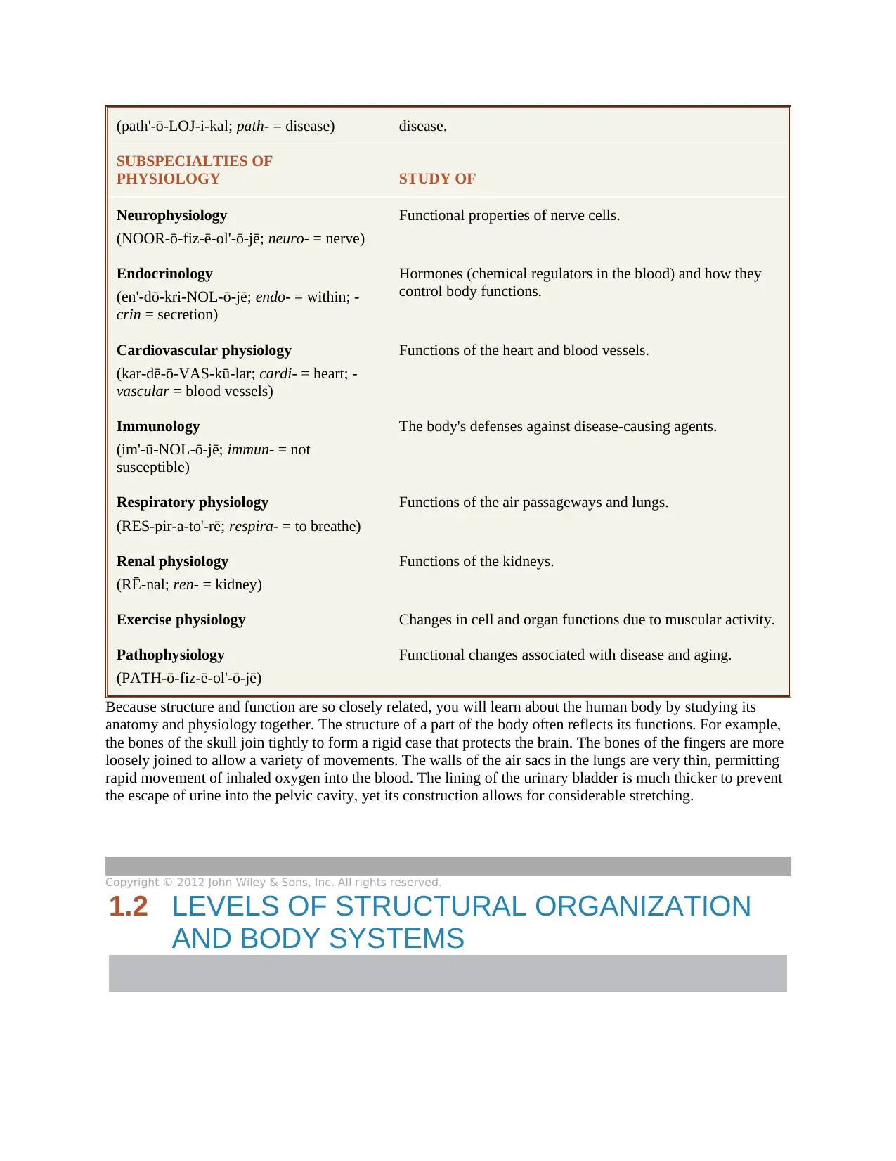
(path'‐ō‐LOJ‐i‐kal; path‐ = disease) disease.
SUBSPECIALTIES OF
PHYSIOLOGY STUDY OF
Neurophysiology
(NOOR‐ō‐fiz‐ē‐ol'‐ō‐jē; neuro‐ = nerve)
Functional properties of nerve cells.
Endocrinology
(en'‐dō‐kri‐NOL‐ō‐jē; endo‐ = within; ‐
crin = secretion)
Hormones (chemical regulators in the blood) and how they
control body functions.
Cardiovascular physiology
(kar‐dē‐ō‐VAS‐kū‐lar; cardi‐ = heart; ‐
vascular = blood vessels)
Functions of the heart and blood vessels.
Immunology
(im'‐ū‐NOL‐ō‐jē; immun‐ = not
susceptible)
The body's defenses against disease‐causing agents.
Respiratory physiology
(RES‐pir‐a‐to'‐rē; respira‐ = to breathe)
Functions of the air passageways and lungs.
Renal physiology
(RĒ‐nal; ren‐ = kidney)
Functions of the kidneys.
Exercise physiology Changes in cell and organ functions due to muscular activity.
Pathophysiology
(PATH‐ō‐fiz‐ē‐ol'‐ō‐jē)
Functional changes associated with disease and aging.
Because structure and function are so closely related, you will learn about the human body by studying its
anatomy and physiology together. The structure of a part of the body often reflects its functions. For example,
the bones of the skull join tightly to form a rigid case that protects the brain. The bones of the fingers are more
loosely joined to allow a variety of movements. The walls of the air sacs in the lungs are very thin, permitting
rapid movement of inhaled oxygen into the blood. The lining of the urinary bladder is much thicker to prevent
the escape of urine into the pelvic cavity, yet its construction allows for considerable stretching.
Copyright © 2012 John Wiley & Sons, Inc. All rights reserved.
1.2 LEVELS OF STRUCTURAL ORGANIZATION
AND BODY SYSTEMS
OBJECTIVES
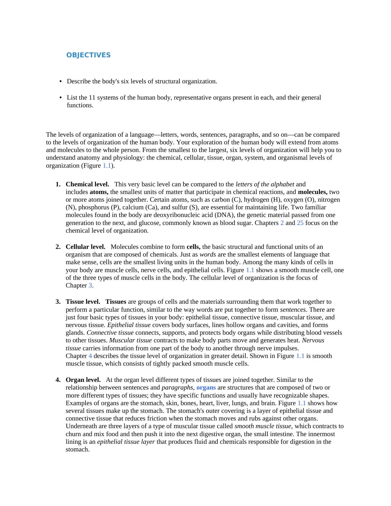
• Describe the body's six levels of structural organization.
• List the 11 systems of the human body, representative organs present in each, and their general
functions.
The levels of organization of a language—letters, words, sentences, paragraphs, and so on—can be compared
to the levels of organization of the human body. Your exploration of the human body will extend from atoms
and molecules to the whole person. From the smallest to the largest, six levels of organization will help you to
understand anatomy and physiology: the chemical, cellular, tissue, organ, system, and organismal levels of
organization (Figure 1.1).
1. Chemical level. This very basic level can be compared to the letters of the alphabet and
includes atoms, the smallest units of matter that participate in chemical reactions, and molecules, two
or more atoms joined together. Certain atoms, such as carbon (C), hydrogen (H), oxygen (O), nitrogen
(N), phosphorus (P), calcium (Ca), and sulfur (S), are essential for maintaining life. Two familiar
molecules found in the body are deoxyribonucleic acid (DNA), the genetic material passed from one
generation to the next, and glucose, commonly known as blood sugar. Chapters 2 and 25 focus on the
chemical level of organization.
2. Cellular level. Molecules combine to form cells, the basic structural and functional units of an
organism that are composed of chemicals. Just as words are the smallest elements of language that
make sense, cells are the smallest living units in the human body. Among the many kinds of cells in
your body are muscle cells, nerve cells, and epithelial cells. Figure 1.1 shows a smooth muscle cell, one
of the three types of muscle cells in the body. The cellular level of organization is the focus of
Chapter 3.
3. Tissue level. Tissues are groups of cells and the materials surrounding them that work together to
perform a particular function, similar to the way words are put together to form sentences. There are
just four basic types of tissues in your body: epithelial tissue, connective tissue, muscular tissue, and
nervous tissue. Epithelial tissue covers body surfaces, lines hollow organs and cavities, and forms
glands. Connective tissue connects, supports, and protects body organs while distributing blood vessels
to other tissues. Muscular tissue contracts to make body parts move and generates heat. Nervous
tissue carries information from one part of the body to another through nerve impulses.
Chapter 4 describes the tissue level of organization in greater detail. Shown in Figure 1.1 is smooth
muscle tissue, which consists of tightly packed smooth muscle cells.
4. Organ level. At the organ level different types of tissues are joined together. Similar to the
relationship between sentences and paragraphs, organs are structures that are composed of two or
more different types of tissues; they have specific functions and usually have recognizable shapes.
Examples of organs are the stomach, skin, bones, heart, liver, lungs, and brain. Figure 1.1 shows how
several tissues make up the stomach. The stomach's outer covering is a layer of epithelial tissue and
connective tissue that reduces friction when the stomach moves and rubs against other organs.
Underneath are three layers of a type of muscular tissue called smooth muscle tissue, which contracts to
churn and mix food and then push it into the next digestive organ, the small intestine. The innermost
lining is an epithelial tissue layer that produces fluid and chemicals responsible for digestion in the
stomach.
⊘ This is a preview!⊘
Do you want full access?
Subscribe today to unlock all pages.
Trusted by 1+ million students worldwide
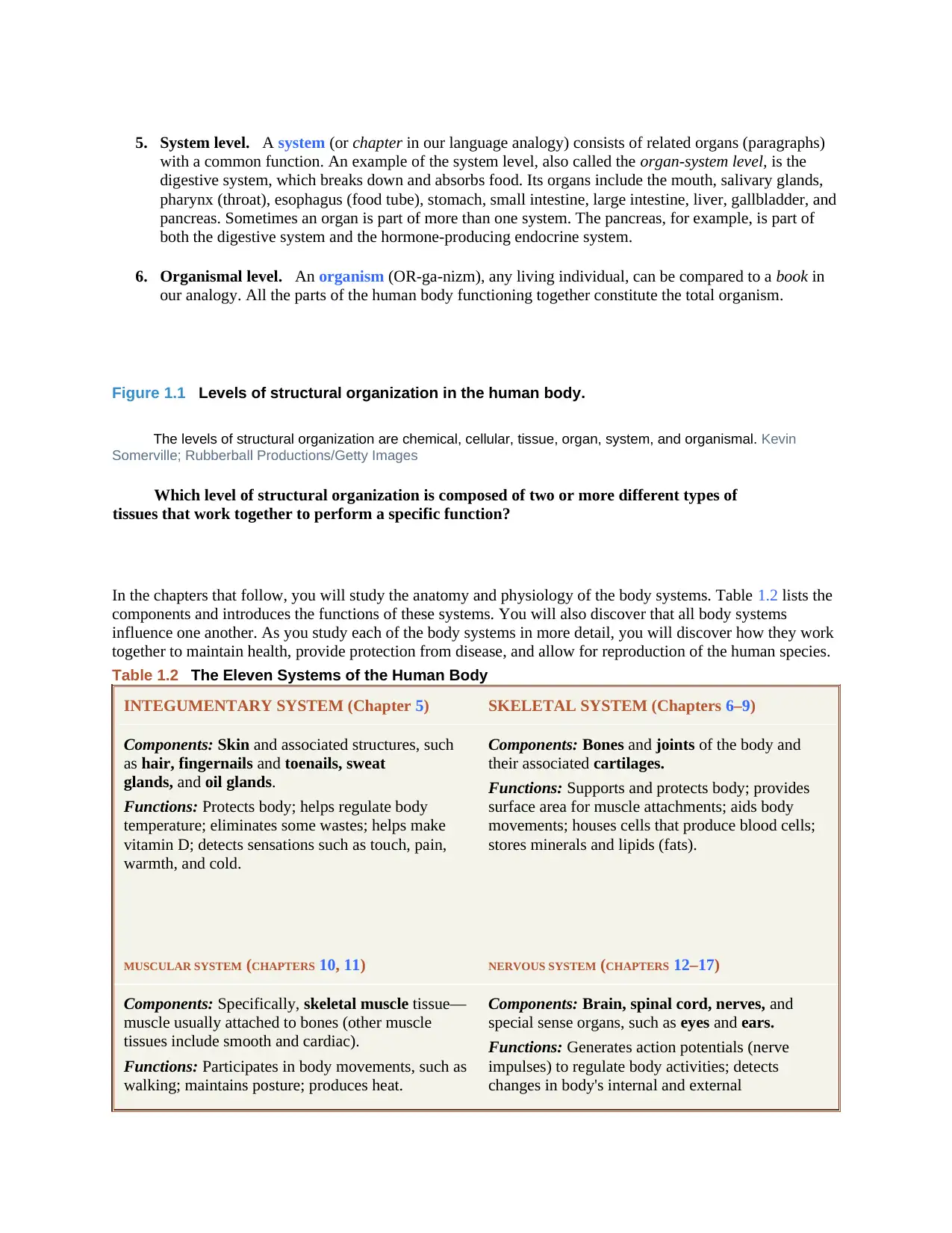
5. System level. A system (or chapter in our language analogy) consists of related organs (paragraphs)
with a common function. An example of the system level, also called the organ‐system level, is the
digestive system, which breaks down and absorbs food. Its organs include the mouth, salivary glands,
pharynx (throat), esophagus (food tube), stomach, small intestine, large intestine, liver, gallbladder, and
pancreas. Sometimes an organ is part of more than one system. The pancreas, for example, is part of
both the digestive system and the hormone‐producing endocrine system.
6. Organismal level. An organism (OR‐ga‐nizm), any living individual, can be compared to a book in
our analogy. All the parts of the human body functioning together constitute the total organism.
Figure 1.1 Levels of structural organization in the human body.
The levels of structural organization are chemical, cellular, tissue, organ, system, and organismal. Kevin
Somerville; Rubberball Productions/Getty Images
Which level of structural organization is composed of two or more different types of
tissues that work together to perform a specific function?
In the chapters that follow, you will study the anatomy and physiology of the body systems. Table 1.2 lists the
components and introduces the functions of these systems. You will also discover that all body systems
influence one another. As you study each of the body systems in more detail, you will discover how they work
together to maintain health, provide protection from disease, and allow for reproduction of the human species.
Table 1.2 The Eleven Systems of the Human Body
INTEGUMENTARY SYSTEM (Chapter 5) SKELETAL SYSTEM (Chapters 6–9)
Components: Skin and associated structures, such
as hair, fingernails and toenails, sweat
glands, and oil glands.
Functions: Protects body; helps regulate body
temperature; eliminates some wastes; helps make
vitamin D; detects sensations such as touch, pain,
warmth, and cold.
Components: Bones and joints of the body and
their associated cartilages.
Functions: Supports and protects body; provides
surface area for muscle attachments; aids body
movements; houses cells that produce blood cells;
stores minerals and lipids (fats).
MUSCULAR SYSTEM (CHAPTERS 10, 11) NERVOUS SYSTEM (CHAPTERS 12–17)
Components: Specifically, skeletal muscle tissue—
muscle usually attached to bones (other muscle
tissues include smooth and cardiac).
Functions: Participates in body movements, such as
walking; maintains posture; produces heat.
Components: Brain, spinal cord, nerves, and
special sense organs, such as eyes and ears.
Functions: Generates action potentials (nerve
impulses) to regulate body activities; detects
changes in body's internal and external
Paraphrase This Document
Need a fresh take? Get an instant paraphrase of this document with our AI Paraphraser

environments, interprets changes, and responds by
causing muscular contractions or glandular
secretions.
ENDOCRINE SYSTEM (CHAPTER 18) CARDIOVASCULAR SYSTEM (CHAPTERS 19–21)
Components: Hormone‐producing glands (pineal
gland, hypothalamus, pituitary gland, thymus,
thyroid gland, parathyroid glands, adrenal
glands, pancreas, ovaries, and testes) and
hormone‐producing cells in several other organs.
Functions: Regulates body activities by releasing
hormones (chemical messengers transported in
blood from endocrine gland or tissue to target
organ).
Components: Blood, heart, and blood vessels.
Functions: Heart pumps blood through blood
vessels; blood carries oxygen and nutrients to cells
and carbon dioxide and wastes away from cells and
helps regulate acid–base balance, temperature, and
water content of body fluids; blood components
help defend against disease and repair damaged
blood vessels.
LYMPHATIC SYSTEM AND IMMUNITY (Chapter 22) RESPIRATORY SYSTEM (CHAPTER 23)
Components: Lymphatic fluid and vessels; spleen,
thymus, lymph nodes, and tonsils; cells that carry
out immune responses (B cells, T cells, and others).
Functions: Returns proteins and fluid to blood;
carries lipids from gastrointestinal tract to blood;
contains sites of maturation and proliferation of B
cells and T cells that protect against disease‐causing
microbes.
Components: Lungs and air passageways such as
the pharynx (throat), larynx (voice box), trachea
(windpipe), and bronchial tubes leading into and
out of lungs.
Functions: Transfers oxygen from inhaled air to
blood and carbon dioxide from blood to exhaled air;
helps regulate acid–base balance of body fluids; air
flowing out of lungs through vocal cords produces
sounds.
DIGESTIVE SYSTEM (CHAPTER 24) URINARY SYSTEM (CHAPTER 26)
Components: Organs of gastrointestinal tract, a
long tube that includes the mouth,
pharynx (throat), esophagus (food tube), stomach,
small and large intestines, and anus; also includes
accessory organs that assist in digestive processes,
such as salivary glands, liver,
gallbladder, and pancreas.
Functions: Achieves physical and chemical
breakdown of food; absorbs nutrients; eliminates
solid wastes.
Components: Kidneys, ureters, urinary
bladder, and urethra.
Functions: Produces, stores, and eliminates urine;
eliminates wastes and regulates volume and
chemical composition of blood; helps maintain the
acid–base balance of body fluids; maintains body's
mineral balance; helps regulate production of red
blood cells.

REPRODUCTIVE SYSTEMS (CHAPTER 28)
Components: Gonads (testes in males
and ovaries in females) and associated organs
(uterine tubes, uterus, vagina, and mammary
glands in females and epididymides, ductus
deferens, seminal vesicles, prostate, and penis in
males).
Functions: Gonads produce gametes (sperm or
oocytes) that unite to form a new organism; gonads
also release hormones that regulate reproduction
and other body processes; associated organs
transport and store gametes; mammary glands
produce milk.
DNA Illustrations
Examples
Anatomy Overview: The Integumentary System
Anatomy Overview: The Skeletal System
Anatomy Overview: The Muscular System
Anatomy Overview: The Nervous System
Anatomy Overview: The Endocrine System
Anatomy Overview: The Cardiovascular System
Anatomy Overview: The Lymphatic and Immune Systems
Anatomy Overview: The Respiratory System
Anatomy Overview: The Digestive System
Anatomy Overview: The Urinary System
Anatomy Overview: The Reproductive System
CLINICAL CONNECTION Noninvasive Diagnostic Techniques
Health‐care professionals and students of anatomy and physiology commonly use several noninvasive
diagnostic techniques to assess certain aspects of body structure and function. A noninvasive diagnostic
technique is one that does not involve insertion of an instrument or device through the skin or a body
opening. In inspection, the examiner observes the body for any changes that deviate from normal. For
⊘ This is a preview!⊘
Do you want full access?
Subscribe today to unlock all pages.
Trusted by 1+ million students worldwide
example, a physician may examine the mouth cavity for evidence of disease. Following inspection, one
or more additional techniques may be employed. In palpation (pal‐PĀ‐shun; palp‐ = gently touching)
the examiner feels body surfaces with the hands. An example is palpating the abdomen to detect enlarged
or tender internal organs or abnormal masses. In auscultation (aws‐kul‐TĀ‐shun; auscult‐ = listening)
the examiner listens to body sounds to evaluate the functioning of certain organs, often using a
stethoscope to amplify the sounds. An example is auscultation of the lungs during breathing to check for
crackling sounds associated with abnormal fluid accumulation. In percussion (pur‐KUSH‐un; percus‐ =
beat through) the examiner taps on the body surface with the fingertips and listens to the resulting echo.
For example, percussion may reveal the abnormal presence of fluid in the lungs or air in the intestines. It
may also provide information about the size, consistency, and position of an underlying structure. An
understanding of anatomy is important for the effective application of most of these diagnostic
techniques.
1.3 CHARACTERISTICS OF THE LIVING HUMAN
ORGANISM
OBJECTIVES
• Define the important life processes of the human body.
• Define homeostasis and explain its relationship to interstitial fluid.
Basic Life Processes
Certain processes distinguish organisms, or living things, from nonliving things. Following are the six most
important life processes of the human body:
1. Metabolism (me‐TAB‐ō‐lizm) is the sum of all the chemical processes that occur in the body. One
phase of metabolism is catabolism (ka‐TAB‐ō‐lizm; catabol‐ = throwing down; ‐ism = a condition),
the breakdown of complex chemical substances into simpler components. The other phase of
metabolism is anabolism (a‐NAB‐ō‐lizm; anabol‐ = a raising up), the building up of complex chemical
substances from smaller, simpler components. For example, digestive processes catabolize (split)
proteins in food into amino acids. These amino acids are then used to anabolize (build) new proteins
that make up body structures such as muscles and bones.
2. Responsiveness is the body's ability to detect and respond to changes. For example, an increase in body
temperature during a fever represents a change in the internal environment (within the body), and
turning your head toward the sound of squealing brakes is a response to a change in the external
environment (outside the body) to prepare the body for a potential threat. Different cells in the body
respond to environmental changes in characteristic ways. Nerve cells respond by generating electrical
signals known as nerve impulses (action potentials). Muscle cells respond by contracting, which
generates force to move body parts.
Paraphrase This Document
Need a fresh take? Get an instant paraphrase of this document with our AI Paraphraser
3. Movement includes motion of the whole body, individual organs, single cells, and even tiny structures
inside cells. For example, the coordinated action of leg muscles moves your whole body from one place
to another when you walk or run. After you eat a meal that contains fats, your gallbladder contracts and
releases bile into the gastrointestinal tract to help digest them. When a body tissue is damaged or
infected, certain white blood cells move from the bloodstream into the affected tissue to help clean up
and repair the area. Inside the cell, various parts, such as secretory vesicles (see Figure 3.20), move
from one position to another to carry out their functions.
4. Growth is an increase in body size that results from an increase in the size of existing cells, an increase
in the number of cells, or both. In addition, a tissue sometimes increases in size because the amount of
material between cells increases. In a growing bone, for example, mineral deposits accumulate between
bone cells, causing the bone to grow in length and width.
5. Differentiation (dif′‐er‐en‐shē‐Ā‐shun) is the development of a cell from an unspecialized to a
specialized state. Such precursor cells, which can divide and give rise to cells that undergo
differentiation, are known as stem cells. As you will see later in the text, each type of cell in the body
has a specialized structure and function that differs from that of its precursor (ancestor) cells. For
example, red blood cells and several types of white blood cells all arise from the same unspecialized
precursor cells in red bone marrow. Also through differentiation, a single fertilized human egg (ovum)
develops into an embryo, and then into a fetus, an infant, a child, and finally an adult.
6. Reproduction (rē‐prō‐DUK‐shun) refers either to (1) the formation of new cells for tissue growth,
repair, or replacement, or (2) the production of a new individual. In humans, the former process occurs
continuously throughout life, which continues from one generation to the next through the latter
process, the fertilization of an ovum by a sperm cell.
When any one of the life processes ceases to occur properly, the result is death of cells and tissues, which may
lead to death of the organism. Clinically, loss of the heartbeat, absence of spontaneous breathing, and loss of
brain functions indicate death in the human body.
CLINICAL CONNECTION Autopsy
An autopsy (AW‐top‐sē = seeing with one's own eyes) or necropsy is a postmortem (after death)
examination of the body and dissection of its internal organs to confirm or determine the cause of death. An
autopsy can uncover the existence of diseases not detected during life, determine the extent of injuries, and
explain how those injuries may have contributed to a person's death. It also may provide more information
about a disease, assist in the accumulation of statistical data, and educate health‐care students. Moreover, an
autopsy can reveal conditions that may affect offspring or siblings (such as congenital heart defects).
Sometimes an autopsy is legally required, such as during a criminal investigation. It may also be useful in
resolving disputes between beneficiaries and insurance companies about the cause of death.
1.4 HOMEOSTASIS
OBJECTIVES
• Define homeostasis.
• Describe the components of a feedback system.
• Contrast the operation of negative and positive feedback systems.
• Explain how homeostatic imbalances are related to disorders.
Homeostasis (hō′‐mē‐ō‐STĀ‐sis; homeo‐ = sameness; ‐stasis = standing still) is the condition of
equilibrium (balance) in the body's internal environment due to the constant interaction of the body's many
regulatory processes. Homeostasis is a dynamic condition. In response to changing conditions, the body's
equilibrium can shift among points in a narrow range that is compatible with maintaining life. For example,
the level of glucose in blood normally stays between 70 and 110 milligrams of glucose per 100 milliliters of
blood.* Each structure, from the cellular level to the system level, contributes in some way to keeping the
internal environment of the body within normal limits.
Homeostasis and Body Fluids
An important aspect of homeostasis is maintaining the volume and composition of body fluids, dilute,
watery solutions containing dissolved chemicals that are found inside cells as well as surrounding them.
The fluid within cells is intracellular fluid (intra‐ = inside), abbreviated ICF. The fluid outside body cells
is extracellular fluid (extra‐ = outside), abbreviated ECF. The ECF that fills the narrow spaces between
cells of tissues is known as interstitial fluid (in′‐ter‐STISH‐al; inter‐ = between). As you progress with
your studies, you will learn that the ECF differs depending on where it occurs in the body: ECF within
blood vessels is termed blood plasma, within lymphatic vessels it is called lymph, in and around the brain
and spinal cord it is known as cerebrospinal fluid, in joints it is referred to as synovial fluid, and the ECF
of the eyes is called aqueous humor and vitreous body.
The proper functioning of body cells depends on precise regulation of the composition of the interstitial
fluid surrounding them. Because of this, interstitial fluid is often called the body's internal environment.
The composition of interstitial fluid changes as substances move back and forth between it and blood
plasma. Such exchange of materials occurs across the thin walls of the smallest blood vessels in the body,
the blood capillaries. This movement in both directions across capillary walls provides needed materials,
such as glucose, oxygen, ions, and so on, to tissue cells. It also removes wastes, such as carbon dioxide,
from interstitial fluid.
Control of Homeostasis
Homeostasis in the human body is continually being disturbed. Some disruptions come from the external
environment in the form of physical insults such as the intense heat of a hot summer day or a lack of
enough oxygen for that two‐mile run. Other disruptions originate in the internal environment, such as a
blood glucose level that falls too low when you skip breakfast. Homeostatic imbalances may also occur due
to psychological stresses in our social environment—the demands of work and school, for example. In most
cases the disruption of homeostasis is mild and temporary, and the responses of body cells quickly restore
balance in the internal environment. However, in some cases the disruption of homeostasis may be intense
and prolonged, as in poisoning, overexposure to temperature extremes, severe infection, or major surgery.
Fortunately, the body has many regulating systems that can usually bring the internal environment back into
balance. Most often, the nervous system and the endocrine system, working together or independently,
provide the needed corrective measures. The nervous system regulates homeostasis by sending electrical
signals known as nerve impulses (action potentials) to organs that can counteract changes from the
⊘ This is a preview!⊘
Do you want full access?
Subscribe today to unlock all pages.
Trusted by 1+ million students worldwide
balanced state. The endocrine system includes many glands that secrete messenger molecules
called hormones into the blood. Nerve impulses typically cause rapid changes, but hormones usually work
more slowly. Both means of regulation, however, work toward the same end, usually through negative
feedback systems.
Examples
Animation: Communication, Regulation and Homeostasis
Feedback Systems
The body can regulate its internal environment through many feedback systems. A feedback
system or feedback loop is a cycle of events in which the status of a body condition is monitored,
evaluated, changed, remonitored, reevaluated, and so on. Each monitored variable, such as body
temperature, blood pressure, or blood glucose level, is termed a controlled condition. Any disruption that
changes a controlled condition is called a stimulus. A feedback system includes three basic components: a
receptor, a control center, and an effector (Figure 1.2).
1. A receptor is a body structure that monitors changes in a controlled condition and sends input to a
control center. This pathway is called an afferent pathway (AF‐er‐ent; af‐ = toward; ‐ferrent =
carried), since the information flows toward the control center. Typically, the input is in the form of
nerve impulses or chemical signals. For example, certain nerve endings in the skin sense
temperature and can detect changes, such as a dramatic drop in temperature.
2. A control center in the body, for example, the brain, sets the range of values within which a
controlled condition should be maintained (set point), evaluates the input it receives from receptors,
and generates output commands when they are needed. Output from the control center typically
occurs as nerve impulses, or hormones or other chemical signals. This pathway is called an efferent
pathway (EF‐er‐ent; ef‐ = away from), since the information flows away from the control center. In
our skin temperature example, the brain acts as the control center, receiving nerve impulses from the
skin receptors and generating nerve impulses as output.
3. An effector (e‐FEK‐tor) is a body structure that receives output from the control center and
produces a response or effect that changes the controlled condition. Nearly every organ or tissue in
the body can behave as an effector. When your body temperature drops sharply, your brain (control
center) sends nerve impulses (output) to your skeletal muscles (effectors). The result is shivering,
which generates heat and raises your body temperature.
Paraphrase This Document
Need a fresh take? Get an instant paraphrase of this document with our AI Paraphraser
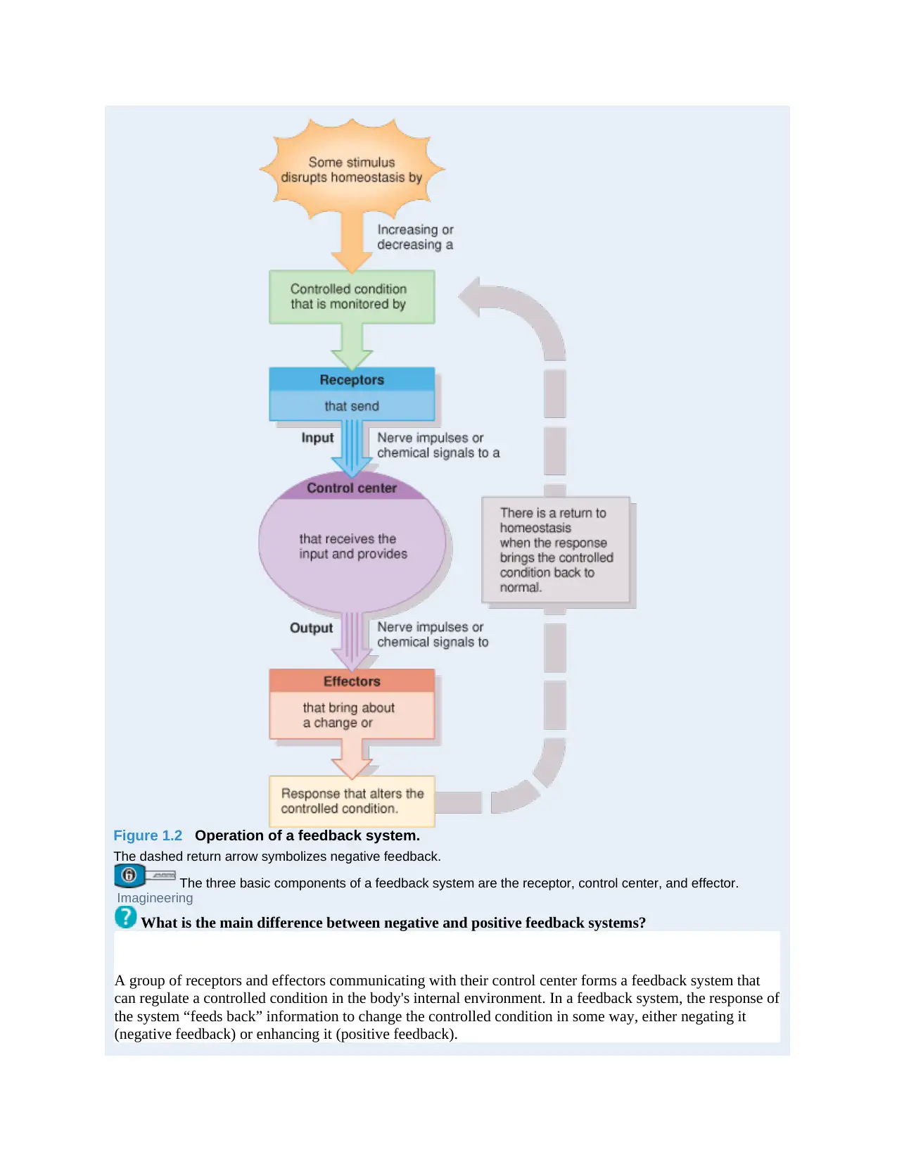
Figure 1.2 Operation of a feedback system.
The dashed return arrow symbolizes negative feedback.
The three basic components of a feedback system are the receptor, control center, and effector.
Imagineering
What is the main difference between negative and positive feedback systems?
A group of receptors and effectors communicating with their control center forms a feedback system that
can regulate a controlled condition in the body's internal environment. In a feedback system, the response of
the system “feeds back” information to change the controlled condition in some way, either negating it
(negative feedback) or enhancing it (positive feedback).
Negative Feedback Systems
A negative feedback system reverses a change in a controlled condition. Consider the regulation of blood
pressure. Blood pressure (BP) is the force exerted by blood as it presses against the walls of blood vessels.
When the heart beats faster or harder, BP increases. If some internal or external stimulus causes blood
pressure (controlled condition) to rise, the following sequence of events occurs
(Figure 1.3). Baroreceptors (the receptors), pressure‐sensitive nerve cells located in the walls of certain
blood vessels, detect the higher pressure. The baroreceptors send nerve impulses (input) to the brain
(control center), which interprets the impulses and responds by sending nerve impulses (output) to the heart
and blood vessels (the effectors). Heart rate decreases and blood vessels dilate (widen), which cause BP to
decrease (response). This sequence of events quickly returns the controlled condition—blood pressure—to
normal, and homeostasis is restored. Notice that the activity of the effector causes BP to drop, a result that
negates the original stimulus (an increase in BP). This is why it is called a negative feedback system.
⊘ This is a preview!⊘
Do you want full access?
Subscribe today to unlock all pages.
Trusted by 1+ million students worldwide
1 out of 110
Related Documents


Your All-in-One AI-Powered Toolkit for Academic Success.
+13062052269
info@desklib.com
Available 24*7 on WhatsApp / Email
![[object Object]](/_next/static/media/star-bottom.7253800d.svg)
Unlock your academic potential
Copyright © 2020–2026 A2Z Services. All Rights Reserved. Developed and managed by ZUCOL.