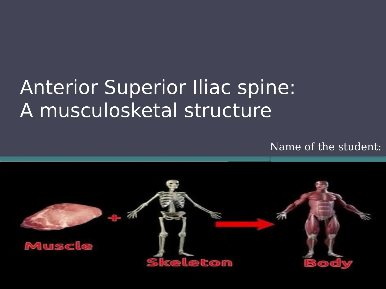
Anterior Superior Iliac Spine: Anatomy and Function Report Analysis

Verified

Added on  2022/09/26

|9
|280
|27
Report
AI Summary
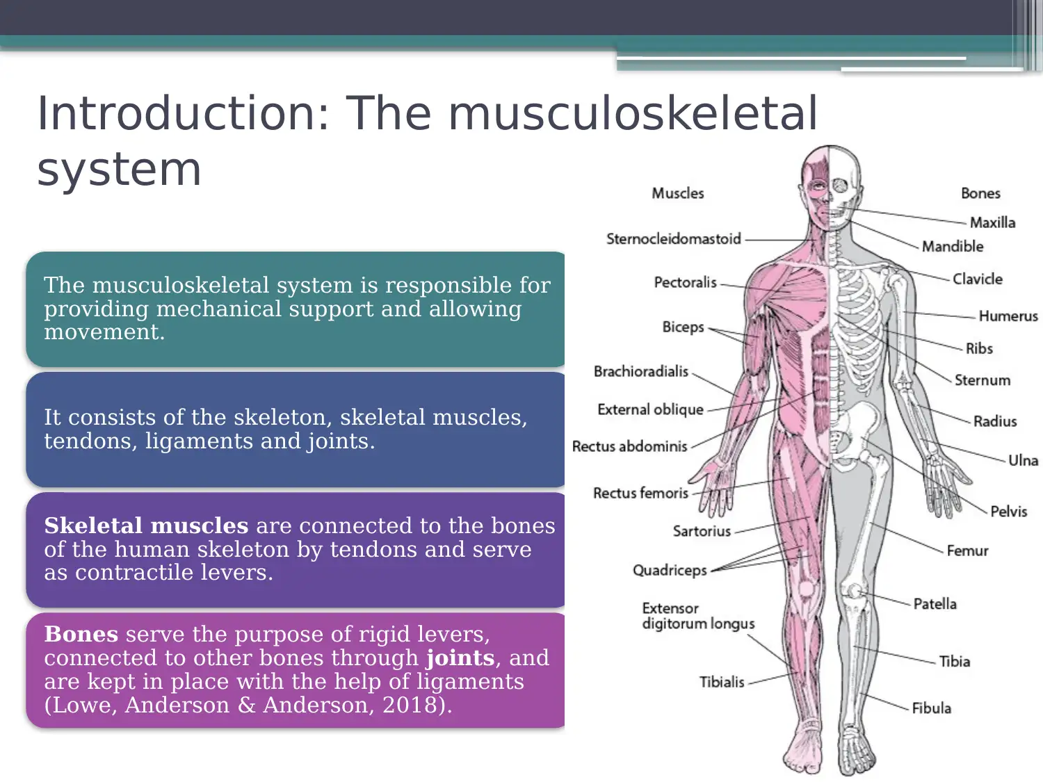
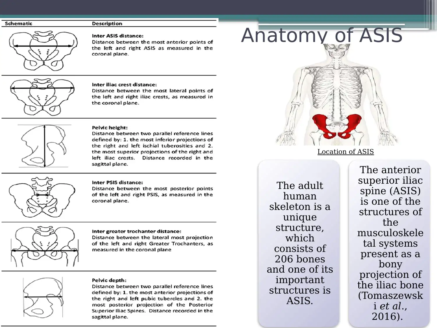
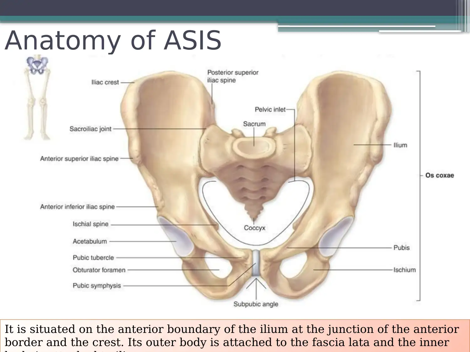
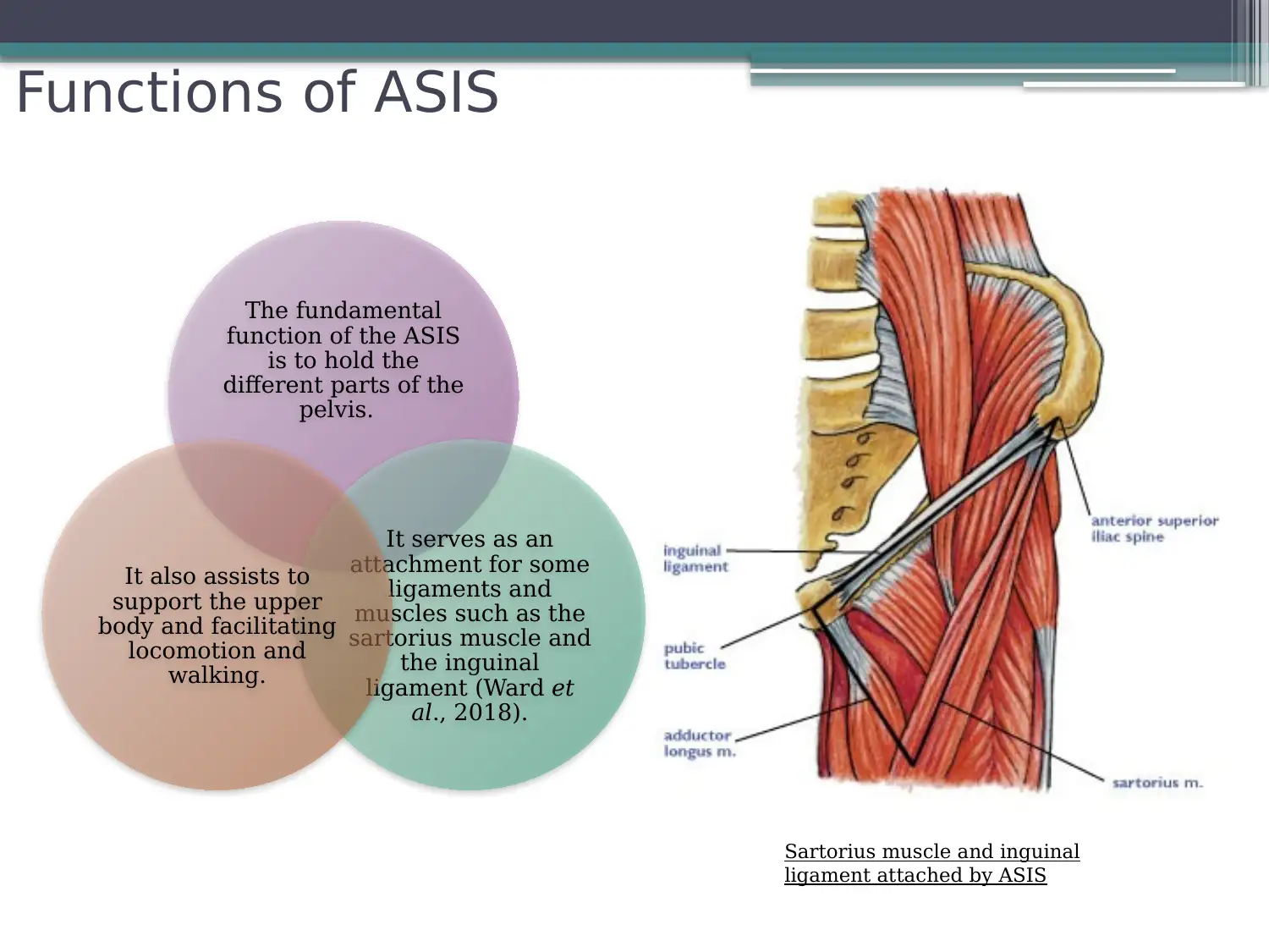
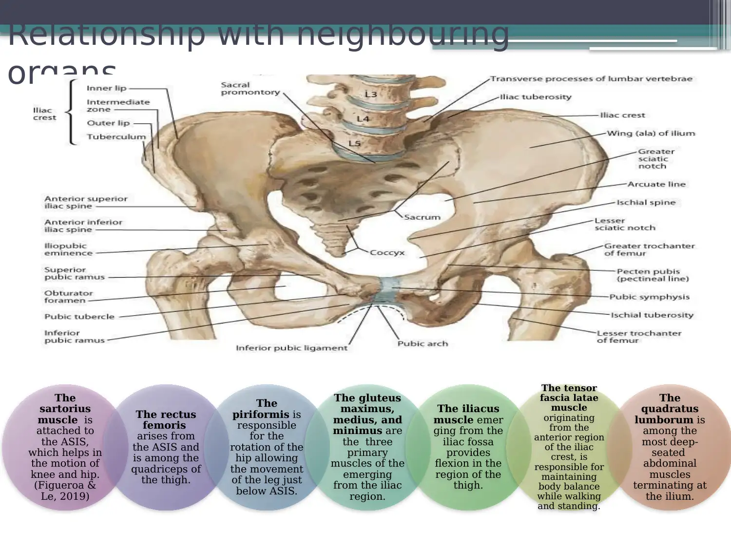
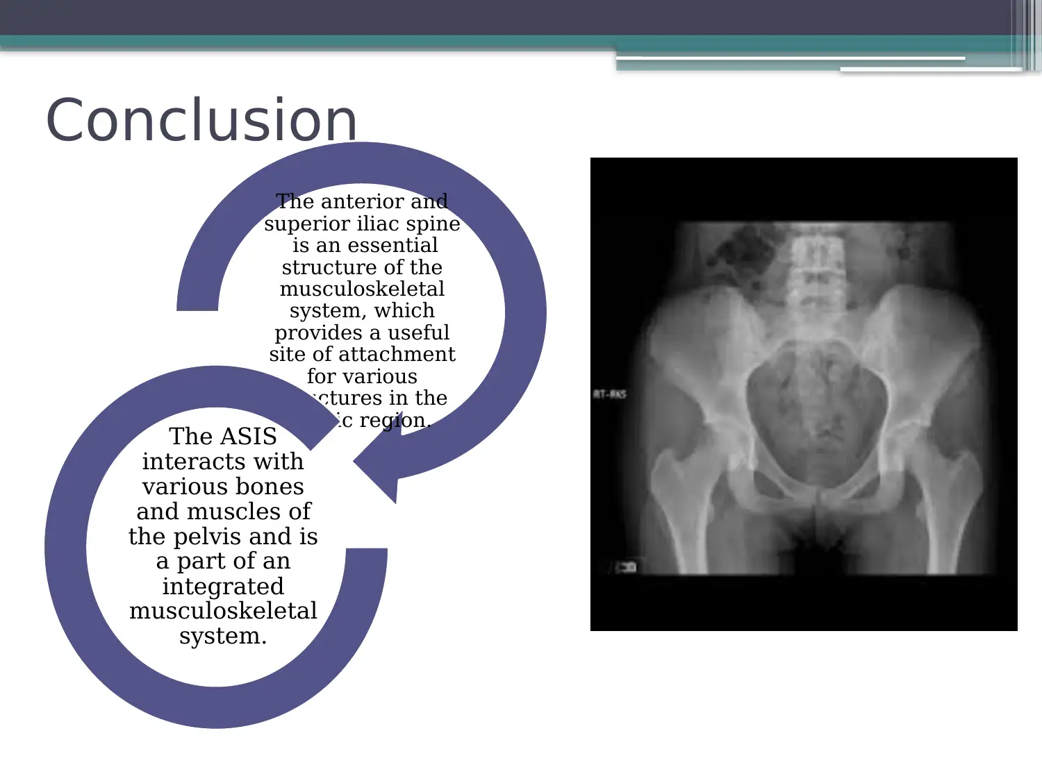
This report delves into the anatomy and function of the Anterior Superior Iliac Spine (ASIS), a key musculoskeletal structure. It begins with an introduction to the musculoskeletal system, followed by a detailed examination of the ASIS's location, and its relationship with surrounding structures. The report highlights the ASIS's connection to the fascia lata, its attachment to the iliacus muscle, and its role in the attachment of the sartorius muscle and inguinal ligament. Furthermore, it explores the ASIS's relationship with neighboring organs. The report concludes with a summary of the key findings and includes a list of relevant references. This report provides a comprehensive understanding of the ASIS and its significance in human anatomy.
Document Page
Anterior Superior Iliac spine:
A musculosketal structure
Name of the student:
tabler-icon-diamond-filled.svg

Paraphrase This Document

Need a fresh take? Get an instant paraphrase of this document with our AI Paraphraser
Document Page
Introduction: The musculoskeletal
system
The musculoskeletal system is responsible for
providing mechanical support and allowing
movement.
It consists of the skeleton, skeletal muscles,
tendons, ligaments and joints.
Skeletal muscles are connected to the bones
of the human skeleton by tendons and serve
as contractile levers.
Bones serve the purpose of rigid levers,
connected to other bones through joints, and
are kept in place with the help of ligaments
(Lowe, Anderson & Anderson, 2018).
Document Page
Anatomy of ASIS
The adult
human
skeleton is a
unique
structure,
which
consists of
206 bones
and one of its
important
structures is
ASIS.
The anterior
superior iliac
spine (ASIS)
is one of the
structures of
the
musculoskele
tal systems
present as a
bony
projection of
the iliac bone
(Tomaszewsk
i et al.,
2016).
Location of ASIS
Document Page
Anatomy of ASIS
It is situated on the anterior boundary of the ilium at the junction of the anterior
border and the crest. Its outer body is attached to the fascia lata and the inner
tabler-icon-diamond-filled.svg

Paraphrase This Document

Need a fresh take? Get an instant paraphrase of this document with our AI Paraphraser
Document Page
Functions of ASIS
The fundamental
function of the ASIS
is to hold the
different parts of the
pelvis.
It serves as an
attachment for some
ligaments and
muscles such as the
sartorius muscle and
the inguinal
ligament (Ward et
al., 2018).
It also assists to
support the upper
body and facilitating
locomotion and
walking.
Sartorius muscle and inguinal
ligament attached by ASIS
Document Page
Relationship with neighbouring
organs
The
sartorius
muscle is
attached to
the ASIS,
which helps in
the motion of
knee and hip.
(Figueroa &
Le, 2019)
The rectus
femoris
arises from
the ASIS and
is among the
quadriceps of
the thigh.
The
piriformis is
responsible
for the
rotation of the
hip allowing
the movement
of the leg just
below ASIS.
The gluteus
maximus,
medius, and
minimus are
the three
primary
muscles of the
emerging
from the iliac
region.
The iliacus
muscle emer
ging from the
iliac fossa
provides
flexion in the
region of the
thigh.
The tensor
fascia latae
muscle
originating
from the
anterior region
of the iliac
crest, is
responsible for
maintaining
body balance
while walking
and standing.
The
quadratus
lumborum is
among the
most deep-
seated
abdominal
muscles
terminating at
the ilium.
Document Page
Conclusion
The anterior and
superior iliac spine
is an essential
structure of the
musculoskeletal
system, which
provides a useful
site of attachment
for various
structures in the
pelvic region.
The ASIS
interacts with
various bones
and muscles of
the pelvis and is
a part of an
integrated
musculoskeletal
system.
tabler-icon-diamond-filled.svg

Paraphrase This Document

Need a fresh take? Get an instant paraphrase of this document with our AI Paraphraser
Document Page
References
Figueroa, C., & Le, P. H. (2019). Anatomy, Bony Pelvis and Lower
Limb, Pelvis Bones. In StatPearls [Internet]. StatPearls
Publishing.
Lowe, J. S., Anderson, P. G., & Anderson, S. I. (2018). Stevens &
Lowe's Human Histology-E-Book. Elsevier Health Sciences.
Tomaszewski, K. A., Popieluszko, P., Henry, B. M., Roy, J., Sanna,
B., Kijek, M. R., & Walocha, J. A. (2016). The surgical anatomy
of the lateral femoral cutaneous nerve in the inguinal region: a
meta-analysis. Hernia, 20(5), 649-657.
Ward, S. H., Blackburn, J. T., Padua, D. A., Stanley, L. E., Harkey,
M. S., Luc-Harkey, B. A., & Pietrosimone, B. (2018). Quadriceps
neuromuscular function and jump-landing sagittal-plane knee
biomechanics after anterior cruciate ligament
reconstruction. Journal of athletic training, 53(2), 135-143.
Document Page
chevron_up_icon
1 out of 9
circle_padding
hide_on_mobile
zoom_out_icon
[object Object]