Case Study: Examining Antibiotic Use and Wound Healing Processes
VerifiedAdded on 2023/06/05
|8
|2408
|303
Case Study
AI Summary
This case study solution analyzes Mary's infected foot laceration, addressing the physiological basis of wound observations like redness, swelling, warmth, pain, and purulent discharge. It identifies potential endogenous (Staphylococcus aureus) and exogenous (Streptococcus pyogenes from a handkerchief) sources of contamination and their modes of transmission. The rationale behind the use of Ceftriaxone (IV), Cephalexin (oral), and Dicloxacillin (oral) antibiotics is explained, including their mechanisms of action and potential adverse effects. The document further describes the four stages of wound healing: haemostasis, inflammation, proliferation, and maturation, detailing the processes involved in each stage. The solution also assesses referencing and presentation based on APA 6th edition guidelines. Desklib provides access to similar case studies and resources for students.
This template must be used to answer the case study.
(Please type your answers within the box underneath each question)
Student name (LAST NAME first name): DHAKAL Shreeya
Student number: 18862443
Campus: Parramatta
Tutorial session (time and room no.):
Tutor’s name:
1. Physiological basis of the wound observations (Total: 10 marks)
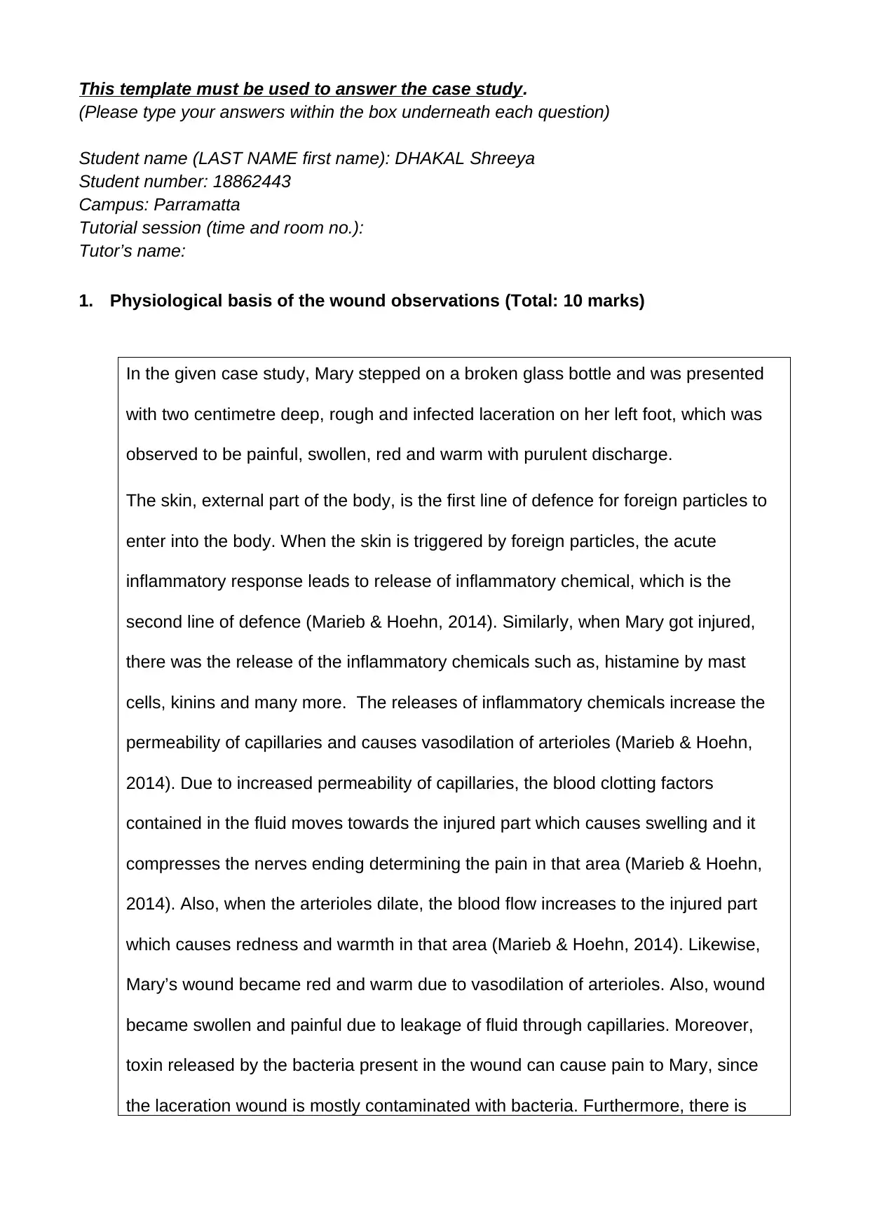
In the given case study, Mary stepped on a broken glass bottle and was presented
with two centimetre deep, rough and infected laceration on her left foot, which was
observed to be painful, swollen, red and warm with purulent discharge.
The skin, external part of the body, is the first line of defence for foreign particles to
enter into the body. When the skin is triggered by foreign particles, the acute
inflammatory response leads to release of inflammatory chemical, which is the
second line of defence (Marieb & Hoehn, 2014). Similarly, when Mary got injured,
there was the release of the inflammatory chemicals such as, histamine by mast
cells, kinins and many more. The releases of inflammatory chemicals increase the
permeability of capillaries and causes vasodilation of arterioles (Marieb & Hoehn,
2014). Due to increased permeability of capillaries, the blood clotting factors
contained in the fluid moves towards the injured part which causes swelling and it
compresses the nerves ending determining the pain in that area (Marieb & Hoehn,
2014). Also, when the arterioles dilate, the blood flow increases to the injured part
which causes redness and warmth in that area (Marieb & Hoehn, 2014). Likewise,
Mary’s wound became red and warm due to vasodilation of arterioles. Also, wound
became swollen and painful due to leakage of fluid through capillaries. Moreover,
toxin released by the bacteria present in the wound can cause pain to Mary, since
the laceration wound is mostly contaminated with bacteria. Furthermore, there is
Paraphrase This Document
Need a fresh take? Get an instant paraphrase of this document with our AI Paraphraser
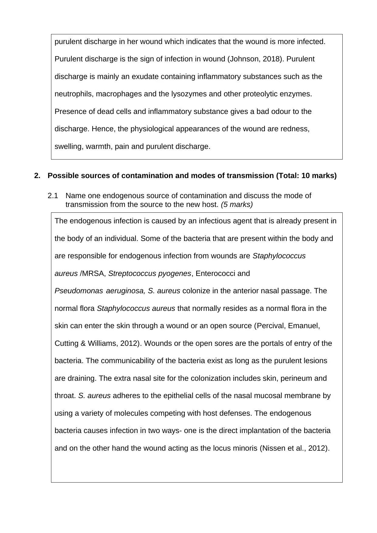
purulent discharge in her wound which indicates that the wound is more infected.
Purulent discharge is the sign of infection in wound (Johnson, 2018). Purulent
discharge is mainly an exudate containing inflammatory substances such as the
neutrophils, macrophages and the lysozymes and other proteolytic enzymes.
Presence of dead cells and inflammatory substance gives a bad odour to the
discharge. Hence, the physiological appearances of the wound are redness,
swelling, warmth, pain and purulent discharge.
2. Possible sources of contamination and modes of transmission (Total: 10 marks)
2.1 Name one endogenous source of contamination and discuss the mode of
transmission from the source to the new host. (5 marks)
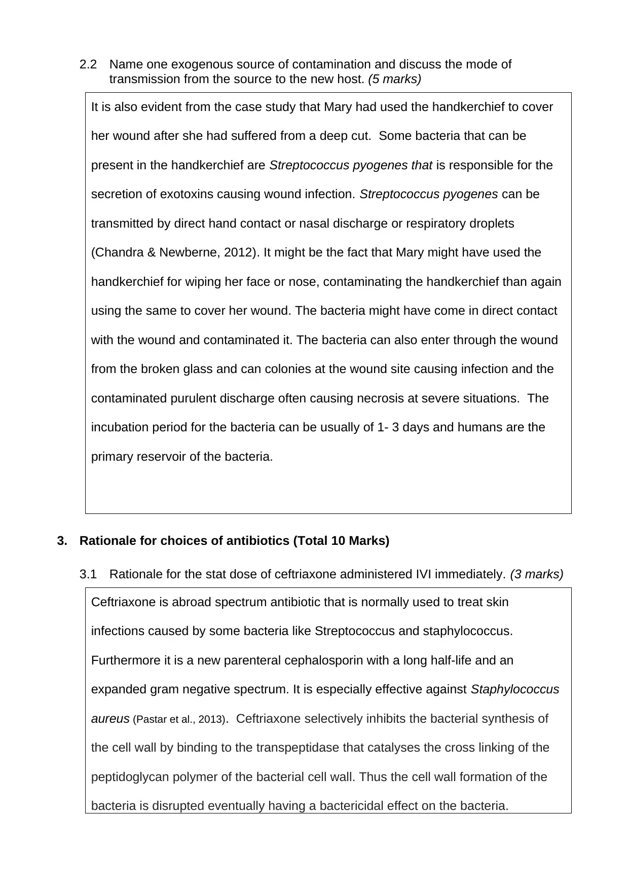
The endogenous infection is caused by an infectious agent that is already present in
the body of an individual. Some of the bacteria that are present within the body and
are responsible for endogenous infection from wounds are Staphylococcus
aureus /MRSA, Streptococcus pyogenes, Enterococci and
Pseudomonas aeruginosa, S. aureus colonize in the anterior nasal passage. The
normal flora Staphylococcus aureus that normally resides as a normal flora in the
skin can enter the skin through a wound or an open source (Percival, Emanuel,
Cutting & Williams, 2012). Wounds or the open sores are the portals of entry of the
bacteria. The communicability of the bacteria exist as long as the purulent lesions
are draining. The extra nasal site for the colonization includes skin, perineum and
throat. S. aureus adheres to the epithelial cells of the nasal mucosal membrane by
using a variety of molecules competing with host defenses. The endogenous
bacteria causes infection in two ways- one is the direct implantation of the bacteria
and on the other hand the wound acting as the locus minoris (Nissen et al., 2012).
2.2 Name one exogenous source of contamination and discuss the mode of
transmission from the source to the new host. (5 marks)
It is also evident from the case study that Mary had used the handkerchief to cover
her wound after she had suffered from a deep cut. Some bacteria that can be
present in the handkerchief are Streptococcus pyogenes that is responsible for the
secretion of exotoxins causing wound infection. Streptococcus pyogenes can be
transmitted by direct hand contact or nasal discharge or respiratory droplets
(Chandra & Newberne, 2012). It might be the fact that Mary might have used the
handkerchief for wiping her face or nose, contaminating the handkerchief than again
using the same to cover her wound. The bacteria might have come in direct contact
with the wound and contaminated it. The bacteria can also enter through the wound
from the broken glass and can colonies at the wound site causing infection and the
contaminated purulent discharge often causing necrosis at severe situations. The
incubation period for the bacteria can be usually of 1- 3 days and humans are the
primary reservoir of the bacteria.
3. Rationale for choices of antibiotics (Total 10 Marks)
3.1 Rationale for the stat dose of ceftriaxone administered IVI immediately. (3 marks)
Ceftriaxone is abroad spectrum antibiotic that is normally used to treat skin
infections caused by some bacteria like Streptococcus and staphylococcus.
Furthermore it is a new parenteral cephalosporin with a long half-life and an
expanded gram negative spectrum. It is especially effective against Staphylococcus
aureus (Pastar et al., 2013). Ceftriaxone selectively inhibits the bacterial synthesis of
the cell wall by binding to the transpeptidase that catalyses the cross linking of the
peptidoglycan polymer of the bacterial cell wall. Thus the cell wall formation of the
bacteria is disrupted eventually having a bactericidal effect on the bacteria.
⊘ This is a preview!⊘
Do you want full access?
Subscribe today to unlock all pages.

Trusted by 1+ million students worldwide
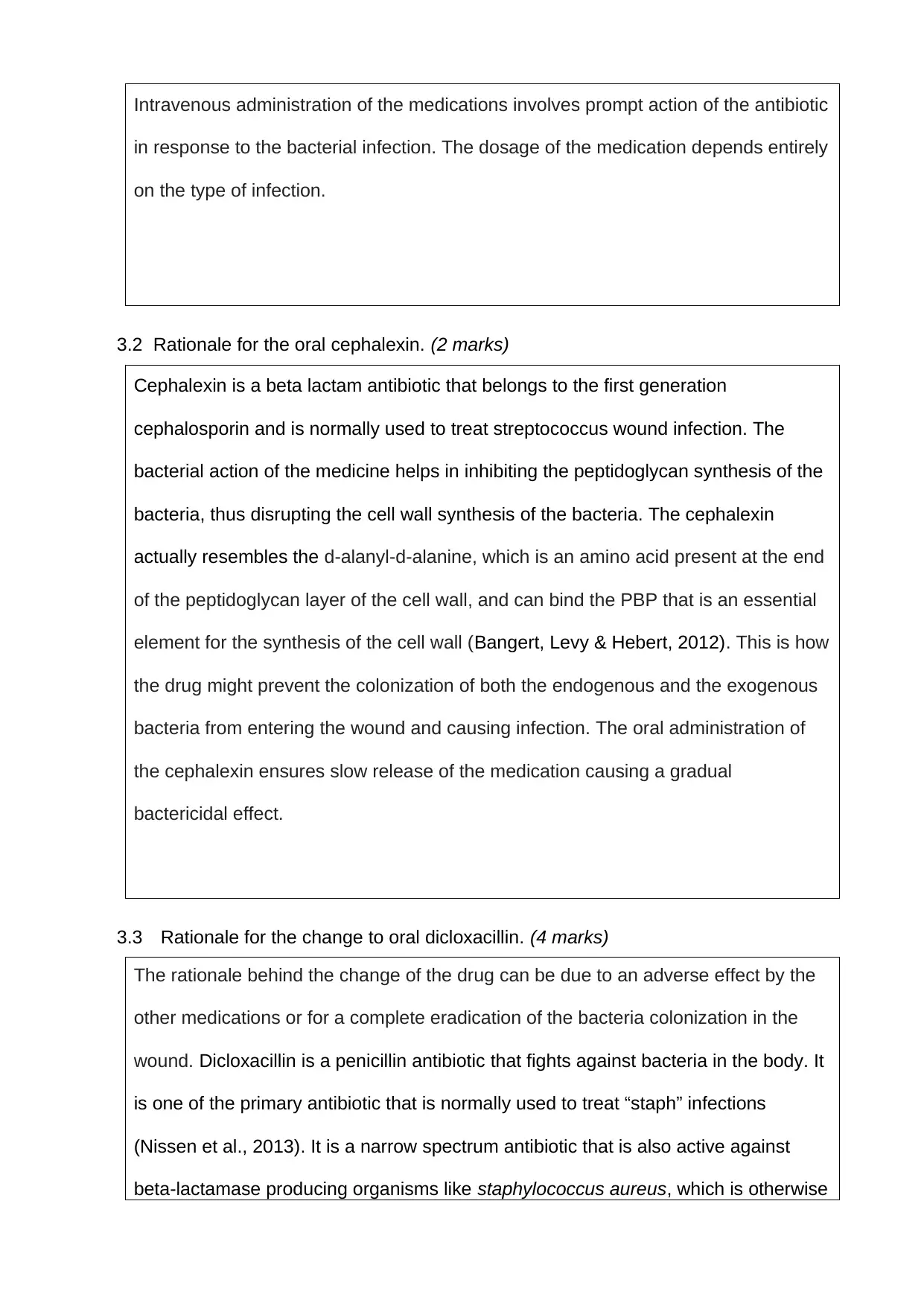
Intravenous administration of the medications involves prompt action of the antibiotic
in response to the bacterial infection. The dosage of the medication depends entirely
on the type of infection.
3.2 Rationale for the oral cephalexin. (2 marks)
Cephalexin is a beta lactam antibiotic that belongs to the first generation
cephalosporin and is normally used to treat streptococcus wound infection. The
bacterial action of the medicine helps in inhibiting the peptidoglycan synthesis of the
bacteria, thus disrupting the cell wall synthesis of the bacteria. The cephalexin
actually resembles the d-alanyl-d-alanine, which is an amino acid present at the end
of the peptidoglycan layer of the cell wall, and can bind the PBP that is an essential
element for the synthesis of the cell wall (Bangert, Levy & Hebert, 2012). This is how
the drug might prevent the colonization of both the endogenous and the exogenous
bacteria from entering the wound and causing infection. The oral administration of
the cephalexin ensures slow release of the medication causing a gradual
bactericidal effect.
3.3 Rationale for the change to oral dicloxacillin. (4 marks)
The rationale behind the change of the drug can be due to an adverse effect by the
other medications or for a complete eradication of the bacteria colonization in the
wound. Dicloxacillin is a penicillin antibiotic that fights against bacteria in the body. It
is one of the primary antibiotic that is normally used to treat “staph” infections
(Nissen et al., 2013). It is a narrow spectrum antibiotic that is also active against
beta-lactamase producing organisms like staphylococcus aureus, which is otherwise
Paraphrase This Document
Need a fresh take? Get an instant paraphrase of this document with our AI Paraphraser
resistant to most of the penicillin. Dicloxacillin binds specifically to the penicillin-
binding proteins (PBPs) that is located in the bacterial cell wall. It inhibits the third
and the last stage of the bacterial cell wall synthesis. The lysis of the cell wall is then
brought about by the bacterial cell wall autolytic enzymes like autolysin. It can be
possible that Dicloxacillin might interfere with the autolysin inhibitor.
3.4 State two adverse reactions to dicloxacillin. (1 mark)
Two adverse effects of Dicloxacillin are- severe stomach pain and diarrhoea or
watery stool and urine retention causing very little or no urination. Diarrhoea can
cause excessive loss of water leading to severe dehydration.
4. Process by which Mary’s wound will heal (Total: 5 marks)
There are normally four stages of wound healing- haemostasis, inflammation,
proliferation and maturation. In spite of these four stages of wound healing the
process can progress forward and backward depending upon the condition of the
patient.
Haemostasis-It is the process of the wound getting closed by clotting. It
occurs when the blood vessels constrict for restricting the blood flow and
coagulation of the platelets to seal the wall of the blood vessels (Bowler, Duerden &
Armstrong, 2012). This phase is followed by the inflammatory phase where the
injured blood vessels leak exudates causing swelling of the wound. Inflammation
controls the bleeding and helps to prevent infection. During this stage the pathogens
and the dead cells are removed from the area of the wound (Clark, 2013). The
redness, pain, swelling and heat found in the injured part of Mary is due to
inflammation. The third stage is the proliferative phase where the new tissues made
up of collagen and extracellular matrix are build. New network of blood vessels are
developed for supplying nutrients to the newly formed tissues. The myofibroblasts
helps in pulling the wound edges together. The final phase of the wound healing
process that Mary has to encounter, is the maturation phase. In this stage the type
III collagen is converted to type I collagen and the wound is closed fully (Clark,
2013). The cells needed to repair the wound are not needed and are generally
removed by apoptosis (Bowler, Duerden & Armstrong, 2012).At this stage the water
is absorbed for the realignment and the cross linking of the collagen fibres. Cross
linking of the collagen makes the skin tough and stronger. Thus it can be said that
the different stages of the wound healing is a complex process and the failure to
progress through this stages can lead to the formation of the chronic wounds.
5. Presentation (Total: 5 marks)
5.1 Referencing in-text and in reference list conforms to APA 6th Ed. referencing
style.
5.2 Critique supported by relevant literature as prescribed.
5.3 Correct sentence structure, paragraph, grammatical construction, spelling,
punctuation and presentation.
https://www.medicalnewstoday.com/articles/320670.php
⊘ This is a preview!⊘
Do you want full access?
Subscribe today to unlock all pages.

Trusted by 1+ million students worldwide
References
Bangert, S., Levy, M., & Hebert, A. A. (2012). Bacterial resistance and impetigo treatment trends: a review.
Pediatric dermatology, 29(3), 243-248. https://doi.org/10.1093/jac/dkt108
Bowler, P. G., Duerden, B. I., & Armstrong, D. G. (2012). Wound Microbiology and Associated Approaches to
Wound Management. Clinical Microbiology Reviews, 14(2), 244–269. DOI: 10.1128/CMR.14.2.244-
269.2012
Bullock, S., & Manias, E. (2017). Fundamentals of pharmacology (8th ed.). Frenchs Forest, Australia: Pearson
Australia.
Chandra, R. K., & Newberne, P. M. (2012). Nutrition, immunity, and infection: mechanisms of interactions.
Springer Science & Business Media. https://books.google.co.in/books?
hl=en&lr=&id=ulnTBwAAQBAJ&oi=fnd&pg=PA1&dq=+Chandra,+R.+K.,+%26+Newberne,+P.+M.
+(2012).+Nutrition,+immunity,+and+infection:+mechanisms+of+interactions.+Springer+Science+
%26+Business+Media.&ots=Y9AHKvFdP3&sig=ioGeAxh-
bQFDj_XCOSJjgDveyxQ#v=onepage&q=Chandra%2C%20R.%20K.%2C%20%26%20Newberne
%2C%20P.%20M.%20(2012).%20Nutrition%2C%20immunity%2C%20and%20infection%3A
%20mechanisms%20of%20interactions.%20Springer%20Science%20%26%20Business
%20Media.&f=false
Clark, R. A. (Ed.). (2013). The molecular and cellular biology of wound repair. Springer Science & Business
Media. https://books.google.co.in/books?
hl=en&lr=&id=HOzcBwAAQBAJ&oi=fnd&pg=PA3&dq=+wound+healing+mechanism&ots=Btg_iIlwAm
&sig=UXdakUEI2Z_DOsutd4oHUlCXGhw#v=onepage&q=wound%20healing%20mechanism&f=false
Nissen, J. L., Skov, R., Knudsen, J. D., Østergaard, C., Schønheyder, H. C., Frimodt-Møller, N., & Benfield, T.
(2013). Effectiveness of penicillin, dicloxacillin and cefuroxime for penicillin-susceptible
Staphylococcus aureus bacteraemia: a retrospective, propensity-score-adjusted case–control and
cohort analysis. Journal of Antimicrobial Chemotherapy, 68(8), 1894-1900.
https://doi.org/10.1093/jac/dkt108
Nissen, J. L., Skov, R., Knudsen, J. D., Østergaard, C., Schønheyder, H. C., Frimodt-Møller, N., & Benfield, T.
(2013). Effectiveness of penicillin, dicloxacillin and cefuroxime for penicillin-susceptible
Staphylococcus aureus bacteraemia: a retrospective, propensity-score-adjusted case–control and
Paraphrase This Document
Need a fresh take? Get an instant paraphrase of this document with our AI Paraphraser
cohort analysis. Journal of Antimicrobial Chemotherapy, 68(8), 1894-1900.
https://doi.org/10.1093/jac/dkt108
Pastar, I., Nusbaum, A. G., Gil, J., Patel, S. B., Chen, J., Valdes, J., ... & Davis, S. C. (2013). Interactions of
methicillin resistant Staphylococcus aureus USA300 and Pseudomonas aeruginosa in polymicrobial
wound infection. PloS one, 8(2), e56846. https://doi.org/10.1371/journal.pone.0056846
Percival, S. L., Emanuel, C., Cutting, K. F., & Williams, D. W. (2012). Microbiology of the skin and the role of
biofilms in infection. International wound journal, 9(1), 14-32. https://doi.org/10.1111/j.1742-
481X.2011.00836.x
1 out of 8
Related Documents
Your All-in-One AI-Powered Toolkit for Academic Success.
+13062052269
info@desklib.com
Available 24*7 on WhatsApp / Email
![[object Object]](/_next/static/media/star-bottom.7253800d.svg)
Unlock your academic potential
Copyright © 2020–2026 A2Z Services. All Rights Reserved. Developed and managed by ZUCOL.


