Exploring Competition Dialysis for Ligand Discovery in Biochemistry

Verified

Added on  2023/06/04

|5
|714
|478
Homework Assignment
AI Summary
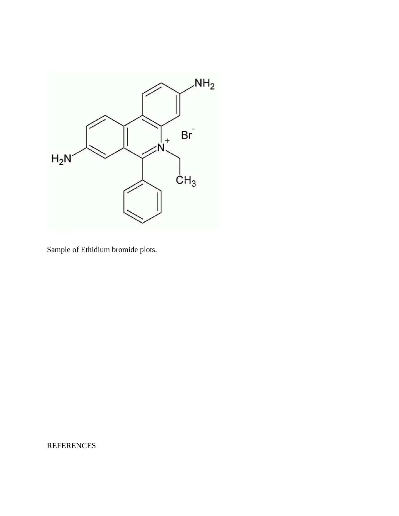
This assignment focuses on competition dialysis, a technique used in biochemistry for discovering ligands that bind to nucleic acids with sequence selectivity. It discusses Brad Chaires' method for competition dialysis, including solution preparation, concentrations used (specifically 1uM and 0.75uM), and the purpose of adding surfactants to improve wetting properties. The assignment identifies fluorescence spectroscopy as the method used for determining ligand concentration due to its parameter adjustability. Data analysis examples include studies with quadruplex forms and ligands like methylene blue and ethidium, detailing binding preferences and energy calculations. The document concludes with examples of selectivity plots, particularly for ethidium bromide, and provides relevant references.
Document Page
BIOCHEMISTRY
By Name
Course
Instructor
Institution
Location
Date
Introduction
tabler-icon-diamond-filled.svg

Paraphrase This Document

Need a fresh take? Get an instant paraphrase of this document with our AI Paraphraser
Document Page
Competition dialysis is a new tool that is considered very powerful for the discovery of ligands.
Such ligands binds to the nucleic acid with sequence selectivity. The sequence selectivity is also
known as structural connectivity. This particular method is based on a very firm principle of
thermodynamic which makes its implementation very simple. In the experiment of competition
dialysis, arrays of the sequence and nucleic acid is dialyzed against a common test ligand
solution(Bewley and Black 2012).
1.How does Brad chaires do the competition dialysis?
This was a procedural process. I t started with the Preparation of the solution of nucleic acid and
dialysate solution. The first and the second generation of the assays utilized 200mL of 1uM
solution. This was introduced into the unit of dialysis with 0.5mL out of 0.75uM of the sample of
the nucleic acid. The upper limit of the concentration was found to be at 5uM.The optimal
concentration that was obtained was1-2uM.The assembly and loading of the dialysis unit took
almost two hours. The solutions of the ligand was mixed with the acid in order to establish the
equilibrium. The establishment of the equilibrium lasted for over 15 hours as the incubation
period. The concentration of the recovered samples was determined using absorbance(Davies
2013).
2. What concentration does he use? Explain and why?
The first and the second generation of the assays utilized 200mL of 1uM solution. This was
introduced into the unit of dialysis with 0.5mL out of 0.75uM of the sample of the nucleic acid.
The final concentration of 75uM was preferred since it could be stored up to a period of 6
months at a temperature of 4 degrees Celsius.
3.Why does he add surfactant? Explain and if there is a reason why?
Document Page
In order to improve the wetting property of the solution that later favor binding,surfuctants were
added. These surfactant lowers the surface tension while improving the wetting and finally
binding characteristics(Stanley and Linskens 2012).
4. Which type of spectroscopy does he use? explain and if there is the reason why?
The experiment exploited the use of florescent spectroscopy in the determination of the
concentration of the ligand. The standard curve was constructed from which the concentration
was determined. The major reason why this spectroscopy was used results from the fact that it
allows for the adjustment of parameters.
5. Give examples of all the data analysis?
The 19-structure that was used contained three quadruplex forms. This included Na+ that exist in
the telomere of human. The ligands that were chosen for the study included methylene
blue,ethidium.NMM,DODC and PIPER. There was binding of Ethidium on DNA.The structural
preference of Methylene blue was not known.Berberine proved to have some activity of binding
towards triplex of DNA and in minor cases of binding to quadruplex DNA.The unit for the
apparent binding is the monomeric unit. The binding free energy can then be calculated as
follows
Change in Gpp=-RTlnKapp
Th binding constants will be considered free energy parameters and will be obtained in a way
that correlate well with some other complete binding constants. Additional metrics may be useful
in the specification of the sum and Turkey box plots(Barrett 2012).
6- show examples of his selectivity plots?
Document Page
Sample of Ethidium bromide plots.
REFERENCES
tabler-icon-diamond-filled.svg

Paraphrase This Document

Need a fresh take? Get an instant paraphrase of this document with our AI Paraphraser
Document Page
Barrett, G. ed., 2012. Chemistry and biochemistry of the amino acids. Springer Science &
Business Media.
Bewley, J.D. and Black, M., 2012. Physiology and biochemistry of seeds in relation to
germination: volume 2: viability, dormancy, and environmental control. Springer Science &
Business Media.
Davies, P.J. ed., 2013. Plant hormones: physiology, biochemistry and molecular biology.
Springer Science & Business Media.
Stanley, R.G. and Linskens, H.F., 2012. Pollen: biology biochemistry management. Springer
Science & Business Media.
chevron_up_icon
1 out of 5
circle_padding
hide_on_mobile
zoom_out_icon
[object Object]