SCIE2004 Biochemistry Assessment Report - Semester 2, 2024
VerifiedAdded on 2023/05/29
|10
|2666
|205
Report
AI Summary
This biochemistry assessment report delves into the intricate world of biomolecules, exploring their structures and functions within biological systems. The report begins by examining the relationship between molecular structure and properties, including boiling points and melting points of various organic compounds. It then progresses to a detailed discussion of carbohydrates, proteins, lipids, and nucleic acids, elucidating their structural classifications and roles in cellular processes. The report further analyzes protein structure, from primary to quaternary levels, and the importance of lipids in maintaining cell membrane structure and function, including the roles of phospholipids, cholesterol, and sphingolipids. The report also explores cellular respiration, the impact of cyanide inhibition on cytochrome c oxidase, and the process of DNA transcription and mRNA translation. Finally, the report examines the role of specific molecules like adenine, guanine, and thymine, and the impact of amino acid sequence variations on protein function, all while providing context for the evolutionary relationships between various species. This report offers a comprehensive overview of essential biochemistry topics, providing valuable insights into the molecular basis of life. Study past papers and solved assignments on Desklib.
Running Head: BIOCHEMISTRY ASSESSMENT
BIOCHEMISTRY ASSESSMENT
Name of student:
Name of university:
Author Note:
BIOCHEMISTRY ASSESSMENT
Name of student:
Name of university:
Author Note:
Paraphrase This Document
Need a fresh take? Get an instant paraphrase of this document with our AI Paraphraser
1BIOCHEMISTRY ASSESSMENT
Part A
1. 1-hexanamine has a linear chain while triethylamine has a branched structure. Boiling point
increases for a linear chain; more compact and branching structures exhibit less interaction
between molecules, thereby having less Vander Waals forces of interaction as in case of
triethylamine. More spread structure causes boiling point to rise in 1-hexanamine (Salammal et
al., 2015).
2. The chemical structure of Propanol has a highly polar OH group which takes part in hydrogen
bonding and its highly electronegative oxygen with lone pairs of electrons contribute to
permanent dipole-dipole interactions.Propanal with CHO functional group lacks the hydrogen
bonding interactions as the highly electronegative oxygen atom is bonded to carbon, only dipole-
dipole interactions result. Vander Waals forces of attractions are present in both propanol and
propanal (Cunningham et al., 2018). Higher number of intermolecular forces contributes to
higher boiling point (97°C) in propanol compared to propanal (48°C).
3. The difference in melting point of fatty acids depends on the molecular weight and the extent
of unsaturation. Stearic acid is a saturated fatty acidwith a linear structure. Linoleic acid has two
unsaturated double bonds positioned at 9 and 12 respectively. Higher the number of unsaturation,
lower will be the melting point of the fatty acid. Linoleic acid with 2 double bonds has a
spherical non-linear structurewith less intermolecular interaction. Resultant decreased Vander
Waals forces lower the melting point of Linoleic acid (Budin et al., 2014).
4. Amides have a functional group CONH2, which performs antibacterial activity by causing
membrane disruption for gram-positive and gram-negative bacteria(Pérez-Peinadoet al., 2018).
The amides and derivatives target the thick peptidoglycan layer in gram-positive bacteria and
outer polysaccharide layer in gram-negative bacteria causing permeabilization and inhibition of
peptidoglycan synthesis.
Amides are synthesized in derivatives to augment the antibacterial activities of
bactericidal agents in response to evolving bacterial infections. They are used as beta-lactam
targets and enzyme inhibitors to dissolve the bacterial cell membrane and inhibit both DNA and
RNA synthesis.
Part B
2BIOCHEMISTRY ASSESSMENT
Biological systems are composed of fundamental units of life termed as cells, whose
structure and function rely on the carbon containing biomolecules: Carbohydrates, Proteins,
Lipids and Nucleic acids. The complex structure of biomolecules are related to the diverse
functions they perform in the living systems.
Structural Classification of Carbohydrates
Carbohydrates serves as one of the major energy sources for living systems. Depending
on the structural units, carbohydrates are classified into three classes namely:
Monosaccharides:The monosaccharides comprise of the simple sugars like glucose and
fructose. They have a short chain length ranging from three to six carbon atoms. They are highly
soluble in water and cannot be further broken down. Glucose is the most abundant
monosaccharide found in living system and major metabolic byproduct. Monosaccharides are
again classified into aldoses and ketoses based on the presence of oxidized functional group.
Glucose is an aldose sugar with aldehyde as the functional group while dihroxyacetone is a
ketose sugar with carbonyl group (Voet, Voet &Pratt, 2016).
Oligosaccharides:Monosaccharides are joined together through covalent linkage called
glycosidic bonds to synthesize short chains varying between two to ten monosaccharide units.
These short chains are termed as oligosaccharides which are easily hydrolysable into their
constituent monosaccharides. Oligosaccharides are further classified depending on the number of
linking monosaccharide units. These are classified as follows:
Disaccharides: These are composed of two units of monosaccharides. Lactose is a
disaccharide composed of glucose and galactose monosaccharides.
Trisaccharides: These comprise of three units of linked monosaccharides. Raffinose is a
trisaccharide consisting of alpha galactosyl derivatives of sucrose.
Tetrasaccharides:These contain four units of monosaccharides linked together.
Stachyose is a tetrasaccharide which hydrolyzes into one unit each of glucose and
fructose and two units of galactose.
Polysaccharides: These comprise of thousands of monosaccharide units linked through
glycosidic bonds. Polysaccharides are insoluble in water but soluble in organic solvents.
Polysaccharides with one type of constituent monosaccharide are termed as
⊘ This is a preview!⊘
Do you want full access?
Subscribe today to unlock all pages.
Trusted by 1+ million students worldwide
3BIOCHEMISTRY ASSESSMENT
homopolysaccharides whereas those with more than one type of monosaccharide unit are
heteropolysaccharides (Lundblad & MacDonald, 2018).Cellulose is a plant homopolysaccharide
with glucose as the constituent unit.Hyaluronic acid composed of glucuronic acid and N-acetyl
glucosamine is a heteropolysaccharide found in synovial fluid of joints.
Organisation of Protein Structure:
Proteins play a pivotal role in relation to genetic synthesis. Understanding their levels of
structural organization help elucidate their functional roles in living systems.Proteins organize
into four fundamental structures which are as follows:
Primary Structure:The sequence of universally occurring twenty amino acids connected by
peptide bonds in the polypeptide chain forms the primary structure of proteins. Different
arrangements of the amino acids in the polypeptide generates a huge diversity among proteins.
The amino acids vary in the side chains which confer differing chemical, physical and structural
properties to the peptide (Kennedy et al., 2016). This amino acid sequence is encoded into the
genetic machinery (DNA) which is transcribed and translated to synthesize proteins. These
proteins undergo modifications to become biologically active inside cells.
Secondary Structure:The polypeptide chains form conformations through hydrogen bonding
into structures called alpha-helix and beta-sheet. Alpha helices are right handed coiled coil
structure with amino acid side chains protruding outward. Alpha helices are stabilized by
hydrogen bonding interactions (Rahal & Waltz, 2018). Beta sheets are composed of either
parallel (both strands are N to C terminus) or antiparallel (N to C terminus for strand and vice-
versa for the other) strands stabilized by inter-strand hydrogen bonds. The antiparallel beta sheets
occur as the most stable form of secondary structure due to less steric hindrance.
Tertiary Structure:Polypeptides fold into random three dimensional shape to remain in the
lowest energy state achieving maximum stability. Under physiological pH, the non-polar amino
acids are buried inside the interior with the polar acidic and basic amino acids facing the outer
hydrophilic aqueous environment.Covalent disulphide bridges, ionic interactions and salt bridge
interactions stabilize the folding of polypeptide chains. The biological activities rely on the
tertiary structure of proteins.
Paraphrase This Document
Need a fresh take? Get an instant paraphrase of this document with our AI Paraphraser
4BIOCHEMISTRY ASSESSMENT
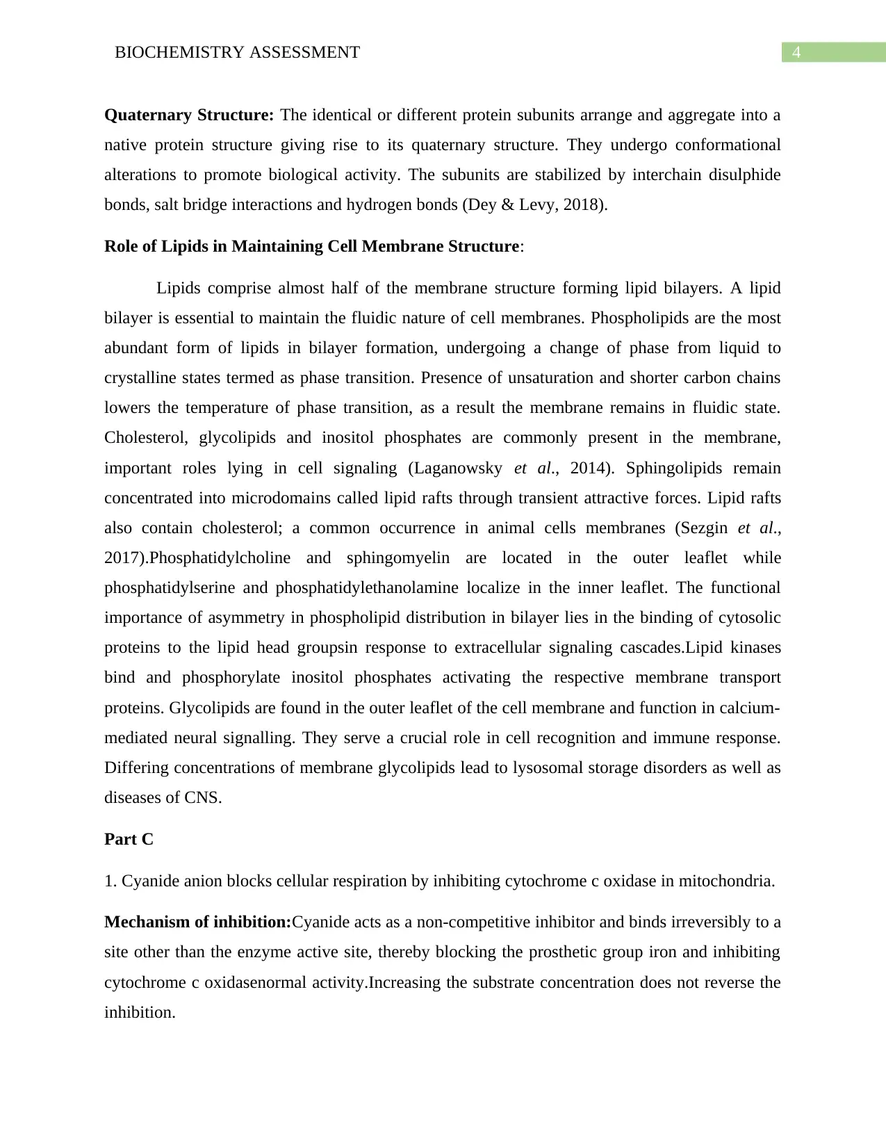
Quaternary Structure: The identical or different protein subunits arrange and aggregate into a
native protein structure giving rise to its quaternary structure. They undergo conformational
alterations to promote biological activity. The subunits are stabilized by interchain disulphide
bonds, salt bridge interactions and hydrogen bonds (Dey & Levy, 2018).
Role of Lipids in Maintaining Cell Membrane Structure:
Lipids comprise almost half of the membrane structure forming lipid bilayers. A lipid
bilayer is essential to maintain the fluidic nature of cell membranes. Phospholipids are the most
abundant form of lipids in bilayer formation, undergoing a change of phase from liquid to
crystalline states termed as phase transition. Presence of unsaturation and shorter carbon chains
lowers the temperature of phase transition, as a result the membrane remains in fluidic state.
Cholesterol, glycolipids and inositol phosphates are commonly present in the membrane,
important roles lying in cell signaling (Laganowsky et al., 2014). Sphingolipids remain
concentrated into microdomains called lipid rafts through transient attractive forces. Lipid rafts
also contain cholesterol; a common occurrence in animal cells membranes (Sezgin et al.,
2017).Phosphatidylcholine and sphingomyelin are located in the outer leaflet while
phosphatidylserine and phosphatidylethanolamine localize in the inner leaflet. The functional
importance of asymmetry in phospholipid distribution in bilayer lies in the binding of cytosolic
proteins to the lipid head groupsin response to extracellular signaling cascades.Lipid kinases
bind and phosphorylate inositol phosphates activating the respective membrane transport
proteins. Glycolipids are found in the outer leaflet of the cell membrane and function in calcium-
mediated neural signalling. They serve a crucial role in cell recognition and immune response.
Differing concentrations of membrane glycolipids lead to lysosomal storage disorders as well as
diseases of CNS.
Part C
1. Cyanide anion blocks cellular respiration by inhibiting cytochrome c oxidase in mitochondria.
Mechanism of inhibition:Cyanide acts as a non-competitive inhibitor and binds irreversibly to a
site other than the enzyme active site, thereby blocking the prosthetic group iron and inhibiting
cytochrome c oxidasenormal activity.Increasing the substrate concentration does not reverse the
inhibition.
5BIOCHEMISTRY ASSESSMENT
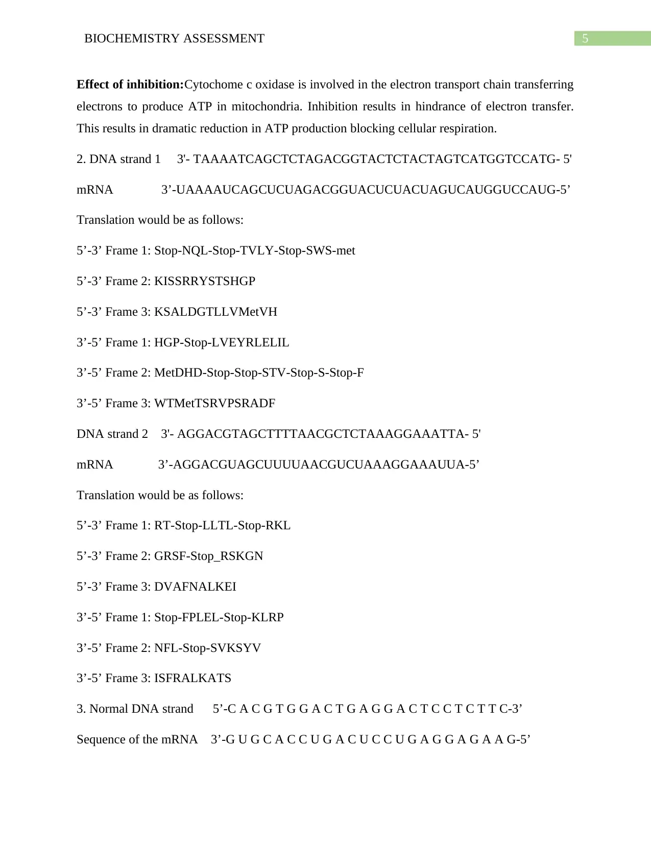
Effect of inhibition:Cytochome c oxidase is involved in the electron transport chain transferring
electrons to produce ATP in mitochondria. Inhibition results in hindrance of electron transfer.
This results in dramatic reduction in ATP production blocking cellular respiration.
2. DNA strand 1 3'- TAAAATCAGCTCTAGACGGTACTCTACTAGTCATGGTCCATG- 5'
mRNA 3’-UAAAAUCAGCUCUAGACGGUACUCUACUAGUCAUGGUCCAUG-5’
Translation would be as follows:
5’-3’ Frame 1: Stop-NQL-Stop-TVLY-Stop-SWS-met
5’-3’ Frame 2: KISSRRYSTSHGP
5’-3’ Frame 3: KSALDGTLLVMetVH
3’-5’ Frame 1: HGP-Stop-LVEYRLELIL
3’-5’ Frame 2: MetDHD-Stop-Stop-STV-Stop-S-Stop-F
3’-5’ Frame 3: WTMetTSRVPSRADF
DNA strand 2 3'- AGGACGTAGCTTTTAACGCTCTAAAGGAAATTA- 5'
mRNA 3’-AGGACGUAGCUUUUAACGUCUAAAGGAAAUUA-5’
Translation would be as follows:
5’-3’ Frame 1: RT-Stop-LLTL-Stop-RKL
5’-3’ Frame 2: GRSF-Stop_RSKGN
5’-3’ Frame 3: DVAFNALKEI
3’-5’ Frame 1: Stop-FPLEL-Stop-KLRP
3’-5’ Frame 2: NFL-Stop-SVKSYV
3’-5’ Frame 3: ISFRALKATS
3. Normal DNA strand 5’-C A C G T G G A C T G A G G A C T C C T C T T C-3’
Sequence of the mRNA 3’-G U G C A C C U G A C U C C U G A G G A G A A G-5’
⊘ This is a preview!⊘
Do you want full access?
Subscribe today to unlock all pages.
Trusted by 1+ million students worldwide
6BIOCHEMISTRY ASSESSMENT
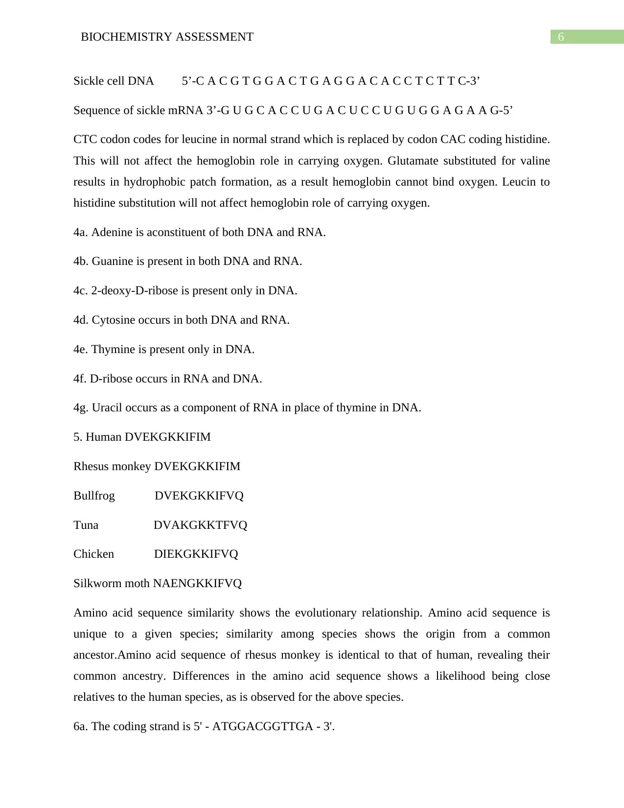
Sickle cell DNA 5’-C A C G T G G A C T G A G G A C A C C T C T T C-3’
Sequence of sickle mRNA 3’-G U G C A C C U G A C U C C U G U G G A G A A G-5’
CTC codon codes for leucine in normal strand which is replaced by codon CAC coding histidine.
This will not affect the hemoglobin role in carrying oxygen. Glutamate substituted for valine
results in hydrophobic patch formation, as a result hemoglobin cannot bind oxygen. Leucin to
histidine substitution will not affect hemoglobin role of carrying oxygen.
4a. Adenine is aconstituent of both DNA and RNA.
4b. Guanine is present in both DNA and RNA.
4c. 2-deoxy-D-ribose is present only in DNA.
4d. Cytosine occurs in both DNA and RNA.
4e. Thymine is present only in DNA.
4f. D-ribose occurs in RNA and DNA.
4g. Uracil occurs as a component of RNA in place of thymine in DNA.
5. Human DVEKGKKIFIM
Rhesus monkey DVEKGKKIFIM
Bullfrog DVEKGKKIFVQ
Tuna DVAKGKKTFVQ
Chicken DIEKGKKIFVQ
Silkworm moth NAENGKKIFVQ
Amino acid sequence similarity shows the evolutionary relationship. Amino acid sequence is
unique to a given species; similarity among species shows the origin from a common
ancestor.Amino acid sequence of rhesus monkey is identical to that of human, revealing their
common ancestry. Differences in the amino acid sequence shows a likelihood being close
relatives to the human species, as is observed for the above species.
6a. The coding strand is 5' - ATGGACGGTTGA - 3'.
Paraphrase This Document
Need a fresh take? Get an instant paraphrase of this document with our AI Paraphraser
7BIOCHEMISTRY ASSESSMENT
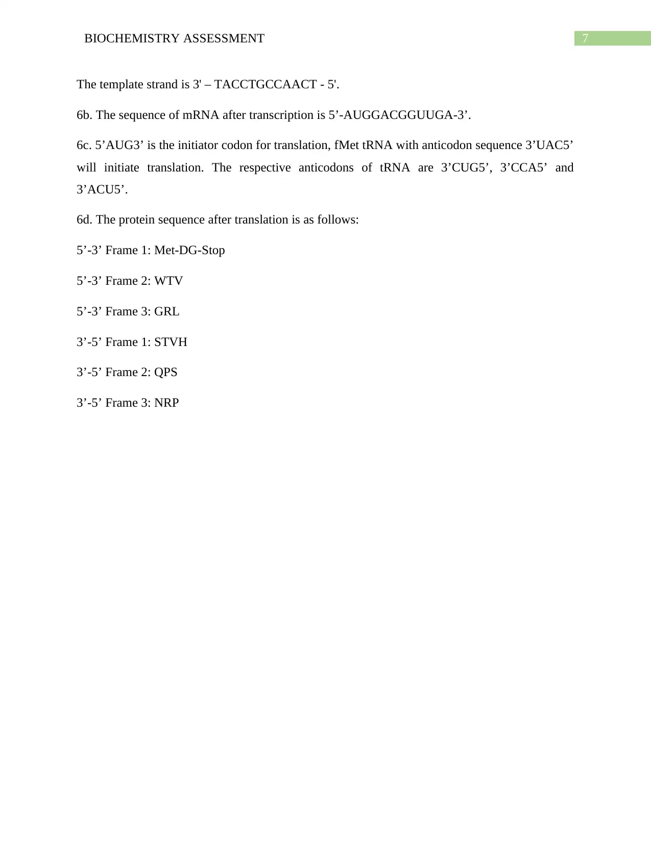
The template strand is 3' – TACCTGCCAACT - 5'.
6b. The sequence of mRNA after transcription is 5’-AUGGACGGUUGA-3’.
6c. 5’AUG3’ is the initiator codon for translation, fMet tRNA with anticodon sequence 3’UAC5’
will initiate translation. The respective anticodons of tRNA are 3’CUG5’, 3’CCA5’ and
3’ACU5’.
6d. The protein sequence after translation is as follows:
5’-3’ Frame 1: Met-DG-Stop
5’-3’ Frame 2: WTV
5’-3’ Frame 3: GRL
3’-5’ Frame 1: STVH
3’-5’ Frame 2: QPS
3’-5’ Frame 3: NRP
8BIOCHEMISTRY ASSESSMENT
References:
Bettleheim, I., Prywes, N., Zhang, N., & Szostak, J. W. (2014). Chain-length heterogeneity
allows for the assembly of fatty acid vesicles in dilute solutions. Biophysical
journal, 107(7), 1582-1590.
Cunningham, W. P., Xia, I., Wickline, K., Garcia Huitron, E. I., & Heo, J. (2018). Studying
Intermolecular Forces with a Dual Gas Chromatography and Boiling Point
Investigation. Journal of Chemical Education, 95(2), 300-304.
Dey, S., & Levy, E. D. (2018). Inferring and Using Protein Quaternary Structure Information
from Crystallographic Data. In Protein Complex Assembly (pp. 357-375). Humana Press,
New York, NY.
Kennedy, E., Dong, Z., Tennant, C., & Timp, G. (2016). Reading the primary structure of a
protein with 0.07 nm 3 resolution using a subnanometre-diameter pore. Nature
nanotechnology, 11(11), 968.
Laganowsky, A., Reading, E., Allison, T. M., Ulmschneider, M. B., Degiacomi, M. T., Baldwin,
A. J., & Robinson, C. V. (2014). Membrane proteins bind lipids selectively to modulate
their structure and function. Nature, 510(7503), 172.
Lundblad, R. L., & Macdonald, F. (Eds.). (2018). Handbook of biochemistry and molecular
biology. CRC Press.
Pérez-Peinado, C., Dias, S. A., Domingues, M. M., Benfield, A. H., Freire, J. M., Rádis-Baptista,
G., ... & Veiga, A. S. (2018). Mechanisms of bacterial membrane permeabilization by
crotalicidin (Ctn) and its fragment Ctn (15–34), antimicrobial peptides from rattlesnake
venom. Journal of Biological Chemistry, 293(5), 1536-1549.
Rahal, I., & Walz, J. (2018). Secondary protein structure prediction combining protein structural
class, relative surface accessibility, and contact number. International Journal of Data
Science, 3(1), 68-85.
Salammal, S. T., Dai, S., Pietsch, U., Grigorian, S., Koenen, N., Scherf, U., ... & Brinkmann, M.
(2015). Influence of alkyl side chain length on the in-plane stacking of room temperature
⊘ This is a preview!⊘
Do you want full access?
Subscribe today to unlock all pages.
Trusted by 1+ million students worldwide
9BIOCHEMISTRY ASSESSMENT
and low temperature cast poly (3-alkylthiophene) thin films. European Polymer
Journal, 67, 199-212.
Voet, D., Voet, J. G., Pratt, C., & BIOCHEMISTRY, F. O. (2016). Life at the molecularlevel.
and low temperature cast poly (3-alkylthiophene) thin films. European Polymer
Journal, 67, 199-212.
Voet, D., Voet, J. G., Pratt, C., & BIOCHEMISTRY, F. O. (2016). Life at the molecularlevel.
1 out of 10

Your All-in-One AI-Powered Toolkit for Academic Success.
+13062052269
info@desklib.com
Available 24*7 on WhatsApp / Email
![[object Object]](/_next/static/media/star-bottom.7253800d.svg)
Unlock your academic potential
Copyright © 2020–2026 A2Z Services. All Rights Reserved. Developed and managed by ZUCOL.