Newcastle University BBF0011 Biology Assignment on Genetic Information
VerifiedAdded on 2022/09/26
|16
|6789
|41
Homework Assignment
AI Summary
This document presents a comprehensive biology assignment focusing on key genetic processes. It begins with an exploration of DNA replication, detailing the semi-conservative model and the Meselson-Stahl experiment. The assignment then delves into transcription in eukaryotic cells, examining the roles of RNA polymerases and transcription factors. Furthermore, the document addresses the process of mRNA modification and explores the concept of mutation, including its classification and types. The provided solution offers clear explanations of genetic information, central dogma, and the mechanisms of inheritance, making it a valuable resource for students studying biology. The document also includes an introduction to biological molecules, including monomers, polymers, carbohydrates, lipids, proteins, nucleic acids, ATP, water, and inorganic ions. The provided assignment also covers mutation, including its classification, types, mutagens, and the impact of mutations on genetic diversity and inheritance. The document provides a detailed understanding of the fundamental principles of molecular biology and genetics.
NEWCASTLE UNIVERSITY
FOUNDATION IN SCIENCE
2019/2020
ASSESSMENT SUBMISSION COVER SHEET
NAME :
STUDENT ID :
MODULE CODE and NAME :
WORD COUNT :
Student Declaration
By signing this, I declare that this assignment meets all the requirement for the
module as detailed in the guidelines, which I have read. It is my own work and I did
not collaborate with or copy from others. I have read and understood my
responsibilities under the university's policy in plagiarism. I have not plagiarised
from published work (including the internet). Where I have used the work of others,
I have referenced it in the text and provided a reference list at the end. I have
understood that plagiarism is the presentation of the work, idea or creation of
another person as though it is your own. It is a form of cheating and is a very serious
academic offence that may lead to expulsion the university. Plagiarism occurs when
the origin of the material used is not appropriately cited. I am aware that late
submission without an authorised extension from the course coordinator will incur a
penalty and after a week the assignment will not be accepted.
Signature:
Date:
Paraphrase This Document
Need a fresh take? Get an instant paraphrase of this document with our AI Paraphraser
Question 1
DNA replication
DNA replication entails a biological process where organisms undergo biological
inheritances aspects. DNA replication takes place in a process referred to as semi-
conservative method which yields double strands of DNA with parental strand and a
new daughter strand. The discovery of DNA by researches Watson and Crick on
double-strand helix ability to offer DNA replication offered a milestone in
understanding the dynamics of DNA action. In the cell division process, each DNA
molecule has to the copied so to produce identical molecules towards the daughter
cells. The double-strand structure of DNA entails that each strand can be reproduced
each acting as a template where it produces a new copy of complementary strand
thus generating two double-strand molecules from the original one (Méchali, 2010).
Various models have been advanced to understand the basis of the DNA replication
process, these models of replication entail conservative, semi-conservative and
dispersive. In the conservative avenue, two originals parental strands undergo base
pairing with each other after template usage in synthesizing new strands and the
new two strands referred to as daughter strands undergoing base pair with each
other. In this process, the outcomes yield two DNA molecules with characteristics of
'all old' and the other being 'all-new'. In semiconservative replication process, each of
the two parental DNA strands is able to act as the templates allowing the new
synthesis of DNA strands, after replication, each of the parental strands undergoes
base pairing with the complementary new synthesized strand. Birth doubles strands
will entail one parental or ‘old strand and one daughter also referred to as ‘new’
strand. In the dispersive process, when both copies have replicated, the new strands
would alternate the segments of the parental DNA somehow and the newly formed
DNA is on every two strands (Masai et al., 2010).
In demonstrating this process as observed in the picture, understanding of the
Meselson and Stahl model of replication is fundamental in this process. In Meselson
and Stahl focus is in their model were keen on understanding the processes of DNA
replication. This process entails the application of growing E. Coli generation in
heavy isotope media of nitrogen 15N which has nitrogen bases and eventually to the
DNA. The E. Coli was shifted into a light isotope medium containing isotope of
nitrogen with 14N and allowed for growth in one generation. After the elapse of this
period, the cells were harvested and the DNA isolated. The next step entailed
centrifugation of DNA at high speeds in a tube which there was known caesium
chloride density gradient. Some of the cells were allowed to go one more generation
further at 14N and centrifuged again (Raghuraman and Brewer, 2010).
During the density gradient ultracentrifugation, the researchers applied the use of
caesium chloride salt and spun at high speeds of about 50,000 to 60,000 rounds per
minute. In the centrifuge tube, the density gradient was created by the salt with
higher density allowing movement farther in the tube. During a point in this process,
2
the molecules stopped the sedimentation process and formed stable bands. The
band molecules allow for closer identification of relative densities, these molecules
that formed in the lowest bands had the highest identities. This is a similar process
illustrated in the picture related to this question, the formation of bands are depicted
(Raghuraman and Brewer, 2010).
The rationale behind this signals that DNA from the 15N produces the lower band
than the DNA from the 14N, thus signalling higher density of the former compared to
the latter, due to the heavy isotope process. The researchers noted that after each
generational growth of 14N the DNA produces single-strand band intermediate in
DNA cells grown from the 15N and DNA cells exclusively in the 14N isotope. This
signalled a semi-conservative or dispersive replication mode. Had it been
conservative replication, then two bands would have been producing each
representing parental DNA with exclusive 14N located in its nitrogen bases, the single
observed demonstrates that there is an equal amount of both 15N and 14N in the DNA
molecules (Raghuraman and Brewer, 2010).
Analysis on this research by Meselson-Stahl’s demonstrates that cells are grown
from two generations in 14N yields two strands, one strand being at the intermediate
position between the 15N and 14N while the other corresponding to the 14N DNA
exclusively. This clearly demonstrates the observation seen in tube 3 contain the
generation in 14N isotope. This depicts a semi-conservative DNA replication. If it had
been a dispersive replication, then the single brand would be produced exclusively in
each new generation with slow movements of the band closer to the top of the 14N
band, thus ruling out dispersive process which is not observed in our figure (Amado,
2010).
Meselson and Stahl’s findings demonstrate that the DNA replication process offers
an avenue for two strands that make up the double helix are serving as the template
where the new strands are synthesized. The new strand will complement to the
parental or 'old while the new strand remains are base-paired on the old strand. Thus
each daughter DNA has old DNA strand and one new synthesized strand. Coping of
the two daughters DNA leads to identical sequence to one another and identical to
the parental DNA while the two daughter cells are further divided equally forming two
daughter cells, which are genetically identical to one another and genetically
identical to the parent cell.
Hence this illustrative depicts the scenarios observed in this question where there is
a semiconservative mode of DNA replication occurring in view of the application of
nitrogen pairs and process of centrifugation. This is an essential aspect in the
production of photocopy cells in organisms allowing duplicate production and genetic
similarities and differences occurrences.
Question 2
3
⊘ This is a preview!⊘
Do you want full access?
Subscribe today to unlock all pages.
Trusted by 1+ million students worldwide
Transcription process in a cell
Transcription process in the eukaryotic cell takes place in the membrane-bound
nucleus and organelles. The genes bound in the nucleus, they transport the mRNA
to the cytoplasm and must be protected from degradation before being translated.
Eukaryotic cells employ three key polymerases to perform the transcribing in
different subset genes and they are monogenetic specifying single protein.
Transcription entails the aspect of DNA information is copied into new molecules
messengers mRNA. The DNA safely has the ability to store genetic material in the
genetic nuclei to be sued as a referenced template. The mRNA used is similar to the
template carrying information as DNA but not used as long term storage and can exit
the nucleus easily. The mRNA has the same information but is not an identical copy
of the DNA due to its complementary sequence to the DNA template (Fatma, 2018).
The transcription process is carried out by the RNA polymerase and other
accessory proteins referred to as transcription factors. These factors are able to bind
to the specific sequence of DNA through the support of enhancers and promoters so
as to recruit the RNA polymerase to a site for the transcription process. The
transcription factors and the RNA polymerase forms complex transcription initiation.
This process begins with the synthesis of mRNA through matching with the original
DNA strand. The mRNA undergoes elongation after synthesization, thus terminating
the transcription. The newly formed mRNA gene copies are the blueprints of the
protein synthesis during the translation process (Puisieux, Brabletz and Caramel,
2014).
The underlying polymerase process in eukaryotic cells entails the aspects described
above entail the transcription factors which are able to bind the promoter area so as
to initiate appropriate polymerase. The eukaryotic mRNA synthesis is often complex,
the polymerase activity has 10 subunits in each of the three polymerases. Each
polymerase has a different transcription factors set to enable it to bring to the DNA
template.
RNA polymerase I is found in the nucleus, a substructure of the nucleus which
allows the ribosomal RNA to transcribed, processes and assembled into many
ribosome’s. The rRNA is categorized as the structural RNAs due to their cellular
roles but do not undergo a translation into the proteins. The ribosomal rRNAs are the
essential components which are fundamental in the translation process. The RNA
polymerase I is able to synthesize the rRNAs except for the 5S rRNA molecule. The
designation of 'S' denotes the 'Svedberg' units which characterize the centrifugation
speeds (Puisieux, Brabletz and Caramel, 2014).
In RNA polymerase II, synthesis of all protein-coding nuclear pre-mRNAs in the
nucleus, the pre-mRNAs undergo processing beyond the transcription phase with
mRNAs being the process for translating the mature processed molecules. RNA
polymerase II is essential in the transcription activity in many eukaryotic genes.
4
Paraphrase This Document
Need a fresh take? Get an instant paraphrase of this document with our AI Paraphraser
In RNA polymerase III, found in the nucleus the polymerase is able to transcribe to
various structural RNAs such as the 5S pre-rRNAs, pre-RNAs and small nucleus
pre-RNAs. The tRNAs have a crucial role in the translation process, as they serve as
adaptive molecules in the process of mRNAs template and the growing chain of the
polypeptide. The small nucleus of the RNA has various functions such as the ‘spicing
activity’ and the transcription factors (Zhang, Shalaby and Buszczak, 2014).
The transcription factors often do not end with the polymerase and promoters, other
transcription factors, silencers and enhancers aid in regulating the frequency of the
pre-mRNAs which are synthesized from the genes. The enhancers and silencers
have the ability to affect the transcription process efficiently but not essential for the
transcription process to proceed. The basal transcription factors are fundamental in
the formation of the pre-initiation of complex DNA template which recruits the RNA
polymerase II transcription initiation (Zhang, Shalaby and Buszczak, 2014).
The basal transcription factors begin with ‘TFII’ which are basically the RNA
polymerase II and specified letters A-J. The transcription factors fall into the DNA
template which each stabilizing to the pre-initiation complex, contributing the
polymerase II RNA recruitment.
The RNA polymerase I and III transportation to the DNA template often entails
complex transcription factors. Each transcription of eukaryotic is regulated hence
requires more proteins variety to allows for interactions with each other and the
strands of DNA. The process of eukaryotic transcription entails greater metabolic
investment compared to that in prokaryotes thus ensuring the precise pre-mRNA in
the protein synthesis.
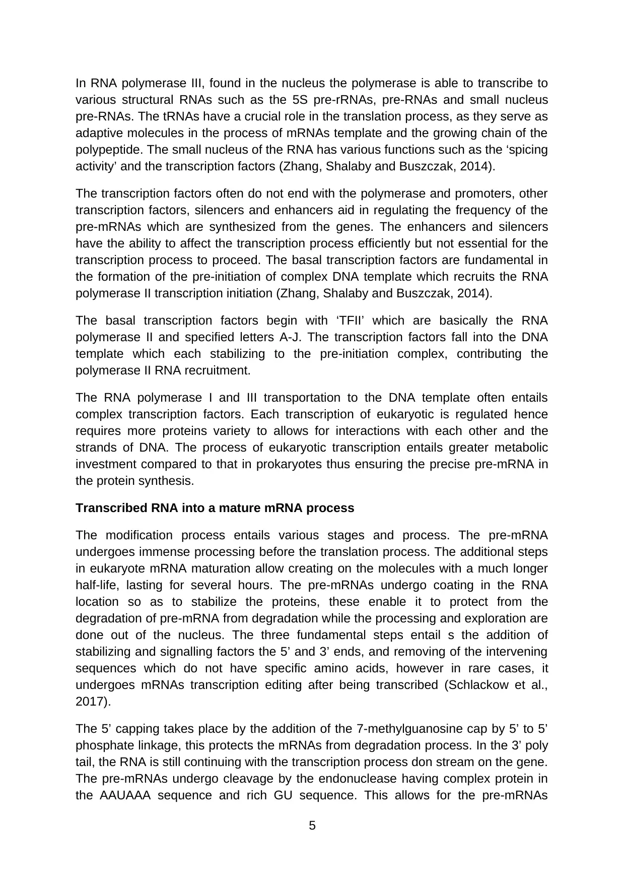
Transcribed RNA into a mature mRNA process
The modification process entails various stages and process. The pre-mRNA
undergoes immense processing before the translation process. The additional steps
in eukaryote mRNA maturation allow creating on the molecules with a much longer
half-life, lasting for several hours. The pre-mRNAs undergo coating in the RNA
location so as to stabilize the proteins, these enable it to protect from the
degradation of pre-mRNA from degradation while the processing and exploration are
done out of the nucleus. The three fundamental steps entail s the addition of
stabilizing and signalling factors the 5’ and 3’ ends, and removing of the intervening
sequences which do not have specific amino acids, however in rare cases, it
undergoes mRNAs transcription editing after being transcribed (Schlackow et al.,
2017).
The 5’ capping takes place by the addition of the 7-methylguanosine cap by 5’ to 5’
phosphate linkage, this protects the mRNAs from degradation process. In the 3’ poly
tail, the RNA is still continuing with the transcription process don stream on the gene.
The pre-mRNAs undergo cleavage by the endonuclease having complex protein in
the AAUAAA sequence and rich GU sequence. This allows for the pre-mRNAs
5
various structural RNAs such as the 5S pre-rRNAs, pre-RNAs and small nucleus
pre-RNAs. The tRNAs have a crucial role in the translation process, as they serve as
adaptive molecules in the process of mRNAs template and the growing chain of the
polypeptide. The small nucleus of the RNA has various functions such as the ‘spicing
activity’ and the transcription factors (Zhang, Shalaby and Buszczak, 2014).
The transcription factors often do not end with the polymerase and promoters, other
transcription factors, silencers and enhancers aid in regulating the frequency of the
pre-mRNAs which are synthesized from the genes. The enhancers and silencers
have the ability to affect the transcription process efficiently but not essential for the
transcription process to proceed. The basal transcription factors are fundamental in
the formation of the pre-initiation of complex DNA template which recruits the RNA
polymerase II transcription initiation (Zhang, Shalaby and Buszczak, 2014).
The basal transcription factors begin with ‘TFII’ which are basically the RNA
polymerase II and specified letters A-J. The transcription factors fall into the DNA
template which each stabilizing to the pre-initiation complex, contributing the
polymerase II RNA recruitment.
The RNA polymerase I and III transportation to the DNA template often entails
complex transcription factors. Each transcription of eukaryotic is regulated hence
requires more proteins variety to allows for interactions with each other and the
strands of DNA. The process of eukaryotic transcription entails greater metabolic
investment compared to that in prokaryotes thus ensuring the precise pre-mRNA in
the protein synthesis.
Transcribed RNA into a mature mRNA process
The modification process entails various stages and process. The pre-mRNA
undergoes immense processing before the translation process. The additional steps
in eukaryote mRNA maturation allow creating on the molecules with a much longer
half-life, lasting for several hours. The pre-mRNAs undergo coating in the RNA
location so as to stabilize the proteins, these enable it to protect from the
degradation of pre-mRNA from degradation while the processing and exploration are
done out of the nucleus. The three fundamental steps entail s the addition of
stabilizing and signalling factors the 5’ and 3’ ends, and removing of the intervening
sequences which do not have specific amino acids, however in rare cases, it
undergoes mRNAs transcription editing after being transcribed (Schlackow et al.,
2017).
The 5’ capping takes place by the addition of the 7-methylguanosine cap by 5’ to 5’
phosphate linkage, this protects the mRNAs from degradation process. In the 3’ poly
tail, the RNA is still continuing with the transcription process don stream on the gene.
The pre-mRNAs undergo cleavage by the endonuclease having complex protein in
the AAUAAA sequence and rich GU sequence. This allows for the pre-mRNAs
5
release form the transcript attached to the RNA polymerase. The enzyme Poly A
polymerase is a fundamental part if the protein complex. It is part of the protein
forming the PAP. It cleaves the pre-mRNAs and adds the string to the 3' end of the
cleaved section. This A poly tail protects from any degradation and facilitates the
transportation of the mature mRNAs to the cytoplasm (Lemay, et al., 2014).
Question 3
Sexual reproduction process
Sexual reproduction refers to the creation of an organism through the creation of two
genetically identical clones, both the sources contribute half of the genetic materials,
here the offspring will have both parents. Organisms tend to produce sexually by
joining gametes, in a process referred to as fertilization in mechanisms producing
haploid gamete, through a process referred to as meiosis, which the chromosomes
are half into. During the meiosis process, the chromosomes pair separate and
undergo random segregation so as to produce gametes having one chromosome for
each pares. The meiosis process entails two nucleus and cell divisions without the
interphase in between, begin with one diploid generating a total of four haploid cells.
Each division is referred to as meiosis I ad meiosis II entailing four stages of
prophase metaphase, anaphase and telophase (Goodenough and Heitman, 2014).
Meiosis, as referred above, is a process which produces chromosomes by reducing
it to half, it takes place in special cells in an organism, among mammals it occurs in
gamete producing cells located in the gonads. During the meiosis process, the
paired chromosomes separate while the haploid cells forming it have only one
chromosome each. The key phases and of meiosis entails;
Meiosis I
In Prophase I, the nuclear envelope initiates with breakdown condensing the
chromosomes, the centrioles start through moving to the opposite’s side of the cell
and initiate the formation of the spindle, the homologous chromosomes pair up which
is a key characteristic of this phase.
In metaphase I, the spindle fibres are attached to the paired homologous
chromosomes. The paired chromosomes are able to line up in the equator cell,
occurring in metaphase I. In anaphase I, the spindle fibres tend to shorten and each
homologous chromosome separate from each other. The chromosomes pairs move
towards the pole of the cell while the chromosomes move in opposite pole direction.
In telophase I and cytokines, there is breaking down of the spindle and the formation
of nuclear membranes is initiated. The cytoplasm of the cell divides leading to the
production of two haploid cells. The daughter cells entail chromosomal random
assortment having one each homologous pair (Hillers et al., 2017).
Meiosis II
6
⊘ This is a preview!⊘
Do you want full access?
Subscribe today to unlock all pages.
Trusted by 1+ million students worldwide
In prophase II, there is a breakdown of the nucleus enveloped proceeded with the
formation of the spindle in each of the haploid daughter cells formed from meiosis I,
further there is a separation of the centrioles in this process.
In metaphase II, the spindle fibres tend to line the sister chromatids in each of the
chromosomes moving along the equator cells. In anaphase II there is the separation
of sister chromatids moving in opposite poles. In telophase II and cytokines, there is
a breakdown of the spindle and formation of new nuclear membranes formed; each
cell cytoplasm divides leading to the production of four haploid cells (Hillers et al.,
2017).
After the meiosis process, four haploid cells have been formed, but the cells are not
yet fully developed to be gametes hence the need for full development before having
the ability to engage in the fertilization process. This process is referred to as
gametogenesis and differs between sexual orientation. The gamete produced by a
male is referred to as sperm with the spermatogenesis process essential in the
production of mature sperm. During this process, the sperm grows with tail aiding its
ability to swim. In females, gametes are referred to as eggs, produced in a process
called oogenesis producing mature eggs. In this process, one egg is produced from
a total of four haploid cells leading to meiosis (Hillers et al., 2017).
The spermatogenesis process is initiated in the tubules of the seminiforeon. They
begin with stem cell the periphery levels and spermatozoa located in the lumen. After
the diploid are in the capsule tubule, having undifferentiated cells. The stem cells
called the spermatogonia undergo one offspring mitosis through the differentiation
phase producing sperm cell, giving rise to new sperm generation.
Meiosis process in spermatogenesis begins with the primary spermatocyte, during
the process of first meiotic division there is production of haploid cells referred to
secondary spermatocyte, it undergoes through meiotic cell division, producing
spermatid. At the tubule lumen end, it produces flagellum referred to as sperm cell,
at the end of the process and phases of meiosis two; there is the production of four
sperm cells. The stem cells disposition takes place in the gestation period and
present at birth through the adolescent stage though it exists as an inactive state. In
the adolescent stage, the gonad tropic hormones in the activation of cells is caused
by the anterior pituitary cause thus producing available sperms (Hillers et al., 2017).
In the oogenesis process, it takes place in the ovary layers. Oogenesis begins with
oogonium which undergoes mitosis leading to an increase in number leading to a
large number of embryo cells.
The meiosis stage begins with primary oocyte progressing to the first prophase
stage, at birth the eggs are in prophase stage. At adolescents stage, development of
follicles is caused by the pituitary hormones in the ovary. This leads to the ending of
the primary oocyte through the meiotic division. There is unequal cell division with
the majority of the cell organelles going to the secondary oocyte and another chosen
7
Paraphrase This Document
Need a fresh take? Get an instant paraphrase of this document with our AI Paraphraser
set and small cytoplasm going through to the other cell. The second cell referred to
as a polar body dies. In the process, a secondary meiotic arrest develops at the
metaphase phase II stage, at the ovulation stage, the secondary oocyte is released
towards the uterus through the oviduct. In case of fertilization on the secondary
oocyte, the cell moves to the meiosis II, successfully completing the meiosis, leading
to the production of second polar body and fertilized egg having 46 chromosomes
having half of them from the sperm (Ohkura, 2015).
Question 4
Colour blindness is a hereditary state which is genetically transferred to offspring.
The red or green colour is usually passed from the mother to the son occurring on
the 23rd chromosome which is referred to as sex chromosomes due to its
determination of sex. Chromosomes contain genes which entail coding on the
development of tissues and organs. Colour blindness signifies that there faulty
instructions on the cone cells development leading to less sensitivity to light (Akhtar,
2015).
Every human egg has single X chromosomes and sperm carries both X or Y
chromosomes. Eggs fertilizing the sperm with an X chromosome are female gender
while eggs fertilizing Y sperm turns to be male.
The 23rd chromosome is made of two sections which are either two chromosomes in
females or X and Y chromosome among males. Blindness among male has to show
faulty gene on the X chromosomes, while among females the greens for colour blind
needs to be present in both of X chromosomes (Akhtar, 2015).
Female having colour blidn genes are carriers, hence they are not blind but having a
child enables him to give the X chromosome with the colour blindness gene to the
son, thus will be blind. A colour blind male cannot get the genes his father, even
though the father will be or is colour blind. The reason being the father has the ability
to pass the X chromosomes to his daughters but not male.
Colour blind daughter thus needs to have a colour blind father and a carrier mother
who has the heterozygous gene. If the father is not having colour blindness defect,
the daughters will not be colour blind, however, she will be a carrier in two avenues;
through acquiring from her mother or from his father who has a colour blind defect.
This depicts the reason why colour blindness in red and green pigments is common
among men than women.
This, in essence, illustrates that the red or green colour defects are due to mutation
being observed on the X chromosomes allele. The X linked colour blindness is
recessive, females will have normal vision, manifesting itself on inheritance from
both parents, while the male inherits the X chromosomes from the mother hence the
red and green colour blindness (Commons n.d).
8
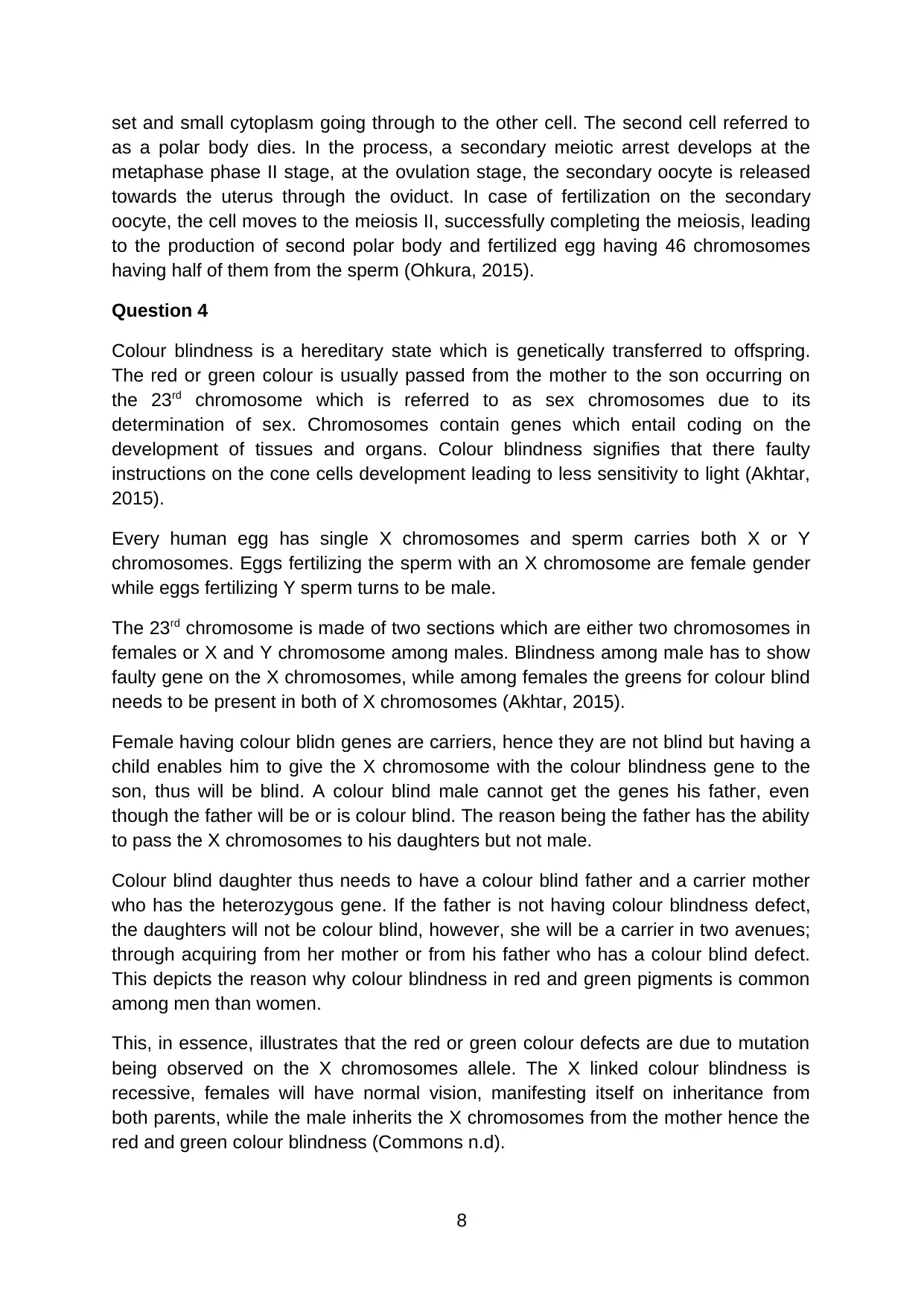
Thus as observed in the diagram the father is colour blind signalling presence of X
recessive trait while the mother is a carrier signalling the presence of homozygous
recessive allele. The daughter is female colour blind due to the recessive X
chromosomes which are inherited while the other daughter is a carrier having both
XX chromosomes, thus due to the presence of the mutant allele she will pass to her
daughter whom will be a carrier. The son is healthy since the mutant alleles do not
have the recessive gene for blindness.
Colour blindness pertaining to blue colour often affects both genders due to its ability
to be accrued in the non-sex chromosome. The colour blindness gene is attached on
the X chromosome, thus men have one X chromosomes. The X chromosome
carriers the blind gene, illustrating that he will be colour blinding in that are displaying
XY. Among women, it occurs in three avenues; two normal X chromosomes thus
having no defects of colour blindness or being a carrier-XX; occurrence of one
normal X and Y colour blind chromosomes thus indicating being a carrier and the
final instance entail inheritance of colour blindness chromosomes X from the father’s
X and colour blind mother X and herself being colour blind with XX (Wong, 2011).
More studies have shown that colour blindness is an inherited genetic disorder
affecting the X chromosomes. The mapping of human in human genomics has
illustrated various causative issues which are linked to causing blindness which
emanate from at least about 19 chromosomes and 56 other different genes.
The most prevalent forms of this inheritance are the protanomaly and protanopia
which is rare to occur, which is combined formed is referred to as potans and
deuteranomaly, they are both accounting to a larger proportion of red-green colour
blindness. The affected persons often exhibit difficulties in differentiating the red and
green hues occurring on the red and green photoreceptor retinal. The sex linkages
traits of the green and red blindness have the ability to affect more males due to its
location on the X chromosomes, where males have one while females have 2
(Wong, 2011).
The red/green colour blindness has the ability to be transmitted from males who are
colour blind to all the daughters whom they are heterozygous, thus unaffected. A
woman has a 50% of crossing the mutated X chromosome to the male offspring
while the son’s offspring will not transfer the trait since they only receive the Y
chromosomes and unlike the defective X chromosome. If the affected father has a
carrier woman or colour blinding their daughters may experience inheritance of the
affected X chromosome from both parents.
Question 5
The three recognizable colours in Retrievers dogs have been shown to result from
the genetic loci which affect the expression pigment. The initial of this colour entail
the dark pigment referred to brown colour represents by B locus. In dogs, TYRP1
has been shown to be responsible for these loci, an enzyme localized in the
9
⊘ This is a preview!⊘
Do you want full access?
Subscribe today to unlock all pages.
Trusted by 1+ million students worldwide
melanosomes. This mutation in this enzyme leads to protein truncation on the amino
acid sequencing in the protein chain. This mutation has the ability to reduce the
ability f the phenotype colourization which is visible traits in 3 mutations (Christopher
et al., 2013). The inherent recessive mutations on the enzyme gene lead to two
copies of gene each from the parent which is represented by B loci displaying brown
pigmentation with both copies having mutant alleles represented by ‘b’. This signifies
a dog with BB or Bb genotypes are able to express the black colour while the brown
colour will be observed among dogs with the bb genotype.
In a further analysis of the effects of the second gene, the colour pigments will be
expressed by the fur referred to as extension E trait directed by the melanocortin 1
receptor. They signal the cell producing pigments leading to fur colour disposition.
These mutations have been demonstrated to entail red or pale colour common
among various species (Lucy et al., 2016).
In dogs, the melanocortin receptor is modulated by molecule signals which are
present in A locus and activator referred to as K locus. A recessive mutation in the E
gene is truncated by the proteins by producing functional receptors which are
capable of directing the colour disposition in the fur. In B locus, the E gene single
receptor leads to a dominant phenotype which is present in the fur. When both
genes are recessive to mutant gene 'e' there will be no colourisation in its fur,
appearing yellow evidence ton the skin nose, footpads and lips determined by the B
locus. The variance of the melanocortin receptor allele has can display facial masks
in other dog breeds (Reese et al., 2011).
Gene interactions
The underlying interplay between the genes impacts the colour of the dogs.
Possession of a dominant phenotype in extension allele that is genotype EE or Ee,
leads to the display of fur colourization which is accustomed by the brown genotype
locus while the recessive extension trait displays yellow colourisation with either
display of BB, Bb or brown bb exposed in the skin, leading to various colours as
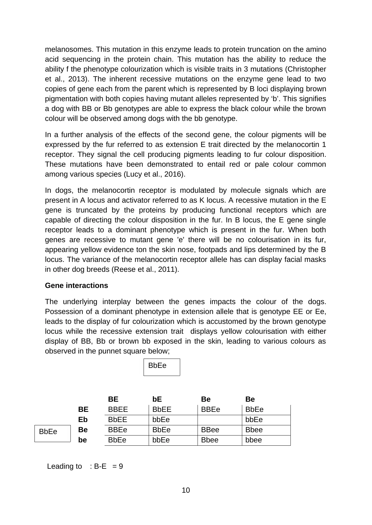
observed in the punnet square below;
BE bE Be Be
BE BBEE BbEE BBEe BbEe
Eb BbEE bbEe bbEe
Be BBEe BbEe BBee Bbee
be BbEe bbEe Bbee bbee
Leading to : B-E = 9
10
BbEe
BbEe
acid sequencing in the protein chain. This mutation has the ability to reduce the
ability f the phenotype colourization which is visible traits in 3 mutations (Christopher
et al., 2013). The inherent recessive mutations on the enzyme gene lead to two
copies of gene each from the parent which is represented by B loci displaying brown
pigmentation with both copies having mutant alleles represented by ‘b’. This signifies
a dog with BB or Bb genotypes are able to express the black colour while the brown
colour will be observed among dogs with the bb genotype.
In a further analysis of the effects of the second gene, the colour pigments will be
expressed by the fur referred to as extension E trait directed by the melanocortin 1
receptor. They signal the cell producing pigments leading to fur colour disposition.
These mutations have been demonstrated to entail red or pale colour common
among various species (Lucy et al., 2016).
In dogs, the melanocortin receptor is modulated by molecule signals which are
present in A locus and activator referred to as K locus. A recessive mutation in the E
gene is truncated by the proteins by producing functional receptors which are
capable of directing the colour disposition in the fur. In B locus, the E gene single
receptor leads to a dominant phenotype which is present in the fur. When both
genes are recessive to mutant gene 'e' there will be no colourisation in its fur,
appearing yellow evidence ton the skin nose, footpads and lips determined by the B
locus. The variance of the melanocortin receptor allele has can display facial masks
in other dog breeds (Reese et al., 2011).
Gene interactions
The underlying interplay between the genes impacts the colour of the dogs.
Possession of a dominant phenotype in extension allele that is genotype EE or Ee,
leads to the display of fur colourization which is accustomed by the brown genotype
locus while the recessive extension trait displays yellow colourisation with either
display of BB, Bb or brown bb exposed in the skin, leading to various colours as
observed in the punnet square below;
BE bE Be Be
BE BBEE BbEE BBEe BbEe
Eb BbEE bbEe bbEe
Be BBEe BbEe BBee Bbee
be BbEe bbEe Bbee bbee
Leading to : B-E = 9
10
BbEe
BbEe
Paraphrase This Document
Need a fresh take? Get an instant paraphrase of this document with our AI Paraphraser
:bbE- =3
:- - ee = 4
P generation
Crossing true black BB and true brown bb produces the genotypes ;
BbEe and BbEe
The allele of b masks the effect of the E allele, thus in all the instances where the b
is demonstrated by the Ee aspect. Dogs having this ee at their extension locus and
bb at the brown are likely to show brown noses, eyes or colourisation on footpads.
F1 generation
Crossing the P generation that is
BbEe and BbEe
Offers the genotypes
BE, bE Be and be all being black retrievers however they have an extension of the E
or e loci in their extension showcasing red or brown colourisation.
F2 generation
Crossing the F1 generations yields that is
BE, bE Be be and BE, bE Be be
Gives the ; 9 B-E
; 3 bbE-
; 4 - -ee
Black retriever
The retriever will have the genotype with at least one domain allele showcasing at
the B and E loci
Brown retriever
The retrievers with at least one dominant allele E are having the recessive allele’s b
and e that is bbEE, bbEe, BBee Oor Bbee.
Golden retriever
The retriever with golden pigment or absence o f skin pigment will have only the
recessive alleges at bbee loci.
11
There is an assortment of genes independently in this demonstration, thus a single
gene cross will entail two black retrievers with recessive alleles both B and E locus,
having the potential of producing all possible colour. The crosses entailing brown
retrievers will never produce black because there is no dominant B allele among the
parents but can produce golden retrievers.
Question 6
Sickle cell disease
Sickle cell diseases are a recessive trait, hereby an individual who has two copies of
the sickle cell allele often referred to as HbS. Persons or individuals have either of
the two copies of beta allele HbA will get the disease. The heterozygous individual
often does not demonstrate the signs of the disease despite having one HbS and
one allele of HbA, however, they have traits due to the ability to produce abnormal
beta-globin. Among these persons, they have red blood cells which are abnormal,
normal and molecule shaving both versions of the normal and sickle cell subunit. In
this state the hydrophobic valine located in the abnormal beta haemoglobin is
exposed, leading to clumping together just in the same manner sickle cell persons
developed, however, because there is normal beat globulin, long chains responsible
for the disease will not occur among this persons. Without these long chains person
with the disease do not show any signs and symptoms (Piel, Steinberg and Rees,
2017).
The difference between the sickle cell and normal beta-globin entails difference of
one single amino acid. In the beta-globin, the nucleotide sequence also differs by
one amnion acid compared to the normal and sickle cell disease alleles.
The prevalence trends of sickle cell anaemia are widely found in sub-Saharan Africa
or those with roots in this region. The sickle-cell allele distribution shows the
historical distribution of infectious diseases with an established relationship on
malaria disease (Garnier-Géré and Chikhi, 2013).
The testing of the disease entails birth tests using prickle test, where the blood is
measured on the levels of haemoglobin. If there are low levels of blood, the patients
especially the infants are referred to further testing by a haematologist who will
perform genetic testing.
The most simple and common method of testing sickle cell anaemia is by application
of polymerase chain reaction and digestion restriction. PCR entails an avenue in
which copies of specific DNA sequence are made. The restriction digest is a process
of DNA cutting as per the defined DNA sequence.
In performing the restriction digest, the enzyme is used to locate the specific
sequence. The enzymes then slice the DNA pieces into tow, the mutant gene is
located in the middle of the restriction enzymes. The normal beta goblin allele
sequence has the CTGAG sequence in its allele; this is a recognition sequence for
12
⊘ This is a preview!⊘
Do you want full access?
Subscribe today to unlock all pages.
Trusted by 1+ million students worldwide
1 out of 16
Related Documents


Your All-in-One AI-Powered Toolkit for Academic Success.
+13062052269
info@desklib.com
Available 24*7 on WhatsApp / Email
![[object Object]](/_next/static/media/star-bottom.7253800d.svg)
Unlock your academic potential
Copyright © 2020–2026 A2Z Services. All Rights Reserved. Developed and managed by ZUCOL.