Biology 1 Assignment: Human Body Organisation and Cell Biology
VerifiedAdded on 2020/05/04
|25
|5946
|66
Homework Assignment
AI Summary
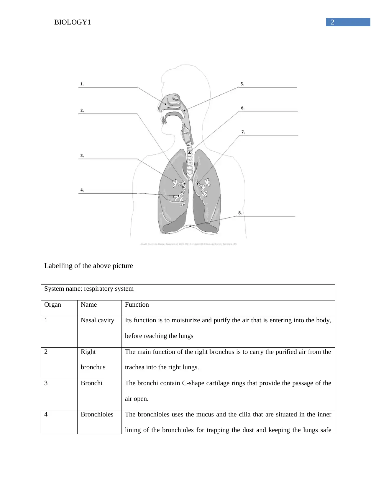
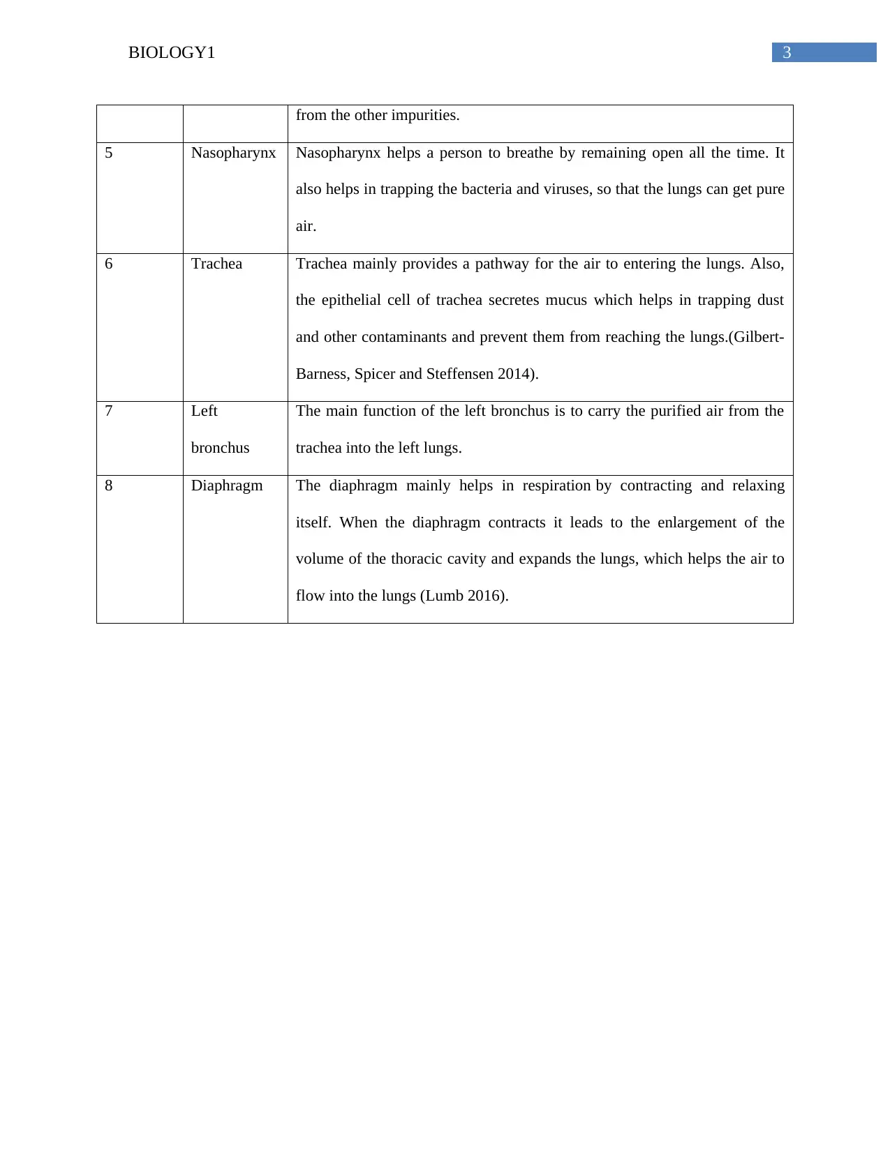
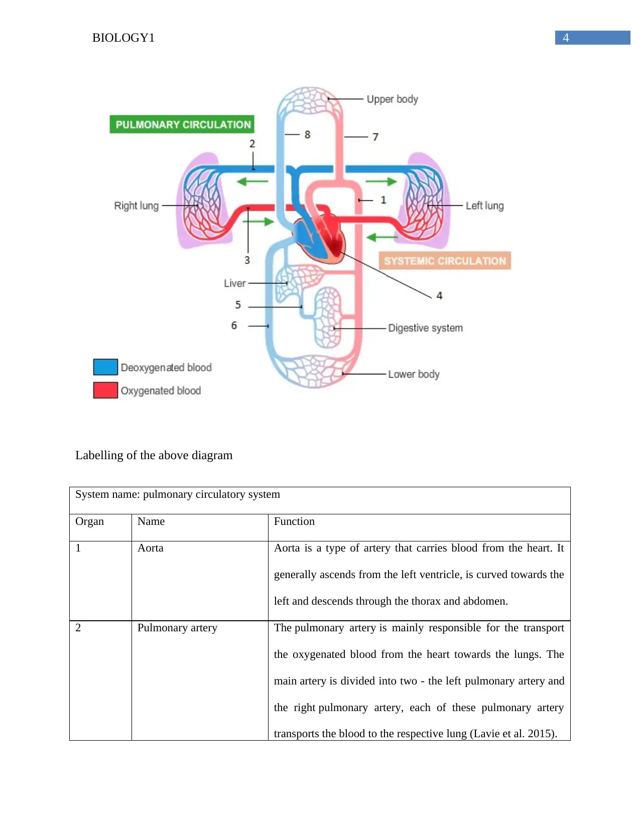
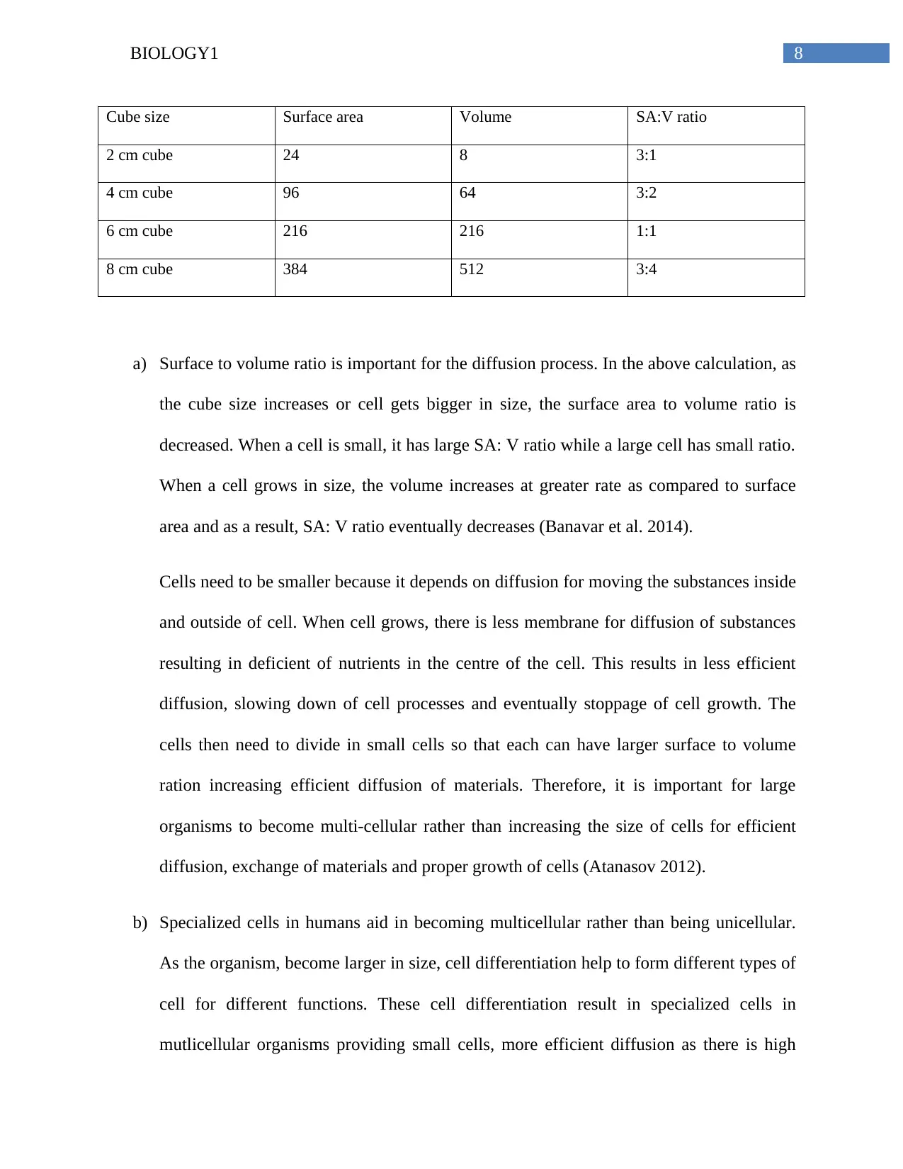
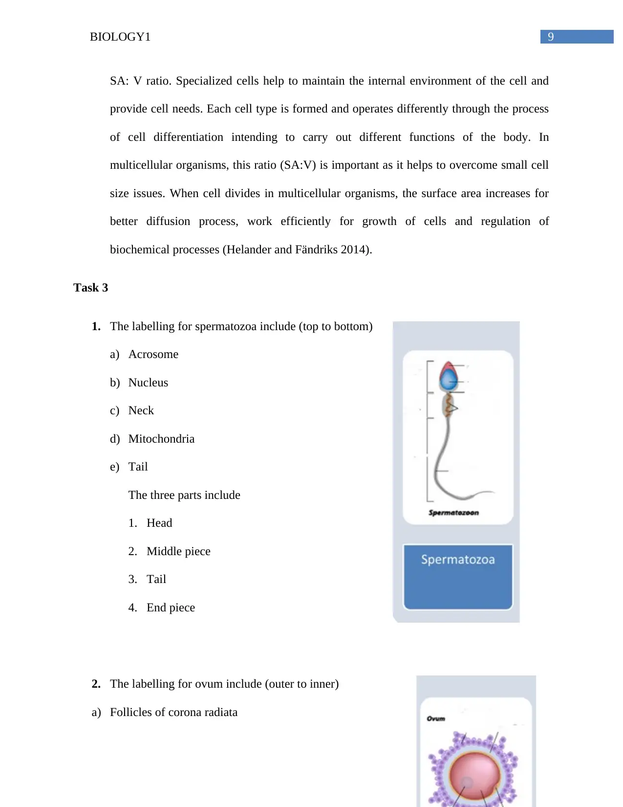
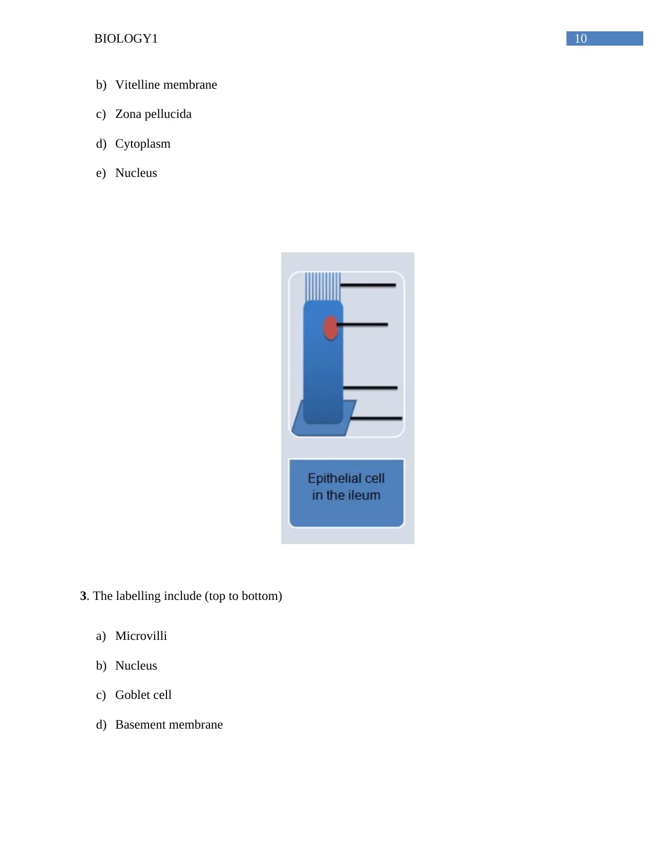
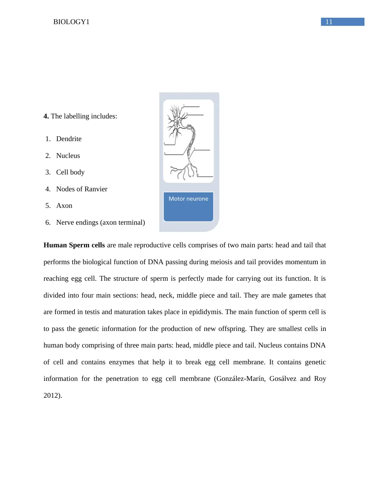
This Biology 1 assignment comprehensively covers the level of organization of the human body, including the respiratory, pulmonary circulatory, and digestive systems. It requires labeling diagrams, outlining the functions of various organs within each system, and explaining the division of labor. The assignment also delves into cell biology, exploring the surface area to volume ratio and its importance for diffusion and cell size. Furthermore, it includes detailed labeling of sperm and ovum structures, as well as epithelial cells in the ileum, with explanations of their functions. The student demonstrates an understanding of cell differentiation, specialized cells, and their roles in multicellular organisms. The assignment references several scientific articles to support the information provided.

Running head: BIOLOGY1
Biology 1
Name of the Student
Name of the University
Author note
Biology 1
Name of the Student
Name of the University
Author note
Paraphrase This Document
Need a fresh take? Get an instant paraphrase of this document with our AI Paraphraser

1BIOLOGY1
Task1
Part 1
Cells-Thesearethebasicunitsoftheformoflife.Itconsistofallthestructuralandfunctionalaspectsneedeforthegrowthanddevelopmentoftheorganism.Tissues-Tissuesarethegroupofcellswhichareofsimilarstructure.Eachofthetissuehassomedifferentfunctionwhichisdonebythewrokingofallthecellstogether.Organs-Organsareformedbytheassociationofthetissuestoserveaspecificfunction.Theorgansaremadeupoffourtypesoftissue;epithelialtissue,whichformstheinnerlining,connectivetissuewhichhelpstotransportthenutrients;muscletissuethatprovidesupporttotheorgansandnervetissuewhichsendstheelectricimpulsetothebrainanddifferentorgans.Organsystem-Agroupoforganstogethermakeanorgansystem.Theorgansofourbodyareoftwotypes-externalorgansandinternalorgans.Organism-Anorganismismadeupoftheorgansystemandtheycancarryoutallthelifeprocessnecessayfordailylife.LEVEL OF ORGANISATION OF THE HUMAN BODY

2BIOLOGY1
Labelling of the above picture
System name: respiratory system
Organ Name Function
1 Nasal cavity Its function is to moisturize and purify the air that is entering into the body,
before reaching the lungs
2 Right
bronchus
The main function of the right bronchus is to carry the purified air from the
trachea into the right lungs.
3 Bronchi The bronchi contain C-shape cartilage rings that provide the passage of the
air open.
4 Bronchioles The bronchioles uses the mucus and the cilia that are situated in the inner
lining of the bronchioles for trapping the dust and keeping the lungs safe
⊘ This is a preview!⊘
Do you want full access?
Subscribe today to unlock all pages.

Trusted by 1+ million students worldwide

3BIOLOGY1
from the other impurities.
5 Nasopharynx Nasopharynx helps a person to breathe by remaining open all the time. It
also helps in trapping the bacteria and viruses, so that the lungs can get pure
air.
6 Trachea Trachea mainly provides a pathway for the air to entering the lungs. Also,
the epithelial cell of trachea secretes mucus which helps in trapping dust
and other contaminants and prevent them from reaching the lungs.(Gilbert-
Barness, Spicer and Steffensen 2014).
7 Left
bronchus
The main function of the left bronchus is to carry the purified air from the
trachea into the left lungs.
8 Diaphragm The diaphragm mainly helps in respiration by contracting and relaxing
itself. When the diaphragm contracts it leads to the enlargement of the
volume of the thoracic cavity and expands the lungs, which helps the air to
flow into the lungs (Lumb 2016).
Paraphrase This Document
Need a fresh take? Get an instant paraphrase of this document with our AI Paraphraser

4BIOLOGY1
Labelling of the above diagram
System name: pulmonary circulatory system
Organ Name Function
1 Aorta Aorta is a type of artery that carries blood from the heart. It
generally ascends from the left ventricle, is curved towards the
left and descends through the thorax and abdomen.
2 Pulmonary artery The pulmonary artery is mainly responsible for the transport
the oxygenated blood from the heart towards the lungs. The
main artery is divided into two - the left pulmonary artery and
the right pulmonary artery, each of these pulmonary artery
transports the blood to the respective lung (Lavie et al. 2015).

5BIOLOGY1
3 Pulmonary vein Pulmonary veins are the type of veins that are mainly
responsible for carrying of the oxygenated blood to the left
atrium of heart from the lungs. This distinguishes
the pulmonary veins from the other veins of the body.
4 Left ventricle During the systole, the ventricles used to contract and pumps
the blood into the body, but during diastole, the ventricles gets
relaxed and are filled with blood again (Sherwood 2015).
5 Portal vein The portal vein is a vessel that transports the blood from
spleen and the gastrointestinal tract to the liver.
6 Inferior vena cava The inferior vena cava is a type of vein that generally carries
the deoxygenated blood to the right atrium of heart from the
lower part of the body (Rizzo 2015).
7 Aorta Aorta is a type of artery that carries blood from the heart. It
generally ascends from the left ventricle, is curved towards the
left and descends through the thorax and abdomen (Miller
2013).
8 Superior vena cava SVC is a type of vein that carries the deoxygenated blood from
the upper part of the body to the heart.
⊘ This is a preview!⊘
Do you want full access?
Subscribe today to unlock all pages.

Trusted by 1+ million students worldwide

6BIOLOGY1
Labelling of the above diagram
System name: digestive system
Organ Name Function
1 Mouth It helps in chewing, which breaks the food into pieces
making it easier for the digestion (Chapman 2013).
2 Esophagus It performs the contraction and relaxation known as
peristaltic movement, so that it can deliver the food to the
stomach.
3 Stomach Stomach used to secrete some strong and powerful acid that
help in the breakdown of the food so that they can be easily
digested (Hall 2015).
4 Small intestine It is made up of duodenum, jejunum and ileum. Duodenum
is responsible for the breakdown of the food. Jejunum and
the ileum help in absorbing the nutrients into the blood
Paraphrase This Document
Need a fresh take? Get an instant paraphrase of this document with our AI Paraphraser

7BIOLOGY1
stream (Zorn 2017).
5 Large intestine It absorbs the water from the undigested food and transfer
the waste of the body to the anus.
6 Anus Its generally eliminate the unwanted semisolid matter,
known as faeces that is produced while digestion of the food
7 Rectum It receives the faeces from the colon and store there until
defecation (Shier, Butler and Lewis 2015).
8 Pancreas It secretes digestive enzyme for the breakdown of the
biochemical.
10 Liver It processes the nutrients that are absorbed from the mall
intestine.
Part 3
The division of labour for respiratory system is the admit to the progress of clan in clan down
patient increase its lengths; having d rest of the words
The division of labour for the circulatory system is transport of blood and nutrients throughout
the body.
The division of labour of the digestive system is the breaking of the food and absorbing the find.
of energy
Task 2

8BIOLOGY1
Cube size Surface area Volume SA:V ratio
2 cm cube 24 8 3:1
4 cm cube 96 64 3:2
6 cm cube 216 216 1:1
8 cm cube 384 512 3:4
a) Surface to volume ratio is important for the diffusion process. In the above calculation, as
the cube size increases or cell gets bigger in size, the surface area to volume ratio is
decreased. When a cell is small, it has large SA: V ratio while a large cell has small ratio.
When a cell grows in size, the volume increases at greater rate as compared to surface
area and as a result, SA: V ratio eventually decreases (Banavar et al. 2014).
Cells need to be smaller because it depends on diffusion for moving the substances inside
and outside of cell. When cell grows, there is less membrane for diffusion of substances
resulting in deficient of nutrients in the centre of the cell. This results in less efficient
diffusion, slowing down of cell processes and eventually stoppage of cell growth. The
cells then need to divide in small cells so that each can have larger surface to volume
ration increasing efficient diffusion of materials. Therefore, it is important for large
organisms to become multi-cellular rather than increasing the size of cells for efficient
diffusion, exchange of materials and proper growth of cells (Atanasov 2012).
b) Specialized cells in humans aid in becoming multicellular rather than being unicellular.
As the organism, become larger in size, cell differentiation help to form different types of
cell for different functions. These cell differentiation result in specialized cells in
mutlicellular organisms providing small cells, more efficient diffusion as there is high
⊘ This is a preview!⊘
Do you want full access?
Subscribe today to unlock all pages.

Trusted by 1+ million students worldwide

9BIOLOGY1
SA: V ratio. Specialized cells help to maintain the internal environment of the cell and
provide cell needs. Each cell type is formed and operates differently through the process
of cell differentiation intending to carry out different functions of the body. In
multicellular organisms, this ratio (SA:V) is important as it helps to overcome small cell
size issues. When cell divides in multicellular organisms, the surface area increases for
better diffusion process, work efficiently for growth of cells and regulation of
biochemical processes (Helander and Fändriks 2014).
Task 3
1. The labelling for spermatozoa include (top to bottom)
a) Acrosome
b) Nucleus
c) Neck
d) Mitochondria
e) Tail
The three parts include
1. Head
2. Middle piece
3. Tail
4. End piece
2. The labelling for ovum include (outer to inner)
a) Follicles of corona radiata
Paraphrase This Document
Need a fresh take? Get an instant paraphrase of this document with our AI Paraphraser

10BIOLOGY1
b) Vitelline membrane
c) Zona pellucida
d) Cytoplasm
e) Nucleus
3. The labelling include (top to bottom)
a) Microvilli
b) Nucleus
c) Goblet cell
d) Basement membrane
b) Vitelline membrane
c) Zona pellucida
d) Cytoplasm
e) Nucleus
3. The labelling include (top to bottom)
a) Microvilli
b) Nucleus
c) Goblet cell
d) Basement membrane

11BIOLOGY1
4. The labelling includes:
1. Dendrite
2. Nucleus
3. Cell body
4. Nodes of Ranvier
5. Axon
6. Nerve endings (axon terminal)
Human Sperm cells are male reproductive cells comprises of two main parts: head and tail that
performs the biological function of DNA passing during meiosis and tail provides momentum in
reaching egg cell. The structure of sperm is perfectly made for carrying out its function. It is
divided into four main sections: head, neck, middle piece and tail. They are male gametes that
are formed in testis and maturation takes place in epididymis. The main function of sperm cell is
to pass the genetic information for the production of new offspring. They are smallest cells in
human body comprising of three main parts: head, middle piece and tail. Nucleus contains DNA
of cell and contains enzymes that help it to break egg cell membrane. It contains genetic
information for the penetration to egg cell membrane (González-Marín, Gosálvez and Roy
2012).
⊘ This is a preview!⊘
Do you want full access?
Subscribe today to unlock all pages.

Trusted by 1+ million students worldwide
1 out of 25
Related Documents
Your All-in-One AI-Powered Toolkit for Academic Success.
+13062052269
info@desklib.com
Available 24*7 on WhatsApp / Email
Unlock your academic potential
Copyright © 2020–2025 A2Z Services. All Rights Reserved. Developed and managed by ZUCOL.


