Biology Assignment 14: Bianca Murray's Diet and Body Systems
VerifiedAdded on 2020/04/21
|18
|3985
|97
Homework Assignment
AI Summary
This biology assignment presents a comprehensive analysis of Bianca Murray's case study, examining her dietary habits, digestive system, and the processes of nutrient absorption. The assignment delves into the structure and function of the alimentary canal, including the roles of various organs and enzymes in digestion. Furthermore, it explores the double circulatory system, detailing blood flow and pressure changes within the heart, and the cardiac cycle. The assignment also examines the characteristics of gaseous exchange surfaces, specifically the alveoli and capillaries, and the process of gas exchange. Lastly, it discusses the role of ATP as the energy currency of the cell, including its structure and function in energy transfer. The assignment provides detailed answers to specific questions related to Bianca's health, diet, and physiological processes.
Running head: BIOLOGY ASSIGNMENT
BIOLOGY ASSIGNMENT USING BIANCA’S CASE STUDY
Name of the student
Name of the University
Author note
BIOLOGY ASSIGNMENT USING BIANCA’S CASE STUDY
Name of the student
Name of the University
Author note
Paraphrase This Document
Need a fresh take? Get an instant paraphrase of this document with our AI Paraphraser
1BIOLOGY ASSIGNMENT
Table of Contents
Answer 1..............................................................................................................................2
1. A..................................................................................................................................2
1. B...................................................................................................................................3
1. C...................................................................................................................................6
Answer 2..............................................................................................................................8
2.1....................................................................................................................................8
2.2....................................................................................................................................8
Answer 3..............................................................................................................................9
Answer 4............................................................................................................................12
Answer 5............................................................................................................................14
References..........................................................................................................................15
2BIOLOGY ASSIGNMENT
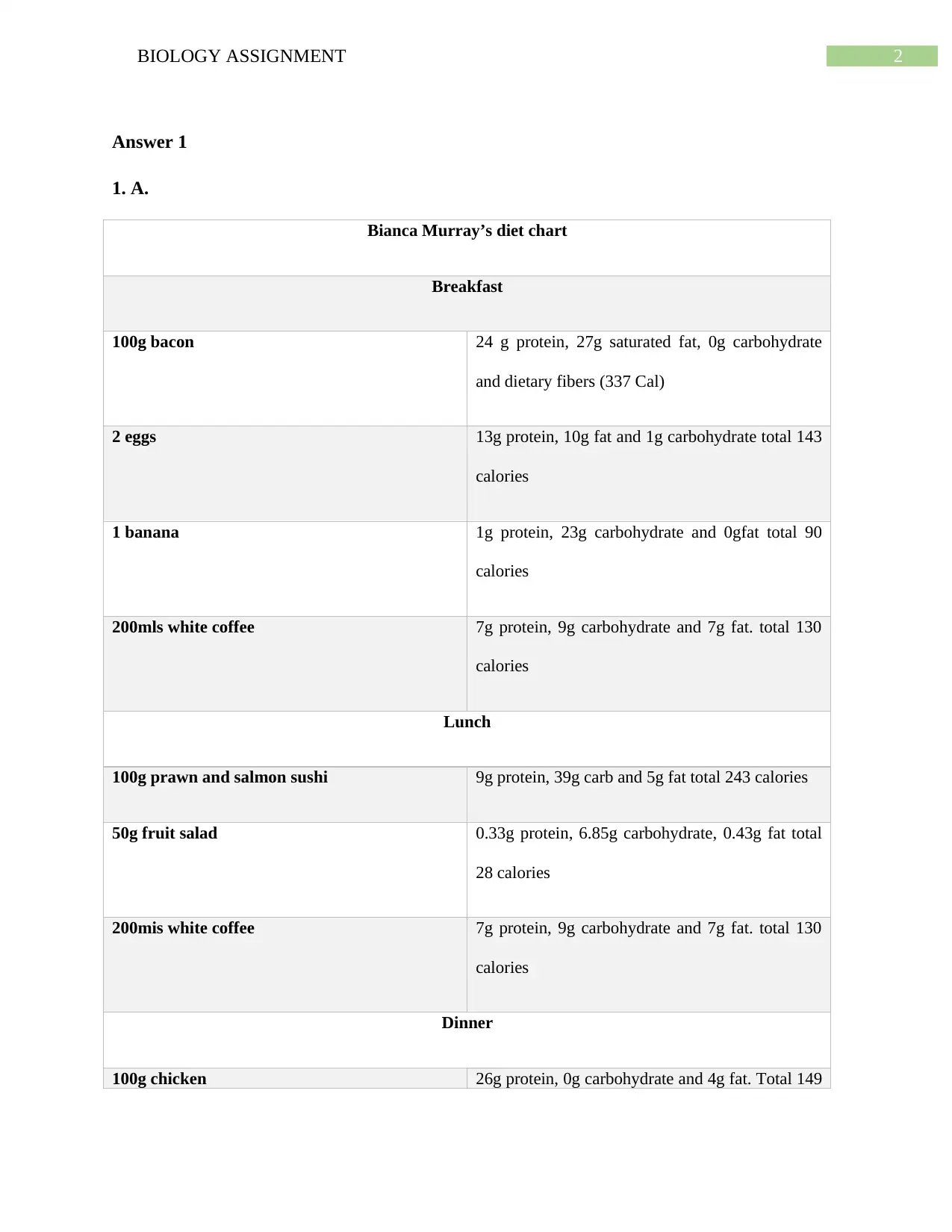
Answer 1
1. A.
Bianca Murray’s diet chart
Breakfast
100g bacon 24 g protein, 27g saturated fat, 0g carbohydrate
and dietary fibers (337 Cal)
2 eggs 13g protein, 10g fat and 1g carbohydrate total 143
calories
1 banana 1g protein, 23g carbohydrate and 0gfat total 90
calories
200mls white coffee 7g protein, 9g carbohydrate and 7g fat. total 130
calories
Lunch
100g prawn and salmon sushi 9g protein, 39g carb and 5g fat total 243 calories
50g fruit salad 0.33g protein, 6.85g carbohydrate, 0.43g fat total
28 calories
200mis white coffee 7g protein, 9g carbohydrate and 7g fat. total 130
calories
Dinner
100g chicken 26g protein, 0g carbohydrate and 4g fat. Total 149
⊘ This is a preview!⊘
Do you want full access?
Subscribe today to unlock all pages.
Trusted by 1+ million students worldwide
3BIOLOGY ASSIGNMENT
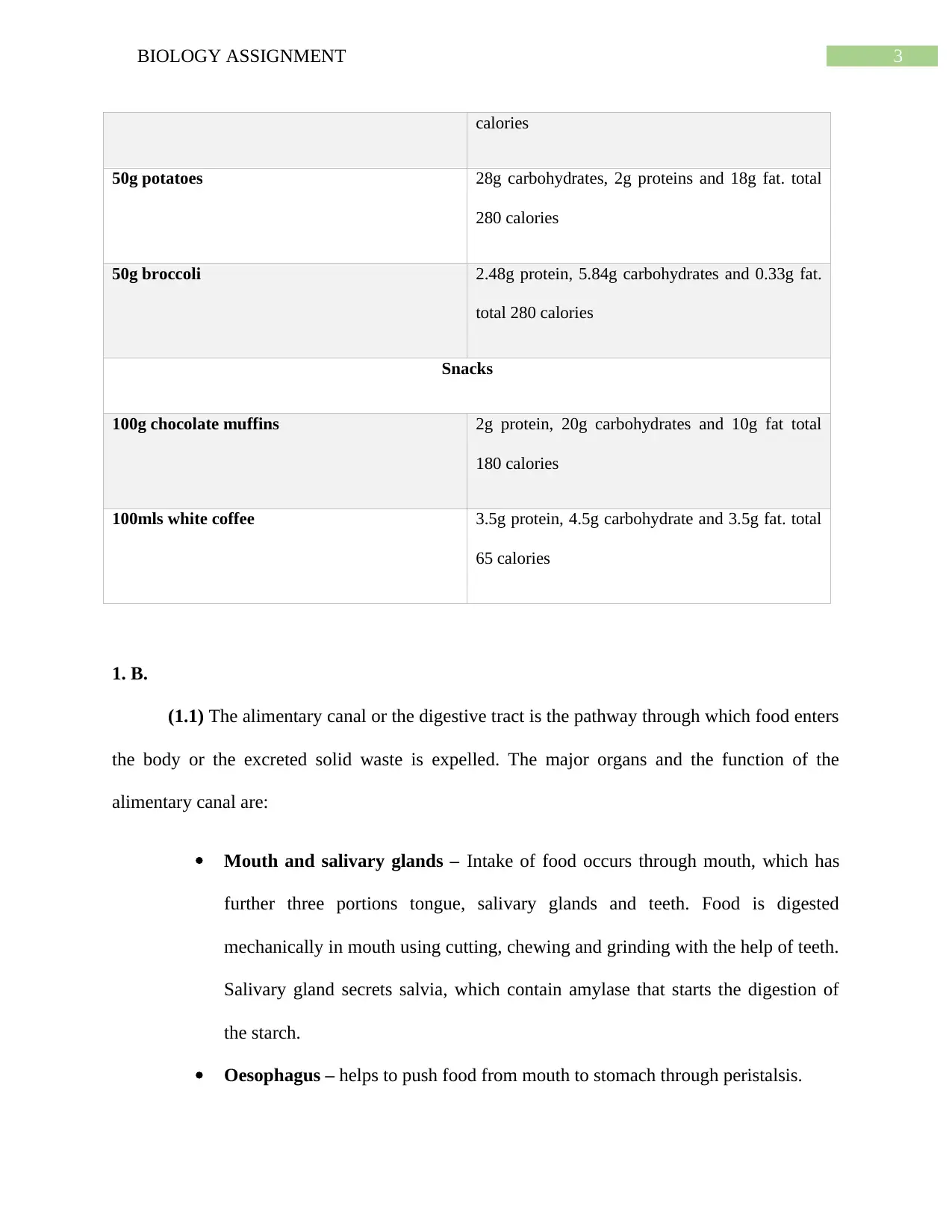
calories
50g potatoes 28g carbohydrates, 2g proteins and 18g fat. total
280 calories
50g broccoli 2.48g protein, 5.84g carbohydrates and 0.33g fat.
total 280 calories
Snacks
100g chocolate muffins 2g protein, 20g carbohydrates and 10g fat total
180 calories
100mls white coffee 3.5g protein, 4.5g carbohydrate and 3.5g fat. total
65 calories
1. B.
(1.1) The alimentary canal or the digestive tract is the pathway through which food enters
the body or the excreted solid waste is expelled. The major organs and the function of the
alimentary canal are:
Mouth and salivary glands – Intake of food occurs through mouth, which has
further three portions tongue, salivary glands and teeth. Food is digested
mechanically in mouth using cutting, chewing and grinding with the help of teeth.
Salivary gland secrets salvia, which contain amylase that starts the digestion of
the starch.
Oesophagus – helps to push food from mouth to stomach through peristalsis.
Paraphrase This Document
Need a fresh take? Get an instant paraphrase of this document with our AI Paraphraser
4BIOLOGY ASSIGNMENT
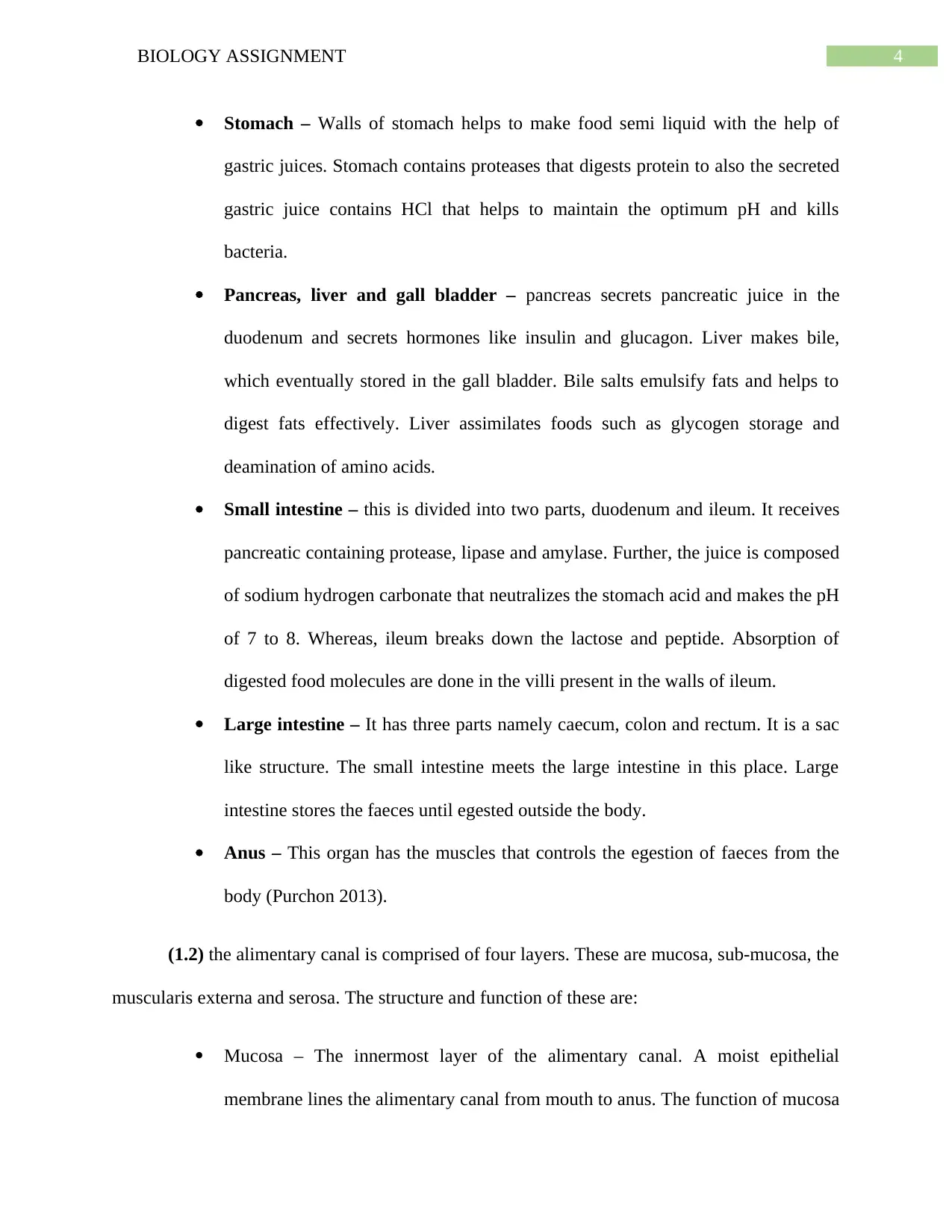
Stomach – Walls of stomach helps to make food semi liquid with the help of
gastric juices. Stomach contains proteases that digests protein to also the secreted
gastric juice contains HCl that helps to maintain the optimum pH and kills
bacteria.
Pancreas, liver and gall bladder – pancreas secrets pancreatic juice in the
duodenum and secrets hormones like insulin and glucagon. Liver makes bile,
which eventually stored in the gall bladder. Bile salts emulsify fats and helps to
digest fats effectively. Liver assimilates foods such as glycogen storage and
deamination of amino acids.
Small intestine – this is divided into two parts, duodenum and ileum. It receives
pancreatic containing protease, lipase and amylase. Further, the juice is composed
of sodium hydrogen carbonate that neutralizes the stomach acid and makes the pH
of 7 to 8. Whereas, ileum breaks down the lactose and peptide. Absorption of
digested food molecules are done in the villi present in the walls of ileum.
Large intestine – It has three parts namely caecum, colon and rectum. It is a sac
like structure. The small intestine meets the large intestine in this place. Large
intestine stores the faeces until egested outside the body.
Anus – This organ has the muscles that controls the egestion of faeces from the
body (Purchon 2013).
(1.2) the alimentary canal is comprised of four layers. These are mucosa, sub-mucosa, the
muscularis externa and serosa. The structure and function of these are:
Mucosa – The innermost layer of the alimentary canal. A moist epithelial
membrane lines the alimentary canal from mouth to anus. The function of mucosa
5BIOLOGY ASSIGNMENT
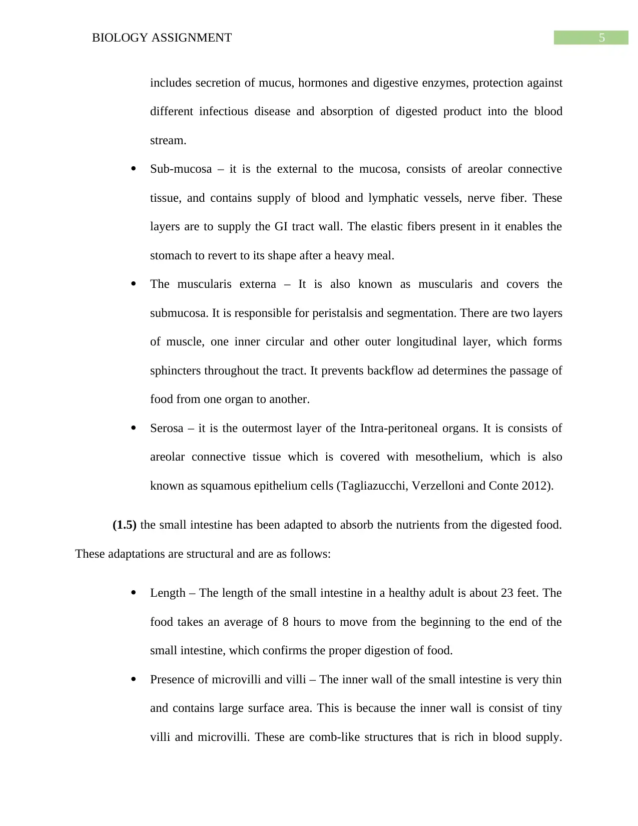
includes secretion of mucus, hormones and digestive enzymes, protection against
different infectious disease and absorption of digested product into the blood
stream.
Sub-mucosa – it is the external to the mucosa, consists of areolar connective
tissue, and contains supply of blood and lymphatic vessels, nerve fiber. These
layers are to supply the GI tract wall. The elastic fibers present in it enables the
stomach to revert to its shape after a heavy meal.
The muscularis externa – It is also known as muscularis and covers the
submucosa. It is responsible for peristalsis and segmentation. There are two layers
of muscle, one inner circular and other outer longitudinal layer, which forms
sphincters throughout the tract. It prevents backflow ad determines the passage of
food from one organ to another.
Serosa – it is the outermost layer of the Intra-peritoneal organs. It is consists of
areolar connective tissue which is covered with mesothelium, which is also
known as squamous epithelium cells (Tagliazucchi, Verzelloni and Conte 2012).
(1.5) the small intestine has been adapted to absorb the nutrients from the digested food.
These adaptations are structural and are as follows:
Length – The length of the small intestine in a healthy adult is about 23 feet. The
food takes an average of 8 hours to move from the beginning to the end of the
small intestine, which confirms the proper digestion of food.
Presence of microvilli and villi – The inner wall of the small intestine is very thin
and contains large surface area. This is because the inner wall is consist of tiny
villi and microvilli. These are comb-like structures that is rich in blood supply.
⊘ This is a preview!⊘
Do you want full access?
Subscribe today to unlock all pages.
Trusted by 1+ million students worldwide
6BIOLOGY ASSIGNMENT
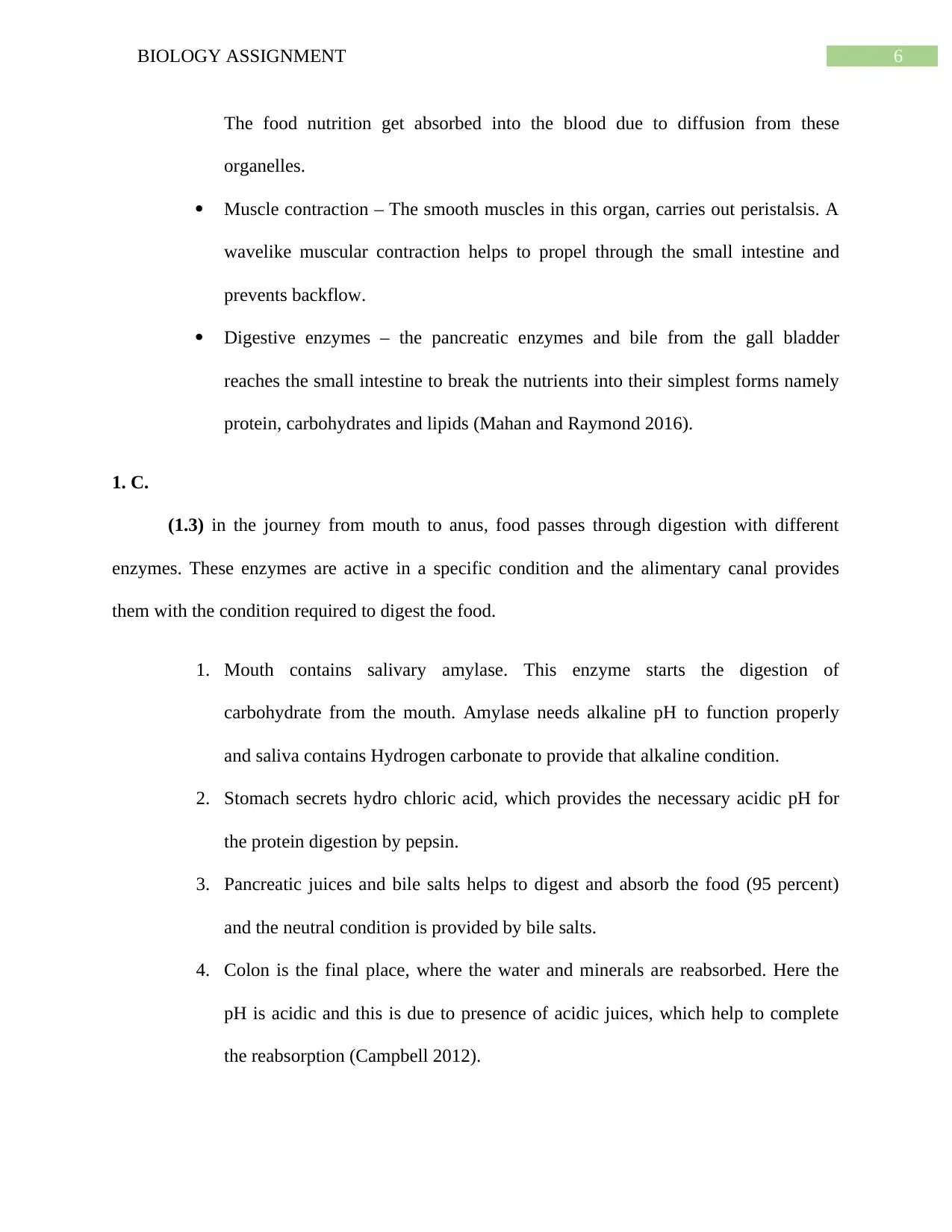
The food nutrition get absorbed into the blood due to diffusion from these
organelles.
Muscle contraction – The smooth muscles in this organ, carries out peristalsis. A
wavelike muscular contraction helps to propel through the small intestine and
prevents backflow.
Digestive enzymes – the pancreatic enzymes and bile from the gall bladder
reaches the small intestine to break the nutrients into their simplest forms namely
protein, carbohydrates and lipids (Mahan and Raymond 2016).
1. C.
(1.3) in the journey from mouth to anus, food passes through digestion with different
enzymes. These enzymes are active in a specific condition and the alimentary canal provides
them with the condition required to digest the food.
1. Mouth contains salivary amylase. This enzyme starts the digestion of
carbohydrate from the mouth. Amylase needs alkaline pH to function properly
and saliva contains Hydrogen carbonate to provide that alkaline condition.
2. Stomach secrets hydro chloric acid, which provides the necessary acidic pH for
the protein digestion by pepsin.
3. Pancreatic juices and bile salts helps to digest and absorb the food (95 percent)
and the neutral condition is provided by bile salts.
4. Colon is the final place, where the water and minerals are reabsorbed. Here the
pH is acidic and this is due to presence of acidic juices, which help to complete
the reabsorption (Campbell 2012).
Paraphrase This Document
Need a fresh take? Get an instant paraphrase of this document with our AI Paraphraser
7BIOLOGY ASSIGNMENT
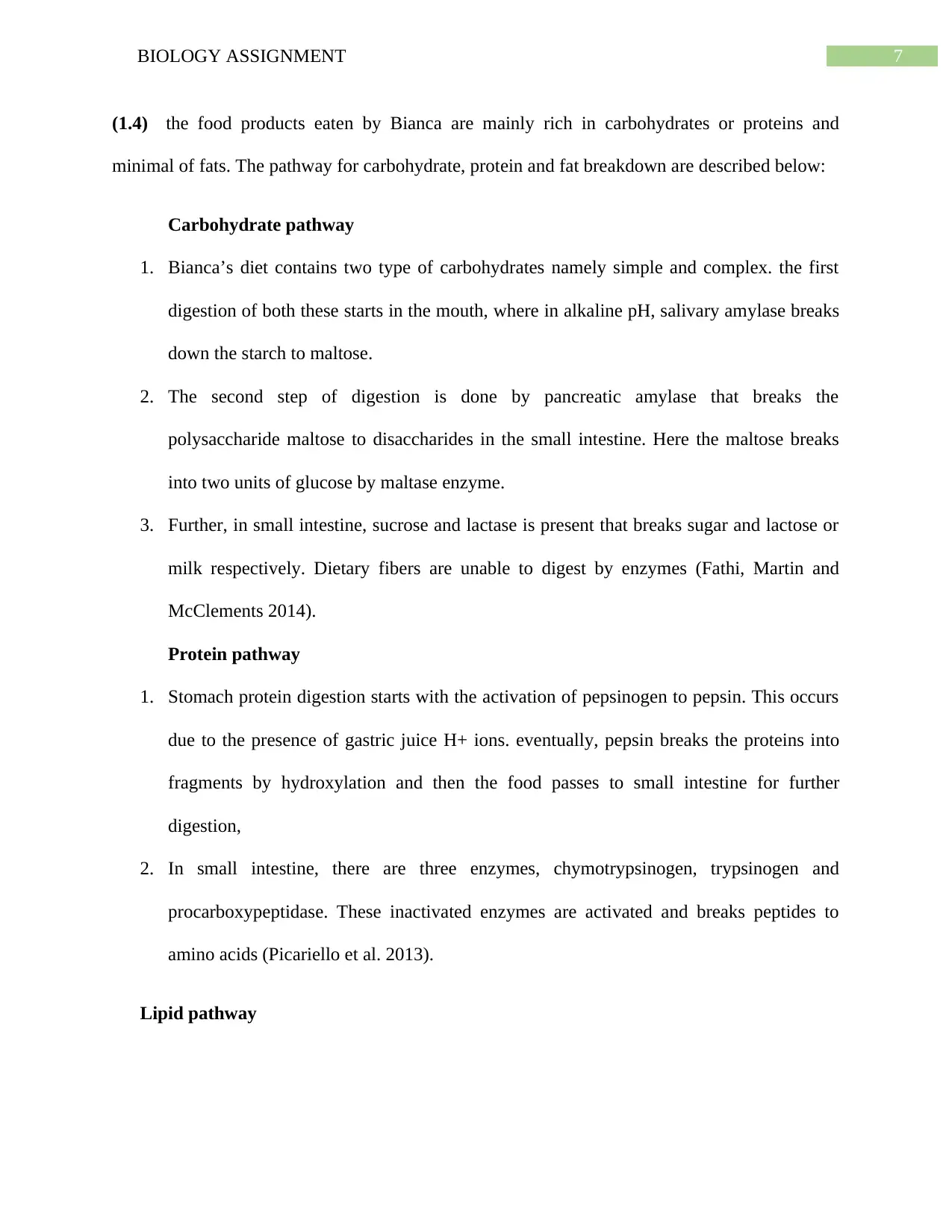
(1.4) the food products eaten by Bianca are mainly rich in carbohydrates or proteins and
minimal of fats. The pathway for carbohydrate, protein and fat breakdown are described below:
Carbohydrate pathway
1. Bianca’s diet contains two type of carbohydrates namely simple and complex. the first
digestion of both these starts in the mouth, where in alkaline pH, salivary amylase breaks
down the starch to maltose.
2. The second step of digestion is done by pancreatic amylase that breaks the
polysaccharide maltose to disaccharides in the small intestine. Here the maltose breaks
into two units of glucose by maltase enzyme.
3. Further, in small intestine, sucrose and lactase is present that breaks sugar and lactose or
milk respectively. Dietary fibers are unable to digest by enzymes (Fathi, Martin and
McClements 2014).
Protein pathway
1. Stomach protein digestion starts with the activation of pepsinogen to pepsin. This occurs
due to the presence of gastric juice H+ ions. eventually, pepsin breaks the proteins into
fragments by hydroxylation and then the food passes to small intestine for further
digestion,
2. In small intestine, there are three enzymes, chymotrypsinogen, trypsinogen and
procarboxypeptidase. These inactivated enzymes are activated and breaks peptides to
amino acids (Picariello et al. 2013).
Lipid pathway
8BIOLOGY ASSIGNMENT
1. Some fats are break down in mouth with lingual lipases and the maximum amount of
fats is digested in the small intestine. Pancreatic lipase and bile salts helps to emulsify
the fats and breaks down these to produce fatty acids (McClements 2013).
The human body goes to extra-ordinary lengths in order to ensure that the normal pH for
the human body is maintained at all circumstances. It has to be mentioned in this context
that the normal pH levels in human blood is 7.4 and it is maintained in various metabolic
pathways in the human body starting from respiration, and maintaining the entire acid
base homeostatis mechanism requires the involvement of various chemical buffers that
are produced in the metabolic pathways as mentioned above. The buffer systems that
function in the human body include bicarbonate buffer system, phosphate buffer system
and protein buffer system however it has to be mentioned in this context that the buffer
systems do not rectify any deviation in the blood pH, they function by reducing and
managing the changes facilitated in the body due to pH deviations (West 2012).
Answer 2
2.1
Double circulatory system
Double circulatory system is circulation of blood in which, blood flows through the heart
twice. In this circulatory system, blood flows between the heart and lungs. It differs from the
single circulation system in which blood flows from heart to the different parts of the body
except the lungs (Noordergraaf 2012).
2.2
The graph represented the pressure change happening in Bianca’s heart.
⊘ This is a preview!⊘
Do you want full access?
Subscribe today to unlock all pages.
Trusted by 1+ million students worldwide
9BIOLOGY ASSIGNMENT
1. This is the first event denoted by number 1 in the graph, where arterial ventricular
valves are closing during arterial diastole. Arterial pressure is lowest at this phase.
After this, the blood is pumped to the ventricles through opening of artrioventricular
valve and the pressure starts to raise.
2. During the second phase, the semilunar valves are opening during ventricular systole.
This occurs when the blood flows from the right ventricle to pulmonary artery and
from the left ventricle to aorta. Therefore, increase in aortic pressure occurs.
3. During the third phase, the pressure is highest as the semilunar valves are closing due
to ventricular diastole. Closing of this valve prevents the backflow of blood into the
ventricles, which are contracting at this phase.
(2.3 and 2.4)
Cardiac cycle is the sequence of electrical and mechanical event happening with each
heartbeat. It consists of relaxation or diastole and contraction or systole. Control of this cardiac
cycle are intrinsic control and extrinsic control.
1. Intrinsic control are due to special cells, which initiates and spreads the electrical impulse
in orderly manner using heart. These cardiac muscles work automatically and contracts as
a unit. The second factor is action potential of pacemaker cells. as well as sequence of
excitation also controls the cardiac cycle.
2. The other factor that is extrinsic factor is consists of external stress or physical actions,
nervous system, depression and different factors such as age, exercise and body
temperature.
Paraphrase This Document
Need a fresh take? Get an instant paraphrase of this document with our AI Paraphraser
10BIOLOGY ASSIGNMENT
Bianca’s heart electro-cardiogram shows that her heart is working normally and the graph
is depicting the ventricular depolarization and arterial systole. Due to her exercise and
healthy diet, the heart is beating perfectly (Beaumont et al. 2013).
Answer 3
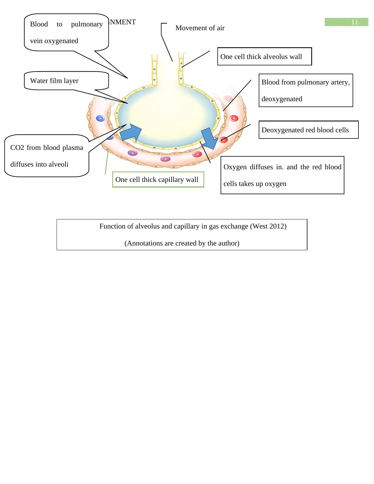
Characteristics of gaseous exchange surfaces (alveoli and capillaries) are as follows:
1. These are highly vascularized and network of capillaries are present.
2. Contains large surface area for gaseous exchange.
3. The membranes are thin and contains moist lining to make the exchange easy
(Goodman 2012).
The annotation diagram has been shown below:
Bianca’s heart electro-cardiogram shows that her heart is working normally and the graph
is depicting the ventricular depolarization and arterial systole. Due to her exercise and
healthy diet, the heart is beating perfectly (Beaumont et al. 2013).
Answer 3
Characteristics of gaseous exchange surfaces (alveoli and capillaries) are as follows:
1. These are highly vascularized and network of capillaries are present.
2. Contains large surface area for gaseous exchange.
3. The membranes are thin and contains moist lining to make the exchange easy
(Goodman 2012).
The annotation diagram has been shown below:
11BIOLOGY ASSIGNMENT
One cell thick alveolus wall
Blood from pulmonary artery,
deoxygenated
Deoxygenated red blood cells
Oxygen diffuses in. and the red blood
cells takes up oxygen
Movement of air
One cell thick capillary wall
Blood to pulmonary
vein oxygenated
Water film layer
CO2 from blood plasma
diffuses into alveoli
Function of alveolus and capillary in gas exchange (West 2012)
(Annotations are created by the author)
⊘ This is a preview!⊘
Do you want full access?
Subscribe today to unlock all pages.
Trusted by 1+ million students worldwide
1 out of 18
Related Documents


Your All-in-One AI-Powered Toolkit for Academic Success.
+13062052269
info@desklib.com
Available 24*7 on WhatsApp / Email
![[object Object]](/_next/static/media/star-bottom.7253800d.svg)
Unlock your academic potential
Copyright © 2020–2026 A2Z Services. All Rights Reserved. Developed and managed by ZUCOL.