Detailed Report on Genetic Skin Diseases: Biology and Treatment
VerifiedAdded on 2022/11/17
|5
|2060
|465
Report
AI Summary
This report provides a detailed overview of genetic skin diseases, also known as genodermatoses. It begins with an introduction to the skin's function and the impact of genetic mutations on its integrity. The report defines genetic diseases and highlights how mutations can disrupt the skin's delicate organization, leading to various conditions. It then discusses several specific genetic skin diseases, including ichthyosis, ectodermal dysplasia, epidermolysis bullosa, progressive symmetrical erythrokeratodermia, and Hailey-Hailey disease, describing their characteristics and causes. The report also covers diagnostic tools such as fetal skin biopsies and next-generation sequencing, as well as potential treatment approaches like gene therapy. It concludes by emphasizing the need for further research to enhance understanding and develop effective treatments for these complex conditions. The report references multiple research papers and studies to support its claims. This assignment is available on Desklib, a platform offering study resources.
Genetic skin diseases
Introduction
The skin constitutes the largest organ of human body and provides protection against various
threats in the form of physical, chemical and biological factors. It also functions to prevent
dehydration and also plays a significant role in thermoregulation. The skin is composed of a
variety of cell types and forms a continuous lining with the mucous membranes (Kanitakis.
2002). The diverse kinds of cells in the skin secrete specific molecules that participate in the
complex interactions and intercellular communication for maintaining the structural and
functional integrity of the skin. Hence, any mutation in one of such molecules holds the potential
to disrupt the delicate organization and function of the essential networks, resulting in cell
separation, blistering, and other lethal and sub-lethal phenotypes observed in inherited skin
diseases (DeStefano & Christiano). A genetic disease may be defined as an abnormality in the
genetic makeup of an individual which can range from a discrete mutation in a single base of a
single gene to a gross chromosome abnormality in the form of an extra or a loss of an entire
chromosome or a part thereof. In some cases, individuals may inherit genetic disorders from
parents, while in some others, acquired changes or mutations in a preexisting gene or group of
genes may lead to the development of a genetic disease. Genetic skin diseases commonly
referred to as genodermatosis, are known to primarily affect the skin and its appendages and
develop as a result of the mutations, which could be either single-gene or polygene in nature.
Substantial progress has been made, in the last couple of decades, in elucidating the genes that
underlie a variety of skin diseases and a wide range of phenotypes. Significant advancements in
histological, ultra-structural, and immunological analyses have provided molecular clues into the
defects underlying the pathologies of several inherited skin diseases. One challenging goal in the
study of inherited skin diseases is to identify the causative gene(s) responsible for the
development of phenotype in a given genodermatosis and understand its role in maintaining the
integrity of the skin. There are an estimated 5,000 genetic diseases in all, of which around one
third involve the skin. In addition to the commonly prevalent genetic skin diseases like-
carcinoma, lupus, acne, psoriasis and other; there are certain others in this category which have
been listed below.
Ichthyosis- It refers to a condition that leads to the development of a persistent thick, dry skin
often called as the "fish-scale" skin. The term Ichthyosis is derived from the Greek, meaning
Paraphrase This Document
Need a fresh take? Get an instant paraphrase of this document with our AI Paraphraser
“fish scales”, with a reference to the characteristic appearance of the skin. The most common
type, which accounts for 95% of cases, is ichthyosis vulgaris affecting 1 in 250-1,000 people. It
is mostly mild and in many cases mistaken for normal dry skin but rare types of ichthyosis can
be severe and even life threatening.
Ectodermal dysplasia ectodermal dysplasia also called as skin fragility syndrome affects the
normal development of ectodermal tissues- hair, nails, teeth, skin and sweat glands. It results
from mutations in one of several genes namely EDA, EDAR, EDARADD, and WNT10A. EDA
gene mutations are the most common cause of the disorder, which accounts for more than 50%
of all cases. EDAR, EDARADD, and WNT10A gene mutations each contribute to a smaller
percentage of cases.
Epidermolysis Bullosa results in blistering at the slightest touch. Then skin becomes so fragile
that a light knock could open a wound or a gentle rub leave it sore and bleeding. There are many
different types and degrees of severity of the rare inherited skin disorder and it is not the skin
only that is affected. The condition develops as a result of mutations in any of 18 different genes
that make proteins that “stick” the top and bottom layer of skin together. As of the present times,
there is no cure and the doctors can only strive to minimize the effects. The big threat to the
patients with severe EB is squamous cell skin cancer. Most start to develop it in their 20s, 30s
and 40s but the youngest case was a child aged six, and there have been several in their early
teens. It is a leading cause of death.
Progressive symmetrical erythrokeratodermia an autosomal dominant genodermatosis with
variable penetrance as described by Darier in 1911. It is a rare condition characterized by
erythematous and keratotic plaques, that are clearly defined and symmetrically distributed
frequently along the extremities, buttocks and rarely, on the face (Guaraldi et al., 2013). Often
referred to as the Gottron syndrome or Darier-Grotton erythrokeratodermia, it was described in
1911 by Darier, however, Grotton, in 1923, coined the name for this particular condition.
According to Darier, progressive symmetrical erythrokeratodermia is a rare keratinization
disorder, marked by non-migratory keratotic plaques on an erythematous base that appear
perniciously and slowly on circinate areas and are surrounded by a delicate hyperpigmented
projection. These may be distributed symmetrically on elbows, knees, hands and feet. Clinically,
hyperkeratosis is observed by the presence of thickened plaques and is more stable than
erythema. The name given to the illness is based on the irregularities in number, shape; size and
duration of the erythematous plaques (Nazarro and Blanchet.1986).
Hailey–Hailey: Often known as the familial benign chronic pemphigus, it is marked by a loss of
cellular adhesion between epidermal cells (acathyolysis) resulting in erosions, blistering and
crusted plaques (Ikeda et al. 1994). A similar autosomal dominant genodermatosis known as the
Darier’s disease is characterized by acanthyolysis (cell separation) of the suprabasal epidermal
cells resulting in plaques, nail abnormalities and warty papules.
Diagnosis and Treatment: The functional cloning approach has, so far, proven to be the most
effective and suitable approach for skin diseases in which information about the defective gene
product, that is the keratin intermediate filament and its general biological function is available
(DeStefano & Christiano). Another important diagnostic tool is the fetal skin biopsy. It has
permitted families with members who carry the abnormal gene for one of these severe skin
diseases, to undertake a pregnancy knowing that the condition of the fetus can be determined.
Samples of skin are obtained from fetus of 16 to 22 weeks' gestation by ultrasound-guided
biopsy and are evaluated using different immunohistochemical, morphological and biochemical
tools (Holbrook et al.1993). In the last few years, the search for genes that causes the diseases
has advanced with the advent of “next generation sequencing” that enabled all 20,000 genes in
the human genome to be sequenced for the same price as a single gene a decade ago. The new
approach includes modeling to filter out what might be significant. Research shows that the skin
possesses qualities that make it a preferred target for gene therapy. Various vectors may be used
to introduce genetic material into the cells to alter gene expression thus altering a pathological
process. This objective may also be accomplished by employing viral vectors. In depth
knowledge of the molecular mechanisms involved in the pathogenesis of genetic skin disorders
is important to design rational genetic therapies. Some of the challenges include- improving the
efficacy and efficiency of cutaneous non-viral vectors and increasing the safety of viral vectors;
ensuring successful genomic integration and negating host’s immune response to accept the
vector.
Conclusion- In consultation with the literature arising from experimentation over past several
decades, significant understanding of the complexity of the structure and function of the skin and
⊘ This is a preview!⊘
Do you want full access?
Subscribe today to unlock all pages.

Trusted by 1+ million students worldwide
its constituents has been achieved. The role of the various molecules and their functions that
maintain the overall structural integrity of the skin have also been explored. However, the
genetics associated with the skin and its diseases are complex and require further in-depth
research. Recent discoveries related to the molecular basis of the genetic disease, has led to the
development of various diagnostic tools such as- prenatal and pre-implantation testing. Based on
the understanding of the basement membrane zone (BMZ) and the genes that are responsible for
it formation and maintenance, effective treatments are evolving and may provide relief to the
patients suffering from the different genetic skin disorders.
References-
DeStefano, Gina. (2014). The Genetics of Human Skin Disease.. 10.1101/cshperspect.a015172..
Lynn M. Boyden, Nicholas G. Vincent, Jing Zhou, Ronghua Hu, Brittany G. Craiglow, Susan J.
Bayliss, Ilana S. Rosman, Anne W. Lucky, Luis A. Diaz, Lowell A. Goldsmith, Amy S. Paller,
Richard P. Lifton, Susan J. Baserga, Keith A. Choate. Mutations in KDSR Cause Recessive
Progressive Symmetric Erythrokeratoderma. The American Journal of Human Genetics, 2017;
100 (6): 978 DOI: 10.1016/j.ajhg.2017.05.003
Yale University. "Cause of, and possible cure for, genetic skin disorder." ScienceDaily.
ScienceDaily, 1 June 2017.
Guaraldi, B., Jaime, T. J., Guaraldi, R., Melo, D. F., Nogueira, O. M., & Rodrigues, N. (2013).
Progressive symmetrical erythrokeratodermia -- case report. Anais brasileiros de dermatologia,
88(1), 109–112. doi:10.1590/S0365-05962013000100016
Nazarro V, Blanchet-Bardon C. Progressive symmetric erythrokeratodermia. Histological and
ultrastructural study of patient before and after treatment with etretinate. Arch
Dermatol. 1986;122:434–40.
DeStefano, G. M., & Christiano, A. M. (). The genetics of human skin disease. Cold Spring
Harbor perspectives in medicine, 4(10), a015172. doi:10.1101/cshperspect.a015172
Kanitakis, J. (2002). Anatomy, histology and immunohistochemistry of normal human skin.
European Journal of Dermatology, 12(4), 390–401 Coulombe PA, Hutton ME, Letai A, Hebert
A, Paller AS,
Fuchs E. 1991. Point mutations in human keratin 14 genes of epidermolysis bullosa simplex
patients: Genetic and functional analyses. Cell 66: 1301–1311.
Paraphrase This Document
Need a fresh take? Get an instant paraphrase of this document with our AI Paraphraser
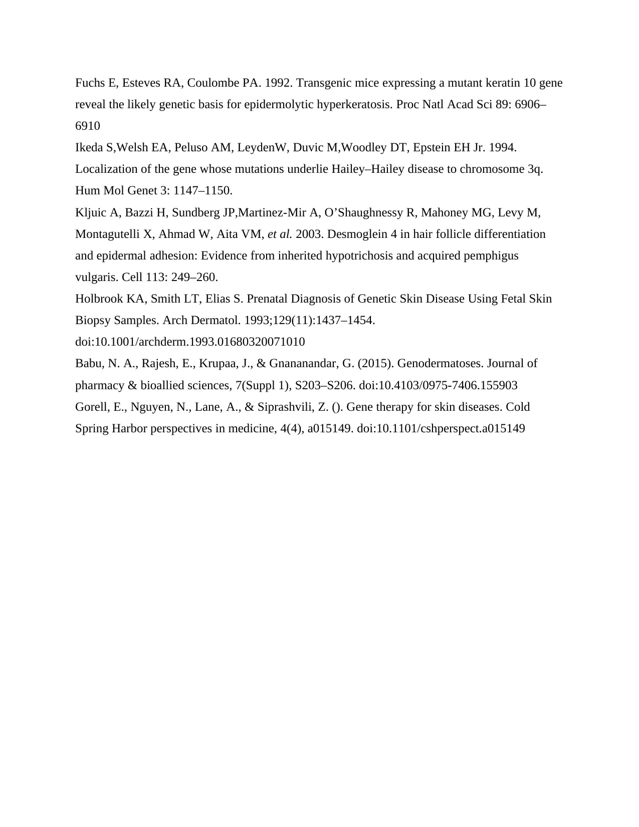
Fuchs E, Esteves RA, Coulombe PA. 1992. Transgenic mice expressing a mutant keratin 10 gene
reveal the likely genetic basis for epidermolytic hyperkeratosis. Proc Natl Acad Sci 89: 6906–
6910
Ikeda S,Welsh EA, Peluso AM, LeydenW, Duvic M,Woodley DT, Epstein EH Jr. 1994.
Localization of the gene whose mutations underlie Hailey–Hailey disease to chromosome 3q.
Hum Mol Genet 3: 1147–1150.
Kljuic A, Bazzi H, Sundberg JP,Martinez-Mir A, O’Shaughnessy R, Mahoney MG, Levy M,
Montagutelli X, Ahmad W, Aita VM, et al. 2003. Desmoglein 4 in hair follicle differentiation
and epidermal adhesion: Evidence from inherited hypotrichosis and acquired pemphigus
vulgaris. Cell 113: 249–260.
Holbrook KA, Smith LT, Elias S. Prenatal Diagnosis of Genetic Skin Disease Using Fetal Skin
Biopsy Samples. Arch Dermatol. 1993;129(11):1437–1454.
doi:10.1001/archderm.1993.01680320071010
Babu, N. A., Rajesh, E., Krupaa, J., & Gnananandar, G. (2015). Genodermatoses. Journal of
pharmacy & bioallied sciences, 7(Suppl 1), S203–S206. doi:10.4103/0975-7406.155903
Gorell, E., Nguyen, N., Lane, A., & Siprashvili, Z. (). Gene therapy for skin diseases. Cold
Spring Harbor perspectives in medicine, 4(4), a015149. doi:10.1101/cshperspect.a015149
1 out of 5
Related Documents
Your All-in-One AI-Powered Toolkit for Academic Success.
+13062052269
info@desklib.com
Available 24*7 on WhatsApp / Email
![[object Object]](/_next/static/media/star-bottom.7253800d.svg)
Unlock your academic potential
Copyright © 2020–2025 A2Z Services. All Rights Reserved. Developed and managed by ZUCOL.


