Practical Biology: Starch Hydrolysis, Human Skeleton & Urine Formation
VerifiedAdded on 2023/06/12
|23
|2792
|391
Practical Assignment
AI Summary
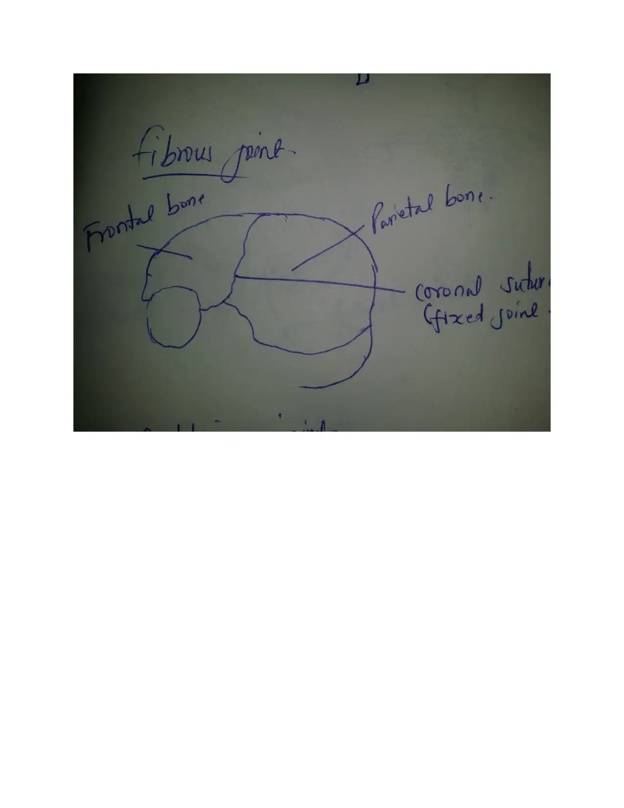
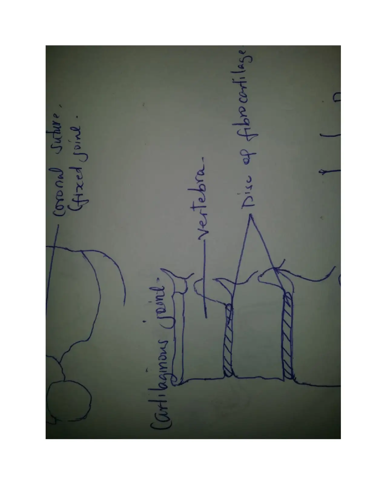
This biology assignment covers several key areas: the effects of temperature on starch hydrolysis by amylase, an overview of the human skeleton including fibrous, cartilaginous, and synovial joints with a focus on the shoulder joint, a comparison of the male and female reproductive systems, and a detailed explanation of urine formation including ultrafiltration, reabsorption, and secretion, as well as the process of osmoregulation and the role of ADH. The experiment on starch hydrolysis investigates how temperature affects the enzymatic breakdown of starch into glucose, while the section on the human skeleton details the axial and appendicular components, different types of joints, and their functions. The reproductive system section outlines the structures and functions of both male and female organs. Finally, the assignment elucidates the steps involved in urine formation and the hormonal control of osmoregulation, demonstrating the body's mechanisms for maintaining fluid balance. Desklib provides this and many more solved assignments for students.

BIOLOGY
PREPARE BY:
DEPARTMENT:
INSTITUTION:
DATE:
FACILITATOR:
Table of Contents
PREPARE BY:
DEPARTMENT:
INSTITUTION:
DATE:
FACILITATOR:
Table of Contents
Paraphrase This Document
Need a fresh take? Get an instant paraphrase of this document with our AI Paraphraser

TASK 1.......................................................................................................................................................1
EFFECTS OF TEMPERATURE ON STARCH HYDROLYSIS BY AMYLASE...............................1
Objectives...............................................................................................................................................1
Introduction...........................................................................................................................................1
Requirements.........................................................................................................................................2
Procedure...............................................................................................................................................2
Observations..........................................................................................................................................3
Discussions.............................................................................................................................................3
Conclusion..............................................................................................................................................4
TASK 2.......................................................................................................................................................4
HUMAN SKELETON...............................................................................................................................4
Fibrous joints.......................................................................................................................................10
Cartilaginous joints.............................................................................................................................10
Synovial joints......................................................................................................................................10
The shoulder joint................................................................................................................................11
TASK 3.....................................................................................................................................................12
TASK 4.....................................................................................................................................................16
A.Diagram.............................................................................................................................................16
B. Urine formation...............................................................................................................................18
C. Osmoregulation...............................................................................................................................19
References................................................................................................................................................21

TASK 1
EFFECTS OF TEMPERATURE ON STARCH HYDROLYSIS BY AMYLASE.
Objectives
To study the effects of temperature on the rate of amylase enzyme hydrolysis of starch
According to (Moore 2014), Starch is the main form of storage in plants. They are synthesized
naturally in plants from simple forms of sugar such as glucose for storage. Starch containing
plant products form a bulk of the human diet across the world. Starch contains thousands of
glucose polymers linked together by alpha glucosides bonds as opposed to beta glucosides bonds
in cellulose.
As (Hanes 2011) states starch is insoluble in water at room temperature hence its stored in small
granules. Human saliva and pancreatic secretions contain amylase enzyme for starch digestion.
Amylase enzyme catalyzes the hydrolysis of starch to form soluble glucose units.
Enzymes, being protein in nature are influenced by factors such as Ph, temperature ,substrate
concentrations and enzyme concentration. The influence of temperature on the enzymatic
hydrolysis of starch will be studied.
Requirements
Test tubes
Test tube racks
Temperature bath
Thermometer
⊘ This is a preview!⊘
Do you want full access?
Subscribe today to unlock all pages.

Trusted by 1+ million students worldwide

Syringe
Graduated cylinder
Starch solution
Amylase enzyme
Iodine reagent
Water
Procedure
Take 3 test tubes and lebel them W, S and SA for water,starch and starch and amylase
respectively.
Pour 10mls of distilled water to each testube.
In the test tube labeled W,pour 10mls of water ,10mls amylase and add 3-4 drops of iodine
solution
In the test tube labeled S, pour 10mls startch,10mls distilled water and add 3-4 drops of iodine
solution
In the 3rd test tube labeled SA,pour 10mls starch and add 3-4 drops iodine solution.
Place all the test tubes in a water bath between 37-42 degrees.
When the temperature reaches 37-42 degrees,in test tube labeled SA add 10mls amylase solution.
Observe for 5 minutes and document your results.
Paraphrase This Document
Need a fresh take? Get an instant paraphrase of this document with our AI Paraphraser

Observations
The test tubes W and S retained the iodine’s color
Test tube SA color changes to colorless when amylase is added and observed for 5 minutes.
Discussions
Starch in nature is stored as small granules that are insoluble in water. This insolubility is due to
the hydrogen bonds within the molecules.
With temperature increase,these hydrogen bonds are weakened and break.
The amylose in preheated start forms helices which alow iodine molecules to assemble forming a
dark blue black color.
As starch is hydrolyzed into smaller carbohydrate units,the blue-black color disappears.
Conclusion
The test is used to confirm that amylase is necessary in the catalyzed hydrolysis of starch into
small carbohyrdrate units in the presence of optimum temperature in accordance to (Konsula
2014).

TASK 2
B.
HUMAN SKELETON
Appendicular skeleton
Cranium.
Comprises of the skull, called the cranium and the facial bones.
cranium is formed from eight plate-shaped bones which fit forming immovable joints called
sutures.
facial bones.14 in number, majorly the mandible, maxilla, the zygomatic or and the nasal
bone
Vertebral column
Made up of 24 separate vertebral bone, the sacrum and the coccyx.
Vertebral column is divided into cervical. thoracic, lumbar sacral vertebrae and the coccyx.
Ribcage
Formed by the 12 paired ribs and the sternum.
The ribs join the vertebra at the back and the sternum in front.
Functions to protect the heart and lungs.
⊘ This is a preview!⊘
Do you want full access?
Subscribe today to unlock all pages.

Trusted by 1+ million students worldwide

Axial skeleton
Shoulder
Made up of the scapula,clavicle and the
humerus
Has a synovial movable joint allowing
360degrees movement
Pelvis
Made up of the ilium,ischium and the femur.
Has a ball and socket synovial joint that
allows 360 degrees movement of the lower
limb
Arm
Made ip of the humerus,ulna,radius wrist
bones and caparls and metacarplas
Leg.
Comprises the femur,tibia,fibula,tarsals and
metatarsals
Paraphrase This Document
Need a fresh take? Get an instant paraphrase of this document with our AI Paraphraser

C. Diagram

⊘ This is a preview!⊘
Do you want full access?
Subscribe today to unlock all pages.

Trusted by 1+ million students worldwide

Paraphrase This Document
Need a fresh take? Get an instant paraphrase of this document with our AI Paraphraser

D.
Fibrous joints
A joint is the location at which two or more bones meet. Fibrous joints are connected by dense
connective tissue of collagen. They have no cavity hence are fused and thus allow no movement.
These joints are found in the cranium forming a union between cranial bones allowing no
movement to protect the brain from impact.
Cartilaginous joints
They occur at bones connected by entirely hyaline cartilage. They allow more movement than
fibrous joints. Such joints include intervertebral disc joints allowing for slight movement of the
vertebral column.
Synovial joints
In these joints the bony surfaces are covered with articular cartilage and connected by ligaments
lined by synovial membrane. synovial membrane that secretes synovial fluid that absorbs shock,
lubricates and provides nutrients to the joint. An example of the synovial joint is the ball and
socket of the femur and the pelvic girdle. It is highly movable allowing for locomotion.

The shoulder joint.
The shoulder joint is made up of the scapula, clavicle and the humerus. The clavicle forms the
roof of the joint by attaching laterally to the scapula and medially to the sternum. The shoulder
joint is a ball and socket synovial joint that allows movement in 360°.
The highly mobile shoulder joint allows the following movements;
Abduction and adduction of the shoulder, in 180 degrees by the muscles attached to the bones of
the joint in the frontal plane. Abduction is by the deltoid and the supraspinatus in the first 90
degrees and trapezius and the serratus anterior to 180 degrees. Abduction is carried out by the
deltoid and the supraspinatus in the first 90 degrees and trapezius and the serratus anterior to 180
degrees.
Internal and external rotation. extension and flexion of the joint of the joint in the sagittal plane.
Extension is by the latissimus dose and posterior fibers of the deltoid. Flexion is made possible
by the fibers of the deltoid, pectoralis major and the coracobrachialis.
Circumduction of the shoulder which involves all the movements of the joints.
⊘ This is a preview!⊘
Do you want full access?
Subscribe today to unlock all pages.

Trusted by 1+ million students worldwide
1 out of 23

Your All-in-One AI-Powered Toolkit for Academic Success.
+13062052269
info@desklib.com
Available 24*7 on WhatsApp / Email
Unlock your academic potential
Copyright © 2020–2025 A2Z Services. All Rights Reserved. Developed and managed by ZUCOL.