Biology Report: Transport and Respiration - Heart and Circulation
VerifiedAdded on 2023/03/17
|27
|5047
|98
Report
AI Summary
This biology report provides a comprehensive overview of transport and respiration, focusing on the heart's structure and function, including its chambers, valves, and blood vessels, and the differences between coronary, pulmonary, and systemic circulation. It explains the cardiac cycle, including electrical stimulation and the role of the pacemaker. The report differentiates between cardiac output and blood pressure, comparing their behavior during rest and exercise. Additionally, it details the components and functions of the respiratory system, including the nasal cavity, larynx, trachea, lungs, and diaphragm, and describes the mechanism of breathing, including pressure changes in the thorax. The report concludes with a summary of key concepts and provides references for further study.
0
Biology
Biology
Transport and Respiration
MAY 28, 2019
Student Details:
Biology
Biology
Transport and Respiration
MAY 28, 2019
Student Details:
Paraphrase This Document
Need a fresh take? Get an instant paraphrase of this document with our AI Paraphraser
1
Biology
Contents
Introduction......................................................................................................................................2
Transport and Respiration................................................................................................................3
Structures of the heart with their function...................................................................................3
Difference between the coronary, pulmonary and systematic circulation...................................4
Comparison of consequences of blockage in the circulatory system..........................................5
Cardiac cycle with electrical stimulation.....................................................................................6
Role of pacemaker.....................................................................................................................10
Difference between cardiac output and blood pressure.............................................................10
Similarities and differences in cardiac output and blood pressure at rest and exercise.............11
Components of Respiratory System..........................................................................................12
Explanation of the structure and function of the components of the respiratory system..........14
Mechanism of breathing............................................................................................................20
Pressure changes in the thorax during breathing at a higher place............................................21
Conclusion.....................................................................................................................................23
References.....................................................................................................................................24
2
Biology
Introduction
The heart is a muscular organ of the body. It consists of muscle fibers, blood vessels named
arteries, veins, and blood capillaries. It works by the help of electric signals generated by sino-
atrial node (SA), atrioventricular node (AV) and Purkinje fibers. It functions for the circulation
of oxygenated blood throughout the parts of the body and deoxygenated blood from the different
parts of the body to the lungs. It is located behind the sternum in the chest above the diaphragm.
The size of the normal heart is equal to fist size and it is of 298 grams or 10.5 ounces in weight.
It has double circulation named pulmonary circulation and systematic circulation. Besides this
one more circulation named coronary circulation is also situated in the heart. Blockage in these
circulatory systems may lead to consequences like heart failure, stroke, arrhythmia, dyspnea,
chest pain and many more diseases (Junior, 2019).
The respiratory system includes many organs of the body like the nasal cavity, larynx, pharynx,
trachea, lungs, and diaphragm. By the help of these organs, inhalation and exhalation process of
breathing takes place (Zimmermann, 2018).
⊘ This is a preview!⊘
Do you want full access?
Subscribe today to unlock all pages.
Trusted by 1+ million students worldwide
3
Biology
Transport and Respiration
Structures of the heart with their function
The heart is the part of the body made up of muscles, 4 chambers, wall, valves, blood vessels,
two electric signal generating nodes and fibers. Four chambers of heart named as left atrium
(LA) carry oxygenated blood from the lungs, “left ventricle (LV)” carry “oxygenated blood from
LA”, right atria take away “deoxygenated blood” from the body parts and right ventricle (RV)
carry deoxygenated blood from right atria (RA) . The heart has four valves. One is “mitral valve
(bicuspid valve)” which is situated in between LA and LV through which oxygenated blood from
left atria to ventricle get passed. Second is “aortic valve” situated in between “left ventricle and
the aorta” by which “oxygenated blood” gets passed to different parts of the body. The third is
“tricuspid valve” stated in between “right atrium and right ventricle” and pass “deoxygenated
blood” from right atria to the right ventricle. The last one is “pulmonary valve” situated in
between pulmonary artery and right ventricle and pass “deoxygenated blood” to lungs to get
“oxygenated” (Newman & Sullivan, 2018). Heart consists of three kinds of blood vessels which
are named to be “arteries which carry out oxygenated blood except for pulmonary artery”, veins
named superior and inferior vena cava which carry out the deoxygenated blood except for
pulmonary vein, and capillaries helps in exchange of compounds like nutrients, water, waste and
oxygen from near about tissues (Lewis & Writer, 2016).
A wall of muscles named septum isolates LA and LV from RA and RV. Three layers of the
tissue named “epicardium, myocardium, and endocardium” combinedly form the wall of the
heart and covered by pericardium tissue layer. Sino-atrial (SA) lies in the top of RA and known
to be a pacemaker. It generates an electric signal for the contraction of atria and passes down of
Paraphrase This Document
Need a fresh take? Get an instant paraphrase of this document with our AI Paraphraser
4
Biology
blood into the ventricles. AV node situated in the bottom of right atrium passed electric signals
to make the gap in the contraction of atria and ventricles. Then Purkinje fibers in the ventricle
walls and generate signal to heart muscle to produce contraction in the ventricles and causes
“diastole (relaxation of atria)”and ventricles filled with blood and “systole (contraction of atria)”,
ventricles get filled by the blood, contraction of ventricles and pumping of blood outside the
heart) to the heart (Beckerman, 2019).
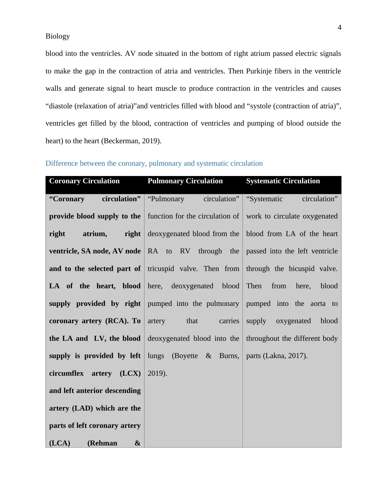
Difference between the coronary, pulmonary and systematic circulation
Coronary Circulation Pulmonary Circulation Systematic Circulation
“Coronary circulation”
provide blood supply to the
right atrium, right
ventricle, SA node, AV node
and to the selected part of
LA of the heart, blood
supply provided by right
coronary artery (RCA). To
the LA and LV, the blood
supply is provided by left
circumflex artery (LCX)
and left anterior descending
artery (LAD) which are the
parts of left coronary artery
(LCA) (Rehman &
“Pulmonary circulation”
function for the circulation of
deoxygenated blood from the
RA to RV through the
tricuspid valve. Then from
here, deoxygenated blood
pumped into the pulmonary
artery that carries
deoxygenated blood into the
lungs (Boyette & Burns,
2019).
“Systematic circulation”
work to circulate oxygenated
blood from LA of the heart
passed into the left ventricle
through the bicuspid valve.
Then from here, blood
pumped into the aorta to
supply oxygenated blood
throughout the different body
parts (Lakna, 2017).
Biology
blood into the ventricles. AV node situated in the bottom of right atrium passed electric signals
to make the gap in the contraction of atria and ventricles. Then Purkinje fibers in the ventricle
walls and generate signal to heart muscle to produce contraction in the ventricles and causes
“diastole (relaxation of atria)”and ventricles filled with blood and “systole (contraction of atria)”,
ventricles get filled by the blood, contraction of ventricles and pumping of blood outside the
heart) to the heart (Beckerman, 2019).
Difference between the coronary, pulmonary and systematic circulation
Coronary Circulation Pulmonary Circulation Systematic Circulation
“Coronary circulation”
provide blood supply to the
right atrium, right
ventricle, SA node, AV node
and to the selected part of
LA of the heart, blood
supply provided by right
coronary artery (RCA). To
the LA and LV, the blood
supply is provided by left
circumflex artery (LCX)
and left anterior descending
artery (LAD) which are the
parts of left coronary artery
(LCA) (Rehman &
“Pulmonary circulation”
function for the circulation of
deoxygenated blood from the
RA to RV through the
tricuspid valve. Then from
here, deoxygenated blood
pumped into the pulmonary
artery that carries
deoxygenated blood into the
lungs (Boyette & Burns,
2019).
“Systematic circulation”
work to circulate oxygenated
blood from LA of the heart
passed into the left ventricle
through the bicuspid valve.
Then from here, blood
pumped into the aorta to
supply oxygenated blood
throughout the different body
parts (Lakna, 2017).
5
Biology
Rehman, 2019).
It helps in coronary
vasodilation.
Inside the lungs
deoxygenated blood gets
oxygenated by releasing
carbon dioxide in the
pulmonary vesicles and by
addition of oxygen into the
bloodstream. Then this
oxygenated blood carried out
to the LA chamber of the
heart from the lungs by
pulmonary veins.
Deoxygenated blood carried
out from the different body
parts to the right atrium
chamber of the heart through
superior vena cava and
inferior vena cava.
It is composed of RCA and
LCA (Rehman & Rehman,
2019).
This circulatory system
consists of the pulmonary
artery and pulmonary vein
(Boyette & Burns, 2019).
In this circulatory system,
superior and inferior vena
cava, aorta takes place
(Lakna, 2017).
Comparison of consequences of blockage in the circulatory system
Coronary Circulation Pulmonary Circulation Systematic Circulation
Due to the occurrence of
blockage in the coronary
artery, narrowing and
Due to blockage in the
pulmonary artery and vein,
pulmonary hypertension takes
Due to blockage in the aorta,
superior and inferior vena
cava, it causes arrhythmia,
⊘ This is a preview!⊘
Do you want full access?
Subscribe today to unlock all pages.
Trusted by 1+ million students worldwide
6
Biology
stiffness would occur to the
artery, blood flow would get
reduced. Due to this,
diseases like acute coronary
syndrome, heart attack,
heart failure, dyspnea, and
arrhythmia may take place
(Staff, 2018).
place. This affects the lungs
and right side of the heart.
Due to which narrowing of
artery and vein, chest pain,
heart failure, arrhythmia,
dyspnea, pulmonary emboli
takes place (Staff, 2017).
heart failure, stroke (Staff,
2018) and dyspnea, swelling
in the body (Fletcher &
Sullivan, 2017).
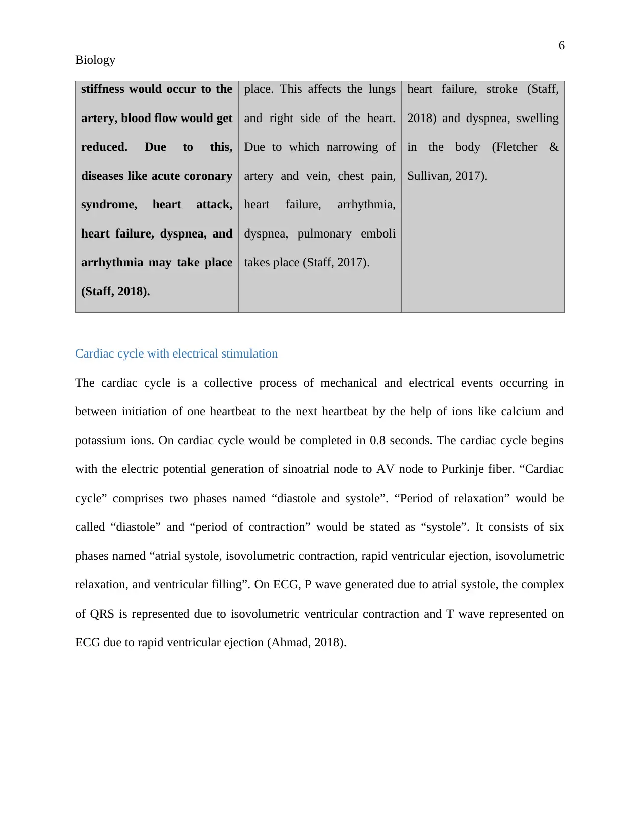
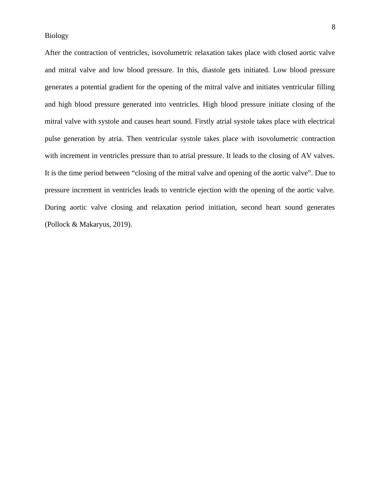
Cardiac cycle with electrical stimulation
The cardiac cycle is a collective process of mechanical and electrical events occurring in
between initiation of one heartbeat to the next heartbeat by the help of ions like calcium and
potassium ions. On cardiac cycle would be completed in 0.8 seconds. The cardiac cycle begins
with the electric potential generation of sinoatrial node to AV node to Purkinje fiber. “Cardiac
cycle” comprises two phases named “diastole and systole”. “Period of relaxation” would be
called “diastole” and “period of contraction” would be stated as “systole”. It consists of six
phases named “atrial systole, isovolumetric contraction, rapid ventricular ejection, isovolumetric
relaxation, and ventricular filling”. On ECG, P wave generated due to atrial systole, the complex
of QRS is represented due to isovolumetric ventricular contraction and T wave represented on
ECG due to rapid ventricular ejection (Ahmad, 2018).
Biology
stiffness would occur to the
artery, blood flow would get
reduced. Due to this,
diseases like acute coronary
syndrome, heart attack,
heart failure, dyspnea, and
arrhythmia may take place
(Staff, 2018).
place. This affects the lungs
and right side of the heart.
Due to which narrowing of
artery and vein, chest pain,
heart failure, arrhythmia,
dyspnea, pulmonary emboli
takes place (Staff, 2017).
heart failure, stroke (Staff,
2018) and dyspnea, swelling
in the body (Fletcher &
Sullivan, 2017).
Cardiac cycle with electrical stimulation
The cardiac cycle is a collective process of mechanical and electrical events occurring in
between initiation of one heartbeat to the next heartbeat by the help of ions like calcium and
potassium ions. On cardiac cycle would be completed in 0.8 seconds. The cardiac cycle begins
with the electric potential generation of sinoatrial node to AV node to Purkinje fiber. “Cardiac
cycle” comprises two phases named “diastole and systole”. “Period of relaxation” would be
called “diastole” and “period of contraction” would be stated as “systole”. It consists of six
phases named “atrial systole, isovolumetric contraction, rapid ventricular ejection, isovolumetric
relaxation, and ventricular filling”. On ECG, P wave generated due to atrial systole, the complex
of QRS is represented due to isovolumetric ventricular contraction and T wave represented on
ECG due to rapid ventricular ejection (Ahmad, 2018).
Paraphrase This Document
Need a fresh take? Get an instant paraphrase of this document with our AI Paraphraser
7
Biology
Time duration to complete cardiac cycle
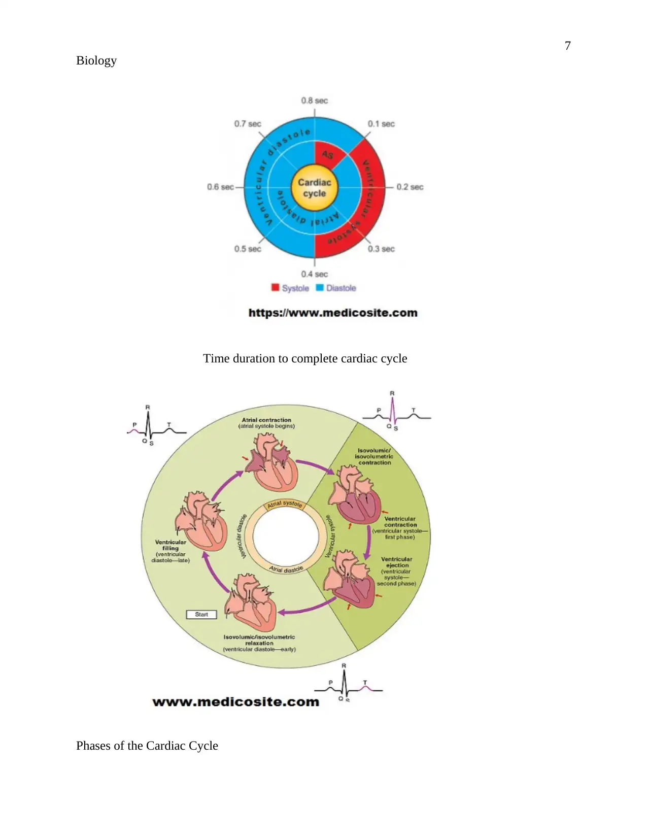
Phases of the Cardiac Cycle
Biology
Time duration to complete cardiac cycle
Phases of the Cardiac Cycle
8
Biology
After the contraction of ventricles, isovolumetric relaxation takes place with closed aortic valve
and mitral valve and low blood pressure. In this, diastole gets initiated. Low blood pressure
generates a potential gradient for the opening of the mitral valve and initiates ventricular filling
and high blood pressure generated into ventricles. High blood pressure initiate closing of the
mitral valve with systole and causes heart sound. Firstly atrial systole takes place with electrical
pulse generation by atria. Then ventricular systole takes place with isovolumetric contraction
with increment in ventricles pressure than to atrial pressure. It leads to the closing of AV valves.
It is the time period between “closing of the mitral valve and opening of the aortic valve”. Due to
pressure increment in ventricles leads to ventricle ejection with the opening of the aortic valve.
During aortic valve closing and relaxation period initiation, second heart sound generates
(Pollock & Makaryus, 2019).
⊘ This is a preview!⊘
Do you want full access?
Subscribe today to unlock all pages.
Trusted by 1+ million students worldwide
9
Biology
Biology
Paraphrase This Document
Need a fresh take? Get an instant paraphrase of this document with our AI Paraphraser
10
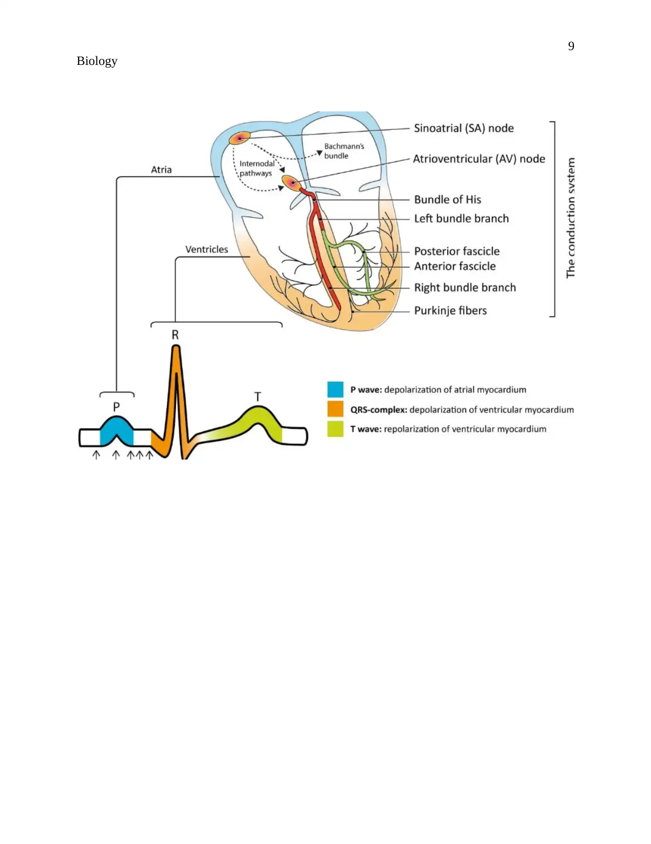
Biology
Components introducing ECG waveforms, Source: (Rawshani, 2017).
Role of pacemaker
Pacemaker cells collectively form the sinus node or pacemaker who generate an electric impulse.
These electric impulses conveyed to the right atrium by perinodal cells or transitional cells to the
AV node and then passed to another electrical conduction system of the heart (Mulpuru, et al.,
2017). Pacemaker generates an electric impulse to introduce myocardial contraction and help in
blood distribution in the different body parts. Regular electrical impulses generation would be
helpful in the introduction of normal rate and rhythm in the healthy heart (Basit, et al., 2019).
Difference between cardiac output and blood pressure
Cardiac output Blood Pressure
“Cardiac output” is “the amount of blood”
pumped by the heart in one minute for
“completing need for oxygen and nutrients”
Blood pressure is the “measurement of force
or pressure on the blood vessels” during the
pumping of blood out of the heart for the
11
Biology
in the parts of the body. It is calculated by
multiplying heart rate and stroke volume. The
measurement unit of cardiac output is litre per
minute (L/min).
body parts. The upper limit of blood pressure
is known to be systolic blood pressure and the
lower limit is known as diastolic blood
pressure. Measuring unit of blood pressure is
millimeters of mercury (mmHg).
During rest, the cardiac output would be 3-4
L/min.
During rest, blood pressure would be 120/80
mmHg.
During exercise, the cardiac output would
be 35L/min (King & Lowery, 2019).
During exercise, systolic blood pressure can
be increased at the range of 160-200mmHg
(Miller, 2018).
Similarities and differences in cardiac output and blood pressure at rest and exercise
Similarities from the graph would be evaluated that cardiac output and right atrial pressure are
directly proportional to each other. It means that when cardiac pressure gets increased then atrial
pressure would also be increased. At the rest cardiac output and pressure of RA remains normal
and increased at a normal rate. It indicates the “normal cardiac output curve” having a resting
cardiac output of 5 L/min at a right atrial pressure of 10 mmHg. During the exercise, the heart is
not pumping normally and cardiac output and blood pressure would get increased as sudden.
Because during the exercise, energy demand would be increased in the body, due to this cardiac
output would be increased and atrial pressure would be increased to “fulfill oxygen and
nutrients” demand inside the body. The uppermost curve shows the cardiac output in hearts that
are pumping well than normal to fulfill body needs. Stationary phase in both rest and exercise
came on the same right atrial pressure at 11 mmHg (Calvert & Lefer, 2012).
⊘ This is a preview!⊘
Do you want full access?
Subscribe today to unlock all pages.
Trusted by 1+ million students worldwide
1 out of 27
Related Documents


Your All-in-One AI-Powered Toolkit for Academic Success.
+13062052269
info@desklib.com
Available 24*7 on WhatsApp / Email
![[object Object]](/_next/static/media/star-bottom.7253800d.svg)
Unlock your academic potential
Copyright © 2020–2026 A2Z Services. All Rights Reserved. Developed and managed by ZUCOL.