Medical Imaging Techniques: X-ray, MRI, and Ultrasound Report

Verified

Added on  2020/03/23

|7
|1113
|69
Report
AI Summary
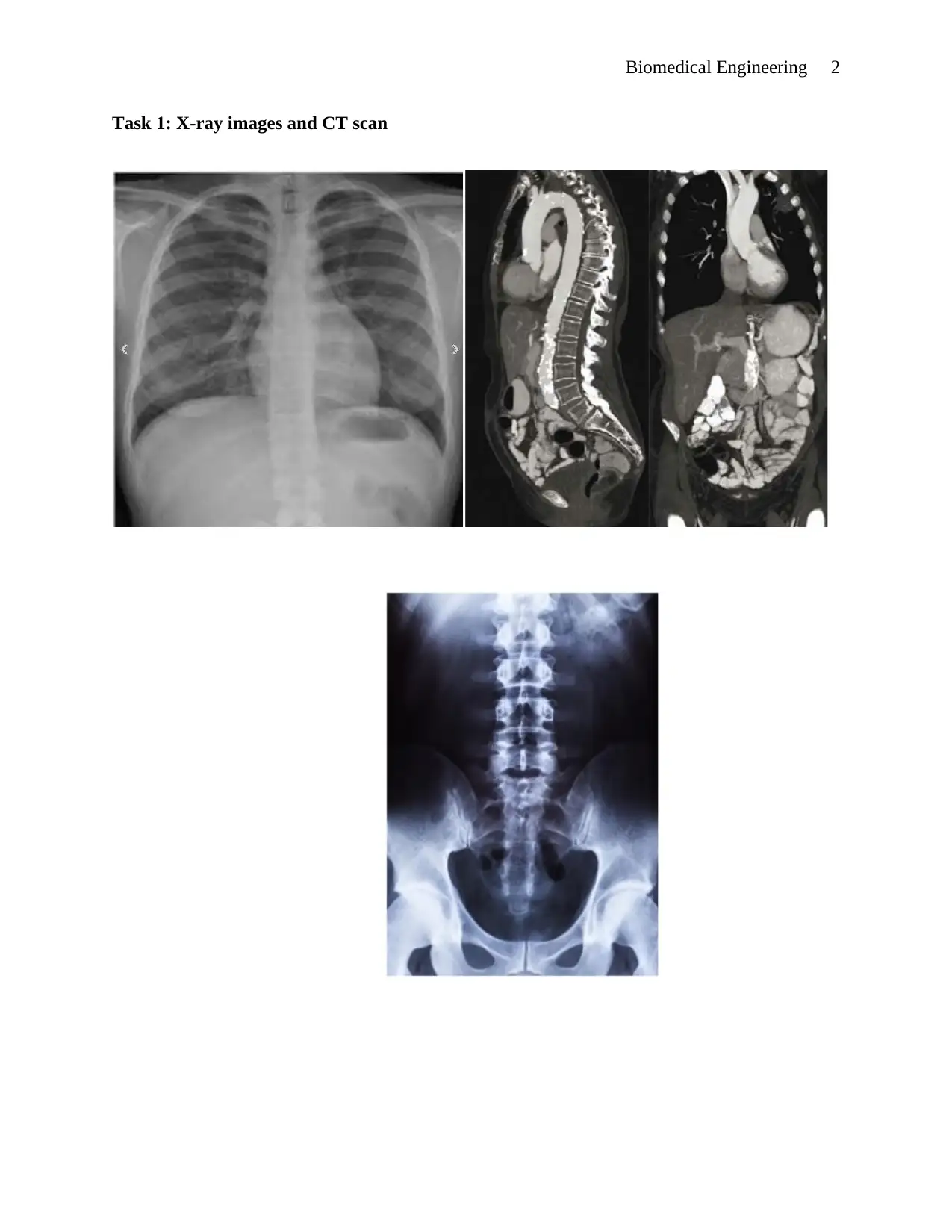
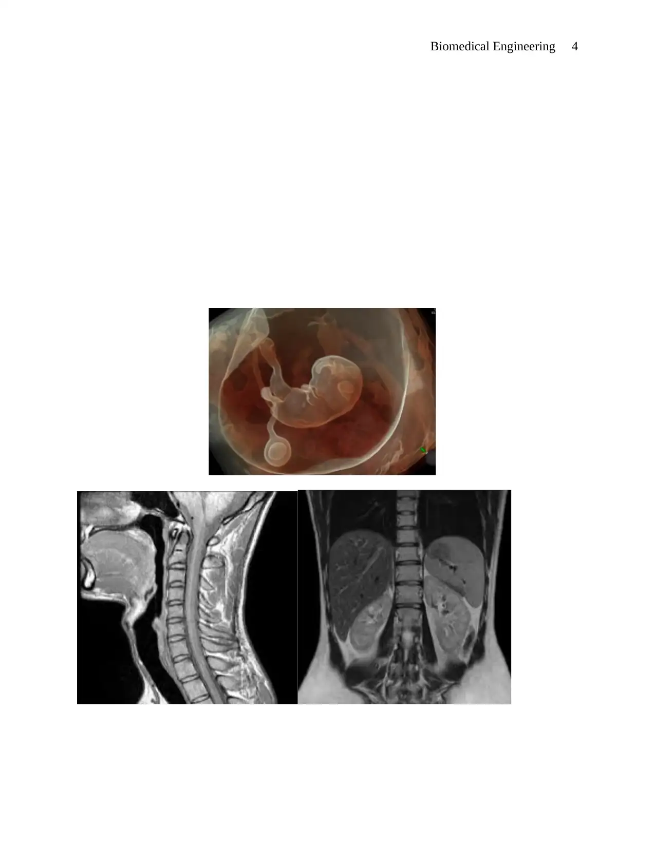
This report provides an analysis of various medical imaging techniques, specifically focusing on X-ray, CT scans, MRI, and ultrasound. The report begins by describing the characteristics and applications of each imaging modality, highlighting their strengths in different diagnostic scenarios. The author selects and discusses specific images, emphasizing their clarity and diagnostic value, including different body angles and views. The report then delves into the advancements and future developments in medical imaging, addressing both the benefits and potential drawbacks of each technology, such as safety concerns related to radiation exposure in X-rays and the need for safer alternatives like improved MRI and ultrasound technologies. The importance of image quality and its impact on the diagnostic process is also emphasized, suggesting a focus on enhancing the quality of medical imaging to improve patient care. The report concludes by underscoring the transformative impact of medical imaging on the field of medicine and the potential for future advancements to improve the quality and safety of these technologies.
Document Page
Running Head: Biomedical Engineering 1
Biomedical Engineering
Institution
Lecturer
Student
Course
Date
tabler-icon-diamond-filled.svg

Paraphrase This Document

Need a fresh take? Get an instant paraphrase of this document with our AI Paraphraser
Document Page
Biomedical Engineering 2
Task 1: X-ray images and CT scan
Document Page
Biomedical Engineering 3
The images selected are that of CT scanning and the X-ray. They all have one thing in
common such that they all use X-ray and CT scan uses computer technology to dissect your body
into slice-like segments that provides accurate assessment of the body organs by the physician.
The images are chosen because they represent the best quality and clarity as needed for easy
examination. Furthermore, the images present different body angles, from the chest, the vertical
spinal view, and the side view. The images provides efficient basis for diagnosis and analysis by
the doctor. The different views also provide alternative analysis and above all the quality of the
images is crystal clear.
Task 2: The MRI and Ultrasound images
Document Page
Biomedical Engineering 4
tabler-icon-diamond-filled.svg

Paraphrase This Document

Need a fresh take? Get an instant paraphrase of this document with our AI Paraphraser
Document Page
Biomedical Engineering 5
The above picture represents to MRI and one ultrasound image. The choice of the images
was influenced by among other reasons, the clarity of the images, and the variation in types and
the quality of the images as well. The ultrasound image is colored and which underlines the
varying range of the choices. Furthermore, the images represent different body parts and
different body views. The different views of the internal body show the spinal cord from rear and
side views. The colored ultrasound image shows a baby’s position in the womb of its mother,
and the quality present across the images is commendable. MRI images are very detailed and can
provide significant information such as the ways blood moves through body organs and enables
any complications about blood flow to be identified, and this include blockages. MRI can also
provide information on structure of the soft tissues including ligaments and can give detailed
analysis about organs such as the eyes, brains among others. All the mentioned qualities of the
MRI make it appropriate image for use in this scenario.
Document Page
Biomedical Engineering 6
Task 3
The imaging development has led to breakthrough in the medical field whereby
diagnosing of internal body conditions has been made easier through the imaging technology
(Bridge et al. 2014). With the effective and successful results that the medical world has
witnessed over the last past few years, the future development promises much better with the
advancement in technology. With the medical imaging making use of equipment to get the
different information about the body, there is room for development of advanced technology.
For instance, although the ultrasound technology has been in use for over 20 years in the
medical imaging, the general assumption that it is entirely safe should be avoided. The
ultrasound imaging safety can only be confirmed by the perfect use of the physician. The waves
from the machine can heat tissues slightly and in some instance produce little amount of gas in
the body fluids. Therefore, in regard to ultrasound, it would be a great breakthrough if the
inventors came up with image technology that cannot cause significant side effects to the body.
Document Page
Biomedical Engineering 7
On the other side, Magnetic Resonance Imaging (MRI) does not use any form of ionizing
radiation which means that patients are not exposed to the subsequent side effects. But MRI
involves strong magnetic pull which means that objects nearby can easily be pulled and cause
injuries to the patient if there are no precautionary measures taken. Therefore, in the near future,
as the technology develops, the development of a much better and advanced MRI will ensure
safety for the patients. However, X-rays imaging is the most debatable of all the medical imaging
technologies. The X-ray is the area that everyone would want to see rapid changes when it comes
to the safety of patients. The desired development in regard to this equipment is to see the review
and upgrade to a more advanced X-ray with less side effects (Groenewal & Groenewald 2016).
X-ray imaging uses ionizing radiation for the generation of images. Ionizing radiation is
too strong enough to cause the damage to the body DNA and too much exposure to it may cause
cancer later on in life, according to many medical researches undertaken in regards to it.
Therefore, as much as there is so much to cheer about the medical imaging and the
transformation the technologies has contributed in advancing the medical field; there is much
room for future improvement in developing this area of medical field. The developments should
be centered on improving the quality rather than quantity. The quality of imaging, the quality of
the results and the safety of patients are the developments anticipated in the future for the
medical images (Party et al. 2004).
Changes in the technological trends, the advancement of technology experienced should
help in transforming the medical field and facilitate in introduction of better medical equipment,
from RMI to the harmless X-ray and the efficient ultrasound are all that the world of medicine
hope to see in the near future, with the change in technology (Norvatis n.d.). It is also important
chevron_up_icon
1 out of 7
circle_padding
hide_on_mobile
zoom_out_icon
[object Object]