Cardiovascular System: Blood Vessels - Lecture Questions Answers

Verified

Added on  2022/08/15

|4
|572
|11
Homework Assignment
AI Summary
This assignment addresses key concepts related to blood vessels, focusing on their structure and function within the cardiovascular system. It begins by defining the three tunica layers (intima, media, and externa) of blood vessels, detailing their composition and roles. The assignment then explores blood flow, its relationship with blood pressure, and the surface area of different blood vessels. It includes the identification of blood vessels, differentiating between arteries and veins based on their structural characteristics, such as wall thickness and lumen shape. The assignment also includes references to support the answers. This resource is designed to help students understand the complexities of the cardiovascular system and its components.
Document Page
Running head: LECTURE QUESTIONS
LECTURE QUESTIONS
Name of the Student:
Name of the University:
Author Note:
tabler-icon-diamond-filled.svg

Paraphrase This Document

Need a fresh take? Get an instant paraphrase of this document with our AI Paraphraser
Document Page
1LECTURE QUESTIONS
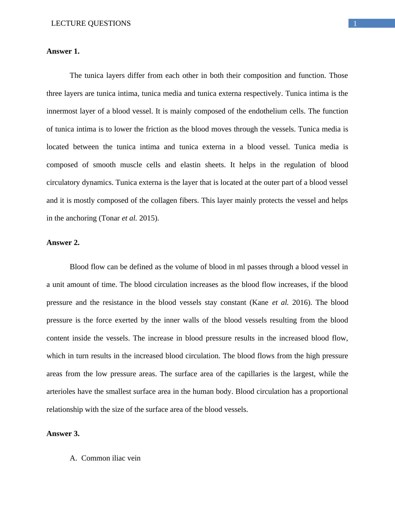
Answer 1.
The tunica layers differ from each other in both their composition and function. Those
three layers are tunica intima, tunica media and tunica externa respectively. Tunica intima is the
innermost layer of a blood vessel. It is mainly composed of the endothelium cells. The function
of tunica intima is to lower the friction as the blood moves through the vessels. Tunica media is
located between the tunica intima and tunica externa in a blood vessel. Tunica media is
composed of smooth muscle cells and elastin sheets. It helps in the regulation of blood
circulatory dynamics. Tunica externa is the layer that is located at the outer part of a blood vessel
and it is mostly composed of the collagen fibers. This layer mainly protects the vessel and helps
in the anchoring (Tonar et al. 2015).
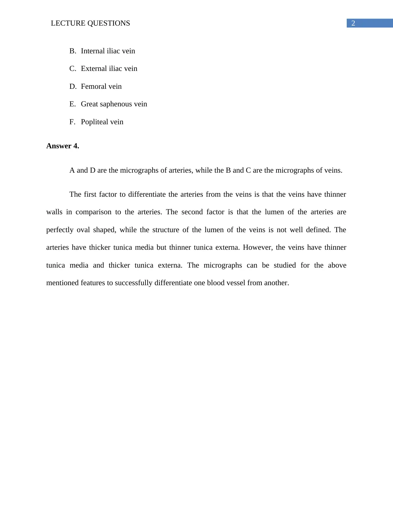
Answer 2.
Blood flow can be defined as the volume of blood in ml passes through a blood vessel in
a unit amount of time. The blood circulation increases as the blood flow increases, if the blood
pressure and the resistance in the blood vessels stay constant (Kane et al. 2016). The blood
pressure is the force exerted by the inner walls of the blood vessels resulting from the blood
content inside the vessels. The increase in blood pressure results in the increased blood flow,
which in turn results in the increased blood circulation. The blood flows from the high pressure
areas from the low pressure areas. The surface area of the capillaries is the largest, while the
arterioles have the smallest surface area in the human body. Blood circulation has a proportional
relationship with the size of the surface area of the blood vessels.
Answer 3.
A. Common iliac vein
Document Page
2LECTURE QUESTIONS
B. Internal iliac vein
C. External iliac vein
D. Femoral vein
E. Great saphenous vein
F. Popliteal vein
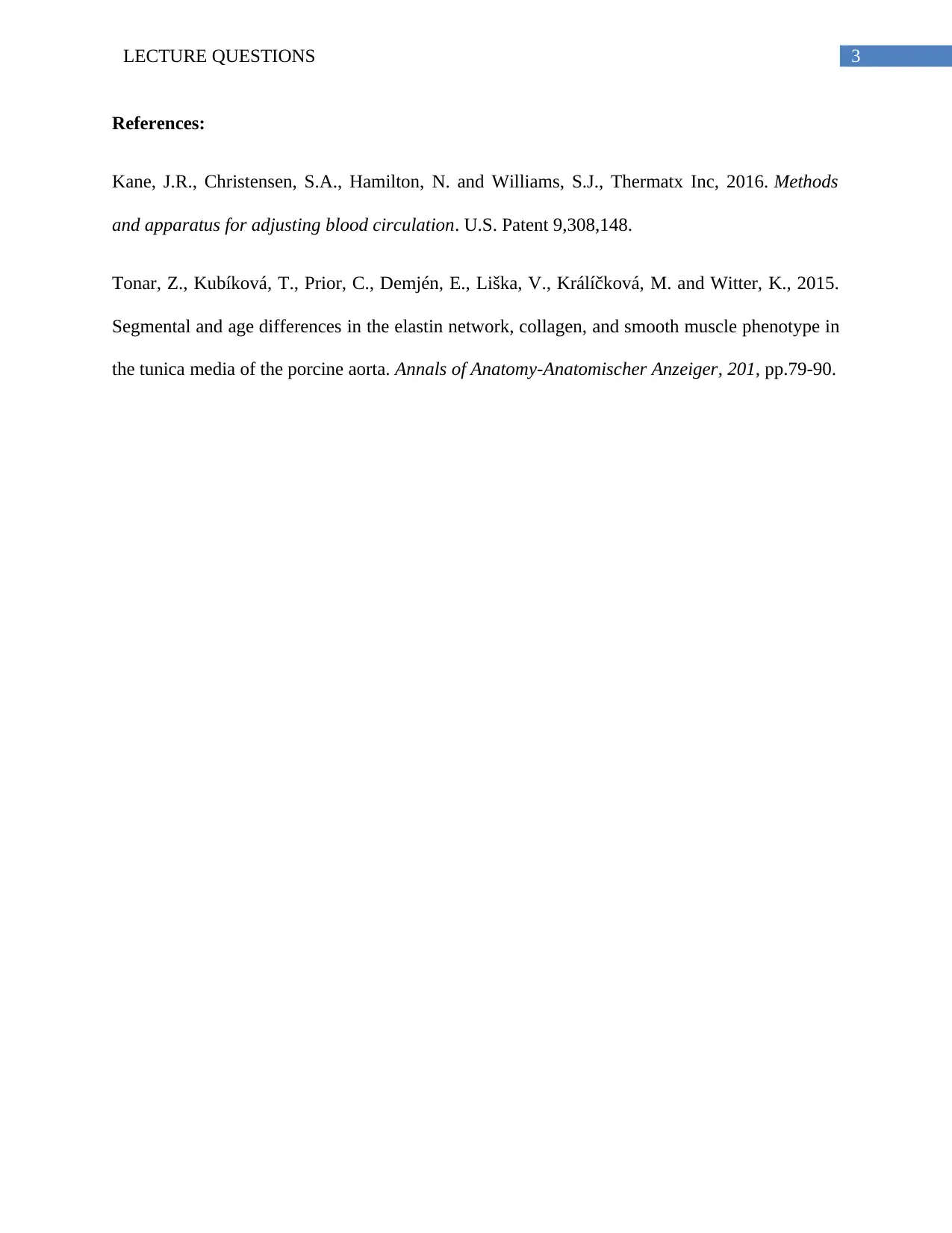
Answer 4.
A and D are the micrographs of arteries, while the B and C are the micrographs of veins.
The first factor to differentiate the arteries from the veins is that the veins have thinner
walls in comparison to the arteries. The second factor is that the lumen of the arteries are
perfectly oval shaped, while the structure of the lumen of the veins is not well defined. The
arteries have thicker tunica media but thinner tunica externa. However, the veins have thinner
tunica media and thicker tunica externa. The micrographs can be studied for the above
mentioned features to successfully differentiate one blood vessel from another.
Document Page
3LECTURE QUESTIONS
References:
Kane, J.R., Christensen, S.A., Hamilton, N. and Williams, S.J., Thermatx Inc, 2016. Methods
and apparatus for adjusting blood circulation. U.S. Patent 9,308,148.
Tonar, Z., Kubíková, T., Prior, C., Demjén, E., Liška, V., Králíčková, M. and Witter, K., 2015.
Segmental and age differences in the elastin network, collagen, and smooth muscle phenotype in
the tunica media of the porcine aorta. Annals of Anatomy-Anatomischer Anzeiger, 201, pp.79-90.
chevron_up_icon
1 out of 4
circle_padding
hide_on_mobile
zoom_out_icon
[object Object]