Evaluation of Bone Scan Techniques for Metastases Detection
VerifiedAdded on 2022/11/28
|8
|2008
|367
Report
AI Summary
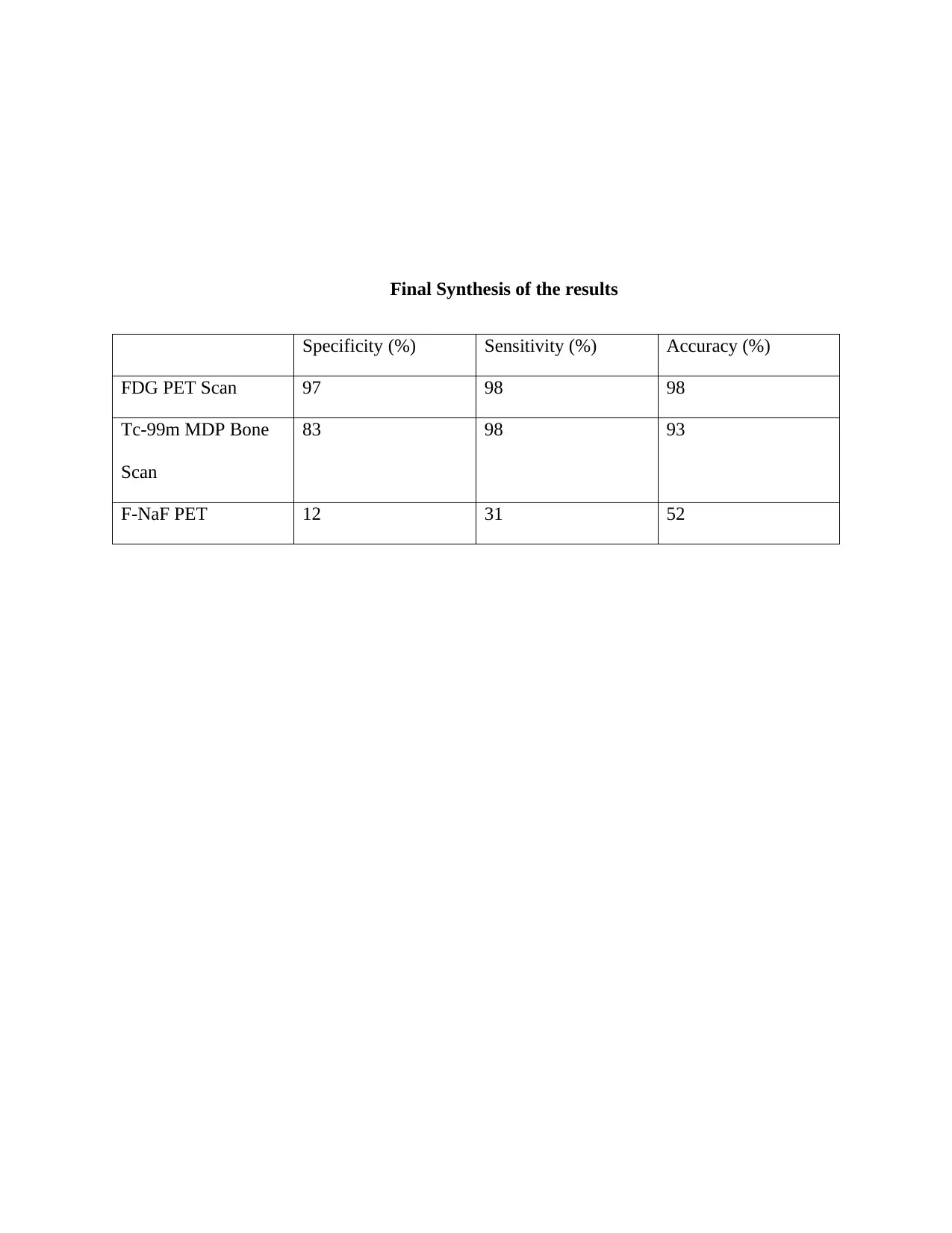
This report provides a detailed analysis of various bone scan techniques used in the diagnosis of bone metastases, particularly in the context of neuroblastoma in children. It begins by explaining the Tc-99m MDP bone scan and then evaluates a review comparing FDG PET, NaF PET, and Tc-99m MDP bone scans, utilizing the QUADAS-2 tool to assess the methodological quality of the review. The report also includes a table summarizing the QUADAS-2 assessment. Furthermore, the report defines bone metastases as identified through Tc-99m bone scan, FDG PET, and NaF PET scans, and presents these definitions in a comparative table. The analysis highlights the advantages and disadvantages of each imaging technique, emphasizing the superior diagnostic capabilities of 18F-NaF-PET in detecting both osteoblastic and lytic lesions, along with its ability to visualize smaller lesions. The report references several studies to support its conclusions, including the use of SPSS software for statistical analysis and a systematic review process involving inclusion, eligibility, screening, and identification of relevant studies. The findings underscore the importance of these imaging techniques in the early detection and management of bone metastases, ultimately improving patient outcomes.

Statistical Analysis
FDG PET scan, Tc-99m MDP and F-NaF PET diagnoses were evaluated with kappa
value. A correlation existing between the above imaging technique and the evaluation was done
on ultimate diagnoses. The accuracies on diagnoses for all the three modalities for imaging were
calculated and equated through comparing. After that, the relationships existing between the
results on the imaging and abnormal lab data were consequentially assessed using Kruskal-
Wallis test. Analysis was then carried out using version 21 of SPSS software. AP value <0.005
was regarded as significant in terms of statistics assessment.
Systematic Review of the Comparison Results
In reviewing, four factors were considered: inclusion, eligibility, screening and
identification. The records were identified through searching through the database (n=1343) and
additional records identified through other sources (n=9). The next stage was to remove
duplicates (n=603) and then allowed screening of the records by the title or the abstract (n=741)
and then 521 records were excluded. Resulting full articles assessed for eligibility were 75, the
other ones that were excluded were 446 because one of the following reasons: they did not meet
inclusion criteria, poster of oral abstract, they had poster of oral abstract only, they were in other
language other than English, had insufficient data for analysis, non-related to the condition in
question and double publication. This resulted to only 10 studies undergoing qualitative
assessment.
The use of the imaging has always played an important role in the diagnosis of the patient
in the paediatric ward with neuroblastoma that often leads to bone metastases in the young
children. 99m-Tc-diphosphate bone scanning is the widely used technique in the imaging of the
Paraphrase This Document
Need a fresh take? Get an instant paraphrase of this document with our AI Paraphraser

bone metastases, however as its lack the specificity in the imaging procedures it has led to a
more improved tool with better resolution and specificity. As the use of 99m-Tc- labelled
disphosphate depends on the uptake of the labelled substance it is not specific for the metastatic
disease and can be taken up by the unresponsive bone cells (Broos et al.,2018) ; (Azad and Cook
2016). Research has showed that the role of 18F-NaF-PET is able to show the lesions that are
both osteoblastic and lytic in nature. It is also much better in the visualization of the smaller
lesions due to the advantage of clearer sectional view because of better resolution and higher
target/background ratio. Research also shows that the uptake of 18F-FDG varies among the
blastic and lytic lesions and that of cranial bone. The 18F-NaF-PET is emerging as a single-step
study for bone metastases after considering the advantages and its potential in the detection of
occult metastases (Evangelista et al., 2016).
The use of single-photon emission computed tomography or SPECT lacks the
quantitative information of the section that is being scanned because of its known limitation (It
can only be used to obtain quantitative information. However, the injection of the 99m-Tc-MDP
along with the SPECT increases the quality of images to some extent. Even after that the
technique is not so successful in the scanning methods. The uptake rate of the radiolabeled
substance is same as that of 18F-PET but the only difference is the ability to differentiate among
the pathological conditions of the various lesions caused at different stages of metastases (Chang
et al. 2016). Research has shown that the standardized uptake of the Tc-99m-methylene
diphosphate using a PET scanner showed variable results in the normal as well as affected area.
The results were different in different individuals who have different height and age. The
difference might be caused due to the sample size, age, tracers, attenuation or PET construction

method. Thus, it signifies that the use of 18-NaF-PET is far better than the use of 99m-Tc-MDP
(Kwee. and Kwee, 2015)
On the other hand, the use of FDG-PET scan also has a lower sensitivity than that of 18-
NaF-PET or even 99m-MDP. There has been several studies that have compared the efficacy of
the FDG-PET with that of 18-NaF-PET scan which shows that the overall accuracy and
specificity of the 18-NaF-PET scan is much more than that of the FDG-PET or 99m-MDP (Ota
et al. 2014). On comparison of FDG-PET with that of 99m-Tc-MDP it has been seen that the
FDG has better diagnostic efficiency than 99m-Tc-MDP. This is due to the use of FDG as a
tracer element that can identify the morphological changes and the metastatic sites of both the
osteoblastic as well as the lytic lesions. The meta-analysis of the FDG-PET has shown that the
sensitivity is about 81% and specificity of about 93% when compared to that of 99m-Tc-MDP.
Thus, the specificity of PET is useful in monitoring the effects of various therapies undertaken
by the patients (O’Sullivan, Carty and Cronin, 2015)
The scanning of bone with 99m-Tc-MDP and 18-NaF-PET provides the information on
the subtle changes in the bone. This information can assist in the management of the clinical
symptoms that causes bone metastases in young children. 18-NaF-PET uses imaging biomarkers
that can capture the dynamic and static images of the bone in order to get better treatment
response (Perez-Lopez et al., 2016). The use of 18-Naf-PET is better than that of the other two
methods in detection and analysis of the neuroblastoma in paediatric patients in such a way that
it captures better images due to higher resolution and it can differentiate better between the lytic
and osteoblastic lesions. Research has also shown that the radiotracer for PET that is 18-NaF use
has low repeatability that means that use of the radiotracer has different variability in the
individual and across the lesions throughout the body. The results also highlight the fact the use
⊘ This is a preview!⊘
Do you want full access?
Subscribe today to unlock all pages.

Trusted by 1+ million students worldwide

of 18-NaF-PET is better in the assessment of the various stages of metastases in the child’s body
(Lutz et al. 2017).
As the Na 18F-PET is a new modality for the imaging and evaluation of the bone diseases,
there is a need to evaluate the accuracies and specificities of the bone PET scan and the detection
of bone metastases. Research reveals that for the acquisition of the PET imaging there is no need
for the specific preparation of the patient and thus, it is a very useful tool for the assessment of
the metastatic stage in the infants as the young children are very hyper during the diagnostic
stage. The use of 18-NaF is far better than the other imaging tools because of the physic-
chemical characters of 18-NaF. The imaging is helpful in the analysis of the malignant bone
diseases but also the benign bone diseases. The technique can also effectively exclude the bone
lesions that are non-metastatic that again reflects the specificity of the tool (Saunders.Macedo et
al., 2017). The more advanced analysis of the bone lesions by that of the PET scan is mainly due
to the tomographic nature of the scan. The planar bone scans like 99m-Tc-MDP often fail in
locating the lesions exactly inside the patient’s body. The specificity of the 18-NaF-PET scan
other than tomographic effect is the referral bias that results in low specificity of the
conventional bone scans. In addition to this there are several advantages of the 18-NaF-PET over
that of normal scans of FDG-PET or 99m-Tc-MDP scans. These advantages include quantitative
imaging used by that of PET scanning which helps in the quantitative determination of the bone
blood flow and the bone turnover rate. The next advantage is the short time required by the tool
to start the imaging process after the acquisition of the injection. The third advantage includes
the non-medical issues that involves the limited supply or the problem in then supply of the
radionuclide 99m-Tc that ultimately leads to the more clinical use of 18-NaF and thus, the 18-NaF-
PET scan becomes much more popular (Jambor et al. 2016).
Paraphrase This Document
Need a fresh take? Get an instant paraphrase of this document with our AI Paraphraser

Final Synthesis of the results
Specificity (%) Sensitivity (%) Accuracy (%)
FDG PET Scan 97 98 98
Tc-99m MDP Bone
Scan
83 98 93
F-NaF PET 12 31 52
Specificity (%) Sensitivity (%) Accuracy (%)
FDG PET Scan 97 98 98
Tc-99m MDP Bone
Scan
83 98 93
F-NaF PET 12 31 52

Bibliography
Azad, G.K. and Cook, G.J., 2016. Multi-technique imaging of bone metastases: spotlight on
PET-CT. Clinical radiology, 71(7), pp.620-631.
Broos, W.A., van der Zant, F.M., Wondergem, M. and Knol, R.J., 2018. Accuracy of 18F-NaF
PET/CT in bone metastasis detection and its effect on patient management in patients with breast
carcinoma. Nuclear medicine communications, 39(4), pp.325-333.
Chang, C.Y., Gill, C.M., Joseph Simeone, F., Taneja, A.K., Huang, A.J., Torriani, M. and
Bredella, M.A., 2016. Comparison of the diagnostic accuracy of 99 m-Tc-MDP bone
scintigraphy and 18 F-FDG PET/CT for the detection of skeletal metastases. Acta
Radiologica, 57(1), pp.58-65.
Evangelista, L., Bertoldo, F., Boccardo, F., Conti, G., Menchi, I., Mungai, F., Ricardi, U. and
Bombardieri, E., 2016. Diagnostic imaging to detect and evaluate response to therapy in bone
metastases from prostate cancer: current modalities and new horizons. European journal of
nuclear medicine and molecular imaging, 43(8), pp.1546-1562.
Jambor, I., Kuisma, A., Ramadan, S., Huovinen, R., Sandell, M., Kajander, S., Kemppainen, J.,
Kauppila, E., Auren, J., Merisaari, H. and Saunavaara, J., 2016. Prospective evaluation of planar
bone scintigraphy, SPECT, SPECT/CT, 18F-NaF PET/CT and whole body 1.5 T MRI, including
DWI, for the detection of bone metastases in high risk breast and prostate cancer patients:
SKELETA clinical trial. Acta oncologica, 55(1), pp.59-67.
⊘ This is a preview!⊘
Do you want full access?
Subscribe today to unlock all pages.

Trusted by 1+ million students worldwide

Kwee, R.M. and Kwee, T.C., 2015. Modern imaging techniques for preoperative detection of
distant metastases in gastric cancer. World Journal of Gastroenterology: WJG, 21(37), p.10502.
Lutz, S., Balboni, T., Jones, J., Lo, S., Petit, J., Rich, S.E., Wong, R. and Hahn, C., 2017.
Palliative radiation therapy for bone metastases: Update of an ASTRO Evidence-Based
Guideline. Practical radiation oncology, 7(1), pp.4-12.
O’Sullivan, G.J., Carty, F.L. and Cronin, C.G., 2015. Imaging of bone metastasis: an
update. World journal of radiology, 7(8), p.202.
Perez-Lopez, R., Mateo, J., Mossop, H., Blackledge, M.D., Collins, D.J., Rata, M., Morgan,
V.A., Macdonald, A., Sandhu, S., Lorente, D. and Rescigno, P., 2016. Diffusion-weighted
imaging as a treatment response biomarker for evaluating bone metastases in prostate cancer: a
pilot study. Radiology, 283(1), pp.168-177.
Saunders.Macedo, F., Ladeira, K., Pinho, F., Saraiva, N., Bonito, N., Pinto, L. and Gonçalves, F.,
2017. Bone metastases: an overview. Oncology reviews, 11(1).
Paraphrase This Document
Need a fresh take? Get an instant paraphrase of this document with our AI Paraphraser

1 out of 8
Related Documents

Your All-in-One AI-Powered Toolkit for Academic Success.
+13062052269
info@desklib.com
Available 24*7 on WhatsApp / Email
Unlock your academic potential
Copyright © 2020–2025 A2Z Services. All Rights Reserved. Developed and managed by ZUCOL.