Restoring Movement: Brain-Spine Interface for Spinal Cord Injury
VerifiedAdded on 2023/06/04
|7
|1561
|399
Report
AI Summary
This report discusses a study on restoring movement in primates with spinal cord injuries using a brain-spine interface. The study involved implanting rhesus monkeys with intracortical microelectrode arrays in the leg area of the motor cortex and using epidural electrical stimulation of the lumbar spin...

RESTORATION OF MOVEMENT IN PRIMATES WITH SPINAL CORD INJURY
By Name
Course
Instructor
Institution
Location
Date
By Name
Course
Instructor
Institution
Location
Date
Paraphrase This Document
Need a fresh take? Get an instant paraphrase of this document with our AI Paraphraser

Summary
Recent research in rats demonstrated that electrical stimulation of the epidural of the lumbar
spinal cord is able to generate natural activation of synergic muscle groups that generate motion.
This study was on interfacing leg motor cortex activity using epidural electrical stimulation
protocols to come up with a brain-sine interface which is alleviated gait deficit following a spinal
cord injury in non-human primates (Schwarz et al., 2014). The study used rhesus monkey which
was implanted using an array of intracortical microelectrode in the area of the leg of the motor
cortex and using a system of stimulation of the spinal cord that was composed of an epidural
implant that is spatially selective as well as a pulse generator that has capabilities of real-time
triggering (Capogrosso et al., 2016).
Wireless systems of control were designed and adopted which joined online extension and motor
states flexion of neural decoding using stimulation proctors enhancing these motions. Such
systems permit the primates to experience a free movement in the absence of any hindrances or
interference with the tied electronics. Upon the confirmation of the interface of the brain and
spine to be in intact of the healthy primates was conducted, a unilateral corticospinal tract lesion
at the level of the thoracic (Capogrosso et al., 2016)
The interface of the brain and spine were able to restore their weight-bearing locomotion, as soon
as after six days of the post-injury alongside excluding prior training of the monkeys, of the
injured leg on an over ground and treadmill. The components that can be implanted which were
included in the interface of the brain and spine were all recommended for use for investigations
in the same study in humans. This illustrated a practical translation pathway that would act as a
proof of the concept studies in people that have injuries of the spinal cord.

The significance of the study in human life and society
The systems that were developed in the study made use of signals that were measured from the
motor cortex of the brain in the triggering of coordinated electrical stimulation of the nerves that
are found in the spine and are responsible for locomotion to movement. When the system was
turned on, the animal in the study experienced normal movement (Jackson, 2016). From such a
study it could be concluded that there was enough evidence to argue that a spinal stimulation
system that is controlled by the brain can lead to the rehabilitation after an injury to the spinal
cord.
Figure 1 below illustrates a neural bridge which is the brain spine interface developed for the
purposes of re-establishing communication following a breakdown of the same after an injury to
the spinal cord. An implant such as this is used in the detection of the spiking activity in the
motor cortex of the brain. As can be observed in the image a pulse generator, a microelectrode
array as well as a silicon model of the brain of a primate is used in the stimulation of the
electrodes that have been implanted on the spinal cord. The final impact of this is noticed when
the primate is able to move.
Figure 1: A Neural Bridge
You're viewing a preview
Unlock full access by subscribing today!

The image is illustrate of the progress of other scientists when it comes to the development of
systems which make use of signals that are measured from the motor cortex of the brain in
triggering coordinated electrical stimulation of the spinal nerves which are tasked with
locomotion (Schwarz et al., 2014). This study in conjunction with the aforementioned summary
are ideal in offering a greener light when it comes to restoration of locomotion in primates after
an injury.
This study thus turned out to be of importance at it would in the restoration of locomotion among
people to normalcy after an injury occurs to the spinal cord. Owing to the currently existing
technology and state of the world, the human race continues to find itself on the wrong side of
life, encountering various injuries through such things as accidents which claim lives in the worst
case scenario or leave them with broken body parts. Breakage or injury of the various parts of the
body significantly paralyzes the normal operations of a person more specifically when it comes
to locomotion (Rajangam et al., 2016). Of special interest for this case is an injury to the spinal
cord which results in great disruption in the communication between the spinal circuits and the
brain part that initiate movement.
Before the study, the lesion was bypassed through the use of a brain-computer interface which
was able to link directly the electrical stimulation of the muscles with the cortical activity
thereby enhancing a restoration of the abilities to grasp following a paralysis of the hand.
Through this study, the challenge is likely to be overcome even further as it offers an opportunity
for conducting further tests on such a possibility (Miranda et al., 2015). A similar system as was
used in the study, when developed can be used in humans who are having injuries of the spinal
cord. An adoption of this study and its full implementation in human life would enhance
restoration of normal movement and locomotion in human beings especially in those who have
Paraphrase This Document
Need a fresh take? Get an instant paraphrase of this document with our AI Paraphraser

undergone spinal cord injury. This would, in turn, serve to restore the hope of value in life to
such people as well as providing them with an opportunity to exploit their potential in life
without the fear of being disabled.
An injury to the spinal cord may result in complete cut off of communication between the lower
spinal cord and the brain despite the fact that the spinal neurons and the motor cortex may both
remain fully functional after the injury (Bensmaia & Miller, 2014). This then calls for a
reestablishment of the communication between the two parts so as to ensure continues harmony
in operation, an achievement that is made in this study. From the findings of the study which
illustrated that upon turning on the system, there was the initiation of a spontaneous motion in
the legs of the animal on a treadmill; a similar challenge in humans is partly solved.
Comparisons of kinematics against the healthy controls illustrated that the lesioned macaques,
with the help of brain stimulation, were capable of generating almost normal patterns of
locomotion (Ajiboye et al., 2016).

References
Ajiboye, A.B., Willett, F.R., Young, D.R., Memberg, W.D., Murphy, B.A., Miller, J.P., Walter,
B.L., Sweet, J.A., Hoyen, H.A., Keith, M.W. and Peckham, P.H., 2017. Restoration of reaching
and grasping movements through brain-controlled muscle stimulation in a person with
tetraplegia: a proof-of-concept demonstration. The Lancet, 389(10081), pp.1821-1830
Bensmaia, S.J. and Miller, L.E., 2014. Restoring sensorimotor function through intracortical
interfaces: progress and looming challenges. Nature Reviews Neuroscience, 15(5), p.313
Capogrosso, M., Milekovic, T., Borton, D., Wagner, F., Moraud, E.M., Mignardot, J.B., Buse,
N., Gandar, J., Barraud, Q., Xing, D. and Rey, E., 2016. A brain–spine interface alleviating gait
deficits after spinal cord injury in primates. Nature, 539(7628), p.284
Jackson, A., 2016. Spinal-cord injury: Neural interfaces take another step
forward. Nature, 539(7628), p.177
Miranda, R.A., Casebeer, W.D., Hein, A.M., Judy, J.W., Krotkov, E.P., Laabs, T.L., Manzo,
J.E., Pankratz, K.G., Pratt, G.A., Sanchez, J.C. and Weber, D.J., 2015. DARPA-funded efforts in
the development of novel brain-computer interface technologies. Journal of neuroscience
methods, 244, pp.52-67
Rajangam, S., Tseng, P.H., Yin, A., Lehew, G., Schwarz, D., Lebedev, M.A. and Nicolelis,
M.A., 2016. Wireless cortical brain-machine interface for whole-body navigation in
primates. Scientific reports, 6, p.22170
You're viewing a preview
Unlock full access by subscribing today!

Schwarz, D.A., Lebedev, M.A., Hanson, T.L., Dimitrov, D.F., Lehew, G., Meloy, J., Rajangam,
S., Subramanian, V., Ifft, P.J., Li, Z. and Ramakrishnan, A., 2014. Chronic, wireless recordings
of large-scale brain activity in freely moving rhesus monkeys. Nature methods, 11(6), p.670
Soekadar, S.R., Birbaumer, N., Slutzky, M.W. and Cohen, L.G., 2015. Brain-machine interfaces
in neurorehabilitation of stroke. Neurobiology of disease, 83, pp.172-179
1 out of 7

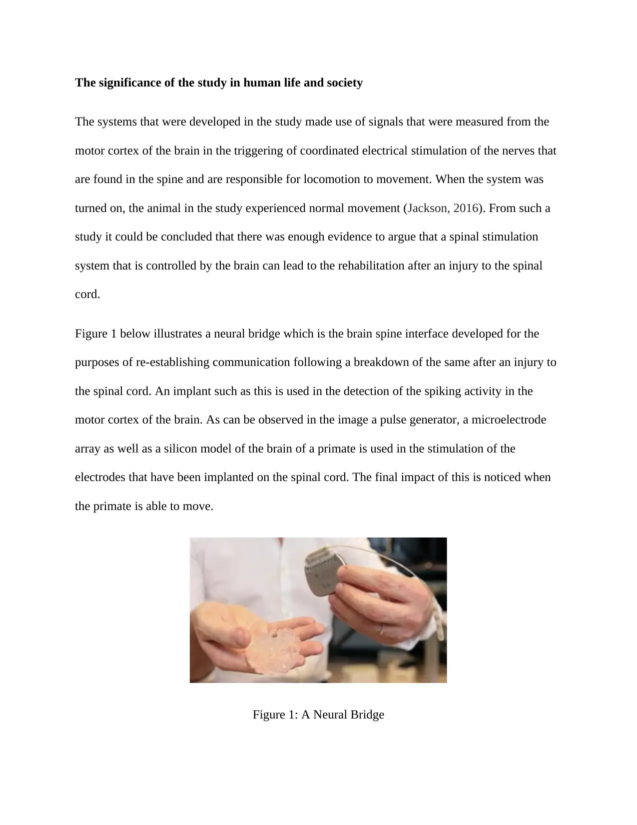
Your All-in-One AI-Powered Toolkit for Academic Success.
+13062052269
info@desklib.com
Available 24*7 on WhatsApp / Email
Unlock your academic potential
© 2024 | Zucol Services PVT LTD | All rights reserved.