AlexNet-DNN for Breast Cancer Detection and Classification Project
VerifiedAdded on 2020/11/23
|16
|14251
|98
Project
AI Summary
This project proposes a robust and efficient Computer-Aided Diagnosis (CAD) system for breast cancer detection and classification using a Deep Neural Network (DNN) based on the AlexNet architecture. The system incorporates pre-processing, an enhanced adaptive learning-based Gaussian Mixture Model (GMM), connected component analysis for region of interest localization, and AlexNet-DNN for feature extraction. Feature selection is performed using Principal Component Analysis (PCA) and Linear Discriminant Analysis (LDA). The model is evaluated on the BreakHis dataset, demonstrating superior performance compared to other state-of-the-art techniques, achieving high accuracy rates. The study highlights the effectiveness of AlexNet-DNN features combined with LDA for dimensional reduction and SVM-based classification, making it suitable for real-world applications. The research emphasizes the importance of early cancer identification and diagnosis using advanced computing techniques.
A Robust Deep Neural Network Based Breast Cancer
Detection And Classification
Abstract— The exponential rise in breast cancer cases across
the globe has alarmed academia-industries to achieve certain more
efficient and robust Breast Cancer Computer Aided Diagnosis (BC-
CAD) system for breast cancer detection. A number of techniques
have been developed with focus on case (i.e., data type) centric
segmentation, feature extraction and classification of breast cancer
Histopathological images. However, rising complexity and accuracy
often demands more robust solution. Recently, Convolutional
Neural Network (CNN) has emerged as one of the most efficient
techniques for medical data analysis and various image
classification problems. In this paper a highly robust and efficient
BC-CAD solution has been proposed. Our proposed system
incorporates pre-processing, enhanced adaptive learning based
Gaussian Mixture Model (GMM), connected component analysis
based region of interest localization, AlexNet-DNN based feature
extraction. The Principle Component Analysis (PCA) and Linear
Discriminant Analysis (LDA) based on feature selection which is
used as dimensional reduction. One of the advantages of the
proposed method is that none of the current dimensional reduction
algorithms employed with SVM to perform breast cancer detection
and classification. The overall results obtained signify that the
AlexNet-DNN based features at fully connected layer; FC6 in
conjunction with LDA dimensional reduction and SVM based
classification outperforms other state-of-art techniques for breast
cancer detection. The proposed BC-CAD system has been
performed over real world data BreakHis having significant
diversity and complexity, and therefore we suggest it to be used for
other real-world applications. The proposed method achieved 96.15
for AlexNet-FC6 and 96.20 for AlexNet-FC7 in term of evaluation
measures.
Keywords—Breast Cancer Detection; Computer Aided
Diagnosis; Convolutional Neural Network; AlexNet-DNN; Linear
Discriminant Analysis.
INTRODUCTION
Cancer is a type of disease involving the development and
growth of abnormal cell that invades or spreads to other parts of a
human body. In last few decades, cancer has emerged as one of
the deadliest heath diseases claiming huge death rate. There are
different types of cancers such as breast cancer, blood cancer,
melanoma or skin cancer…etc. However, in last few years, breast
cancer has emerged as the second most common cancer type after
skin cancer in women. Literatures reveal that almost 50% of the
breast cancer cases are found in the developing countries, while
approximate 58% of deaths take place in less developed
countries. It is estimated that around the world over 5,08,000
women died in 2011 due to breast cancer [1]. Various case
studies [1] have revealed that the earlier cancer identification and
its diagnosis can play vital role in providing success diagnosis or
treatment. A similar survey [2] also indicated high pace rise in
deaths caused due to breast cancer in 2012, which is even
increasing with vast pace [2]. The study revealed that the death
rate can reach up to 27 million till 2030 [3]. The exponential rise
in breast cancer has alarmed academia-industries to develop
certain vision based approach for earlier breast cancer detection
and diagnosis. The rise in advanced computing techniques, vision
based computations and decision process…etc. has given rise to a
new dimension having immense potential to meet major
demands. Non-deniably such development often gives a hope for
better human life, health and decision process. In fact, the
development of science and technology often intends to enable
human life secure, healthy and productive. On the other hand,
gigantically rising population across world requires optimal
healthcare solutions. In fact, the traditional manual diagnosis
processes are either confined or less productive to meet the
demands and enable optimal healthcare solution. This as a result
has motivated academia-industries to achieve more efficient
computer aided diagnosis (CAD) solutions for earlier health-
diagnosis. The development of Histopathological analysis and
molecular biology has made vision based breast cancer CAD
system more efficient [4]. Unlike traditional manual microscopic
analysis based approaches vision based CAD system can be of
paramount significance for breast cancer detection and
classification [5].
In last few years, efforts have been made to exploit X-ray
mammography and ultrasound technique to assist breast cancer
detection and diagnosis; however, mammography usually
exhibits poor sensitivity to the minute size cancers. In addition, it
is insignificant especially for augmented breast or dense breast
conditions [60]. Similarly, poor specificity and the complexity in
whole breast-imaging process make ultrasound method confined
and ineffective. As an enhanced solution, authors proposed
Magnetic Resonance Imagining (MRI) systems that possess
better soft tissue resolution capacity and enhanced sensitivity for
breast cancer detection [6]. The MRI was method used to avoid
any ionization radiation and hence it can be suitable for patients
with implants. The key usefulness of MRI method is its ability of
quantification of tumor volume, multi-focal and multi-centric
breast cancer detection [7]. Vision based CAD systems have been
1
Paraphrase This Document
Need a fresh take? Get an instant paraphrase of this document with our AI Paraphraser
found efficient to perform more time efficient and accurate breast
cancer detection to assist early diagnosis. Exploring in depth of
the CAD systems, it can be found that its efficacy primarily
depends on the efficiency of the Region Of Interest (ROI)
segmentation, feature extraction, feature mapping and
classification techniques. There are a number of techniques, such
as wavelet transform [8][9][10], Gabor transform [11], applied
for feature extraction while classification is done using standard
support vector machine (SVM) or other artificial intelligence
approaches [9][12][13]. However, most of the traditional
approaches are confined due to limited feature extraction
efficiency and classification for lesion of different shape, size,
and orientation density. The low information availability too
confines the efficacy of the traditional feature extraction and
cancer classification models. In addition, there is an inevitable
need to develop efficient approaches to deal with high
dimensional features, feature selection and classification or
prediction. Considering large size unannotated data (i.e., breast
cancer image data), the use of CNN as feature extraction tool can
ensure better breast cancer CAD (BC-CAD) solution.
In this paper, a highly robust and efficient CNN based Deep
Neural Network (DNN) model has been developed for breast
cancer detection and classification. To enable an optimal
solution, our proposed model encompasses various enhancements
including pre-processing, ROI identification, Caffe-enet-AlexNet
DNN based feature extraction, PCA and LDA based feature
selection and SVM based classification. To examine the
effectiveness of the proposed DNN model, a parallel BC-CAD
model using Spatial Invariant Fourier Transform (SIFT) feature
extraction has been developed. In addition, to perform breast
cancer prediction, a two class classification model is developed;
where the overall result affirms that the AlexNet-DNN with LDA
feature extraction outperforms other state-of-art techniques for
BC-CAD purpose.
The other sections of the presented manuscript are divided as
follows: Section two presents the related work, which is followed
by the discussion of the proposed AlexNet DNN based breast
cancer detection and classification in Section three. Section four
presents the results and discussion, while the overall research
conclusion is presented in Section five.
RELATED WORK
This section briefs some of the key literatures pertaining to
the breast cancer detection and related technologies.
Rehman et al. [12] derived a diverse feature based breast
cancer detection model, where they applied phylogenetic trees,
various statistical features and local binary patterns for
generating certain distinctive and discriminative features to
perform classification. Authors applied radial basis function
based SVM for two class classification: cancerous and non-
cancerous. Pramanik et al. [8] assessed the breast thermogram to
perform cancer detection, where they at first applied Initial
Feature point Image (IFI) to perform feature extraction for each
segmented breast thermogram. They applied DWT to perform
feature extraction, which was followed by feature classification
using feed-forward Artificial Neural Network (ANN). A similar
effort was made in [9], where authors applied wavelet and
counterlet transformation for feature extraction and SVM
classifier based classification of the mammograms. Yousefi et al.
[11] used multi-channel Gabor wavelet filter for feature
extraction over mammograms. Authors applied Gabor filter
design to perform feature extraction, which was then followed by
two class classifications using Bayesian classifier. Caorsi et al.
[14] proposed an ANN based radar data processing model for
breast cancer detection where they emphasized not only on the
detection of the cancerous tumour but also its geometric features
such as depth and width. A similar effort was made in [15],
where authors proposed a data-driven matched field model to
assist microwave breast cancer detection. George et al. [16]
developed robust and intelligent breast cancer detection and
classification model using cytological images. Authors applied
different classifiers including back-propagation based multilayer
perceptron, Probabilistic Neural Network (PNN), SVM and
learning vector quantization to perform breast cancer
classification. Considering mitosis as a cancer signifier, Tashk et
al. [17] developed an automatic BC-CAD model, where at first
they applied 2-D anisotropic diffusion for noise removal and
image morphological process. To achieve pixel-wise features
from the ROI (i.e., mitosis region), authors derived a statistical
feature extraction model. In addition, to alleviate the issue of
misclassification of mitosis and non-mitosis objects, authors
developed an object-wise Completed Local Binary Pattern
(CLBP) that enabled efficient textural features extraction. The
predominant novelty of the CLBP model was that it is robust
against positional variation, color changes …etc. Authors applied
SVM to perform feature classification. A space processing model
was developed in [18] that enabled time reversal imaging
approach to perform breast cancer detection. To perform
malignant lesion detection, authors applied FDTD breast model
that comprises dense breast tissues with differing fibro-glandular
tissue composition. To achieve better accuracy, Naeemabadi et
al. [13] applied LS and SMO techniques rather applying
traditional SVM for classification. Li et al. [19] developed an
MFSVM-FKNN ensemble classifier for breast cancer detection,
where authors applied the concept of mixture membership
function. Fuzzy classifier based breast cancer detection and
classification model was proposed in [20]. A bio-inspired
immunological scheme was proposed for mammographic mass
classification that classifies malignant tumors from the benign
ones. Gaike et al. [21] at first focused on retrieving the higher
order (particularly third order) features that was later processed
for clustering to perform malignant breast cancer detection. Ali et
al. [22] developed an automatic segmentation model using the
concept of the data acquisition protocol parameter over the image
statistics of DMR-IR database. A similar effort was made in [23],
where authors developed a two phase BC-CAD system. In their
approach at first they applied Neutrosophic Sets (NS) and
optimized Fast Fuzzy C-Mean (F-FCM) algorithm for ROI
segmentation, which was followed by classification to perform
two class classifications, i.e. normal and abnormal tissue.
2
Ismahan et al. [24] used a mathematical morphology concept to
perform masses detection over digitized mammograms. Texture
analysis based ROI detection for thermal imaging based BC-
CAD was performed in [25]. Considering the significance of
feature extraction for mammograms classification, Sanae et al.
[10] exploited comprehensive statistical Block-Based features,
which were extracted from sub-bands of the discrete wavelet
transformation. Once mapping the extraction features, SVM was
applied to perform classification. Unlike traditional approaches,
Maken et al. [26] examined the efficacy of Multiple Instance
Learning (MIL) algorithm for mammograms classification,
where they developed MIL using tile-based spatio-temporal
features. Rosa et al. [27] and Hatipoglu et al. [28] intended to
exploit MIL in conjunction with SVM for mammograms
classification. Non-deniably, MIL approaches have performed
better, particularly for unannotated data; however the likelihood
of better performance with deep features cannot be ignored.
To enable more efficient feature extraction and accurate
performance, DNN has gained significant attention. CNN has
gained widespread recognition to extract features from the
complex image data, such as mammograms, MRI medical data or
even histopathological datasets. CNN techniques which are
inspired biologically by the organization of human visual cortex
possess robust efficiency due to its ability to learn features
invariant to translation, rotation and shifting is their great
advantage. Spanhol et al. [29] applied CNN approach to classify
breast cancer Histopathological images. Their proposed model
performed learning CNN by means of training patches generated
through varied approaches. Authors obtained the best
classification accuracy of 89.6% for images with 40x
enlargement. To achieve this result, authors considered the
patches of the size 64x64 pixels. Hatipoglu et al. [28] used CNN
to classify cellular and non-cellular structures in breast cancer
histopathological images. Authors applied neural network to
perform classification, where they achieved the best classification
accuracy of 86.88%. Recently, Wang et al. [30] used Googlenet
CNN model to perform Histopathological image classification,
where they achieved the accuracy of 98.4% patch classification.
To detect the invasive ductal carcinoma tissue in histological
images for BC-CAD, Cruz-Roa et al. [31] applied CNN feature
extraction approach. Ertosun et al. [32] developed BC-CAD
model using Deep Learning with three distinct CNN models so as
to localize masses in mammography images. To enhance
accuracy of segmentation or ROI identification, authors
incorporated additional novelties such as cropping, translation,
rotation, flipping and scaling techniques. Arevalo et al. [33]
applied CNN based feature extraction followed by SVM based
classification for BC-CAD solution. Authors achieved Receiver
Operating Characteristics (ROC) of 86%. A similar work was
performed in [34], where authors achieved classification
accuracy of 96.7%. Russakovsky et al. [35] and Zuiderveld et al.
[40] trained CNN over ImageNet to perform breast cancer
classification. Authors Abdel-Zaher et al. [36] and Wang et al.
[41] performed breast cancer classification, developing a
classifier by means of the weights of a previously trained Deep
Belief Network (DBN). They applied Levenberg Marquardt
learning based ANN to perform classification. Amongst the
various researches it has been realized that in addition to the ROI
identification and feature extraction efficiency, selecting optimal
features is equally significant to ensure efficient performance. To
achieve this, dimensional reduction and feature selection
measures are of utmost significance. With these objectives, Olfati
et al. [37] applied Genetic Algorithm (GA) to enhanced PCA by
selecting optimal principal components analysis (GA-SPCA).
They applied PCA for dimension reduction, GA for feature
selection [38] and SVM for classification which facilitates to
achieve better performance in order to gain desired results.
The developed system is much effective as it is prepared by
utilizing advanced technology but it has a risk which may create
major problems due to minor technical issues. Moreover, it is
suggested that they can utilize Computerized tomography (CT)
scan and Positron emission tomography (PET) scan to detrmine
actual situation of breats cancer appropriately.
I. PROPOSED METHODOLOGY
This section primarily discusses the proposed research
work and implementation model to achieve intended BC-CAD
solution. This solution provide an accurate breast cancer
detection to assist early diagnosis in more efficient way. My
contribution is all about to make highly robust and efficient CNN
based on a model named as Deep Neural Network (DNN) model.
Moreover, it has been developed for breast cancer detection and
classification. In the current research work, a novel deep learning
based Breast Cancer detection and classification algorithm is
developed. In our work, the predominant emphasis is made on
developing a novel and robust automated CAD system for Breast
Cancer (BC) detection and classification. Our research method
applied on multi-dimensional shape to increase the performance
of the proposed BC-CAD system, where enhancement has been
made for major functional processes including pre-processing,
ROI detection and segmentation, feature extraction, feature
selection and classification. In addition, a standard breast cancer
dataset named BreaKHis [39], which is the Histopathological
image data for breast cancer, has been considered for our study.
To ensure better efficiency enriching input data or medical image
quality is often a better solution. With this objective, the input
Histopathological images are processed for noise removal and
resizing. Once retrieving the suitable input images, it has been
further processed for ROI segmentation, where an enhanced
GMM approach has been taken into consideration. Unlike
generic GMM algorithm, we have derived an adaptive learning
based Gaussian approach that enables swift and accurate ROI
identification. This as a result can play vital role in accurate and
significant feature retrieval for optimal BC-CAD solution.
Noticeably, our applied datasets BreaKHis [39] possesses key
features as marked cellular atypia, mitosis, disruption of
basement membranes, metastasize …etc. These features have
been applied to perform two class classifications. Realizing the
non-deniable fact that inaccurate ROI localization and
insignificant pixel conjuncture might lead inaccurate
3
⊘ This is a preview!⊘
Do you want full access?
Subscribe today to unlock all pages.
Trusted by 1+ million students worldwide
classification accuracy, we have applied Connected Component
Analysis (CCA) model over segmented region. It eliminates
those image components that percept to be connected with the
target ROI; however, it is insignificant toward BC-CAD
classification. Once obtaining the CCA processed segmented
ROI, we have processed it for feature extraction followed by
feature selection and classification to achieve anticipated BC-
CAD solution. Unlike existing feature extraction models, such as
Gabor filter [11], Wavelet Transform [8][9][10], MIL [26-28] or
even classical CNNs [28-33][35], in our research a CNN model
is derived. In the proposed research work, an enhanced DNN
model named AlexNet DNN has been applied to extract
significant features from the Histopathological image datasets.
Realizing the fact that high dimensional features often provide
more significant information to make precise classification or
analysis; in our research work, we have developed AlexNet-DNN
to extract high dimensional features with 4096 Kernels or
dimensions. The proposed AlexNet-DNN model comprises five
convolutional layers in succession with three Fully Connected
(FC) layers (FC6, FC7 and FC8). The feature extracted at the
higher layer provides more significant information that helps in
achieving accurate BC-CAD performance. Considering major
classical approaches, where the probability of over-fitting and
accuracy is often ignored, we have applied a supplementary
model called Caffe-enet that assists AlexNet-DNN to overcome
existing limitations and enables its (AlexNet-DNN) execution
over general purpose computers without any need of
sophisticated Graphical Processing Units (GPUs). The use of
Caffe-enet in conjunction with AlexNet-DNN plays a vital role in
assuring efficient feature extraction even under large scale
unannotated data. It makes a suitable environment for major
breast cancer detection applications. Specifically, owing to high
unannotated breast cancer data, performing DNN learning and
further classification is a mammoth task. Hence to alleviate such
issues, in our proposed model AlexNet applied multilayered
DNN architecture, where at each layer we retrieve the features
for further mapping and classification. However, to achieve more
efficient results, we have considered 4096-dimensional features
extracted at the Fully Connected (FC) layers, FC-6 and FC-7 of
the AlexNet-DNN. The detailed discussion of the developed
DNN model and its implementation for BC-CAD is given in the
next section. In addition, to the proposed AlexNet-DNN based
BC-CAD solution, in this research we have developed a parallel
feature extraction model, called Scale Invariant Fourier
Transform (SIFT). We have applied Fully-Connected (FC)
layers: FC6 and FC7 features, which are 4096-dimensional
features and hence amounts a huge data to process. To enhance
computational efficiency, in this research work, we have applied
PCA and LDA techniques to perform dimension reduction or
feature selection. Once processing for the dimensional reduction,
the selected features are projected to polynomial kernel based
classifier that performs two class classifications: Malignant and
Benign. To further strengthen the efficiency of our proposed
work, 10-fold cross validation approach is applied that ensures
optimal classification accuracy for BC-CAD solution.
The overall proposed research methodology and
implementation schematic is given in the next section.
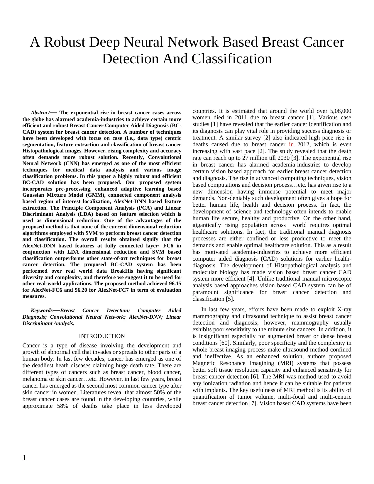
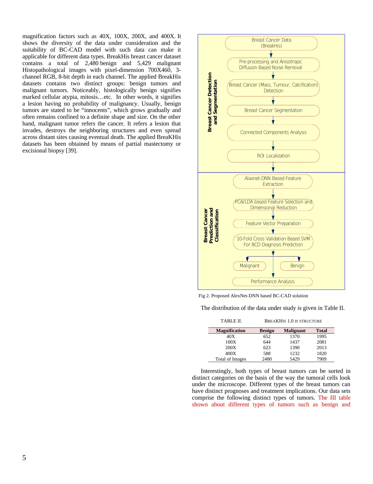
PROPOSED SYSTEM DESIGN
In this section, the detailed discussion of the proposed
research work and implementation schematic is presented. Before
discussing the proposed BC-CAD solution, introducing key
terminologies is must. Table I presents the nomenclature of
different abbreviated terms.
BC Data
Pre/Post
Processing
Breast Cancer
region
Segmentation
SIFT
Caffee-AlexNet
DNN
FC6
FC7
AlexNet-FC6-LDA
AlexNet-FC6-PCA
AlexNet-FC7-LDA
AlexNet-FC7-PCA
SIFT-FV--PCA
SIFT-FV-LDA
Fig. 1. Propsoed BC-CAD System
A detailed discussion of the proposed research model is
given, as presented in Fig. 2, as follows:
.a Breast Cancer Histopathological Image Data
Collection
.b Pre-processing
.c ROI Detection and Localization
.d Feature Extraction
.e Dimensional reduction of Feature Selection,
and
.f BC-CAD Two-Class Classification.
Which will be discussed in details:
1. Breast Cancer Histopathological Image Data
collection
In this research or study, we have used standard datasets
named Breast Cancer Histopathological Image Classification
(BreakHis) [39] that contains a total of 9,109 Histopathological
(say, microscopic) images of breast cancer tissue. The historical
perspective of the BreaKHis states that the microscopic images
are retrieved from 82 patients. BreakHis dataset has been built in
association with the P&D Laboratory - Pathological Anatomy
and Cytopathology, Parana, Brazil (http://www.
prevencaoediagnose.com.br). Unlike generic singe size or single
feature datasets, BreaKHis data are obtained with varied
4
Analysis (CCA) model over segmented region. It eliminates
those image components that percept to be connected with the
target ROI; however, it is insignificant toward BC-CAD
classification. Once obtaining the CCA processed segmented
ROI, we have processed it for feature extraction followed by
feature selection and classification to achieve anticipated BC-
CAD solution. Unlike existing feature extraction models, such as
Gabor filter [11], Wavelet Transform [8][9][10], MIL [26-28] or
even classical CNNs [28-33][35], in our research a CNN model
is derived. In the proposed research work, an enhanced DNN
model named AlexNet DNN has been applied to extract
significant features from the Histopathological image datasets.
Realizing the fact that high dimensional features often provide
more significant information to make precise classification or
analysis; in our research work, we have developed AlexNet-DNN
to extract high dimensional features with 4096 Kernels or
dimensions. The proposed AlexNet-DNN model comprises five
convolutional layers in succession with three Fully Connected
(FC) layers (FC6, FC7 and FC8). The feature extracted at the
higher layer provides more significant information that helps in
achieving accurate BC-CAD performance. Considering major
classical approaches, where the probability of over-fitting and
accuracy is often ignored, we have applied a supplementary
model called Caffe-enet that assists AlexNet-DNN to overcome
existing limitations and enables its (AlexNet-DNN) execution
over general purpose computers without any need of
sophisticated Graphical Processing Units (GPUs). The use of
Caffe-enet in conjunction with AlexNet-DNN plays a vital role in
assuring efficient feature extraction even under large scale
unannotated data. It makes a suitable environment for major
breast cancer detection applications. Specifically, owing to high
unannotated breast cancer data, performing DNN learning and
further classification is a mammoth task. Hence to alleviate such
issues, in our proposed model AlexNet applied multilayered
DNN architecture, where at each layer we retrieve the features
for further mapping and classification. However, to achieve more
efficient results, we have considered 4096-dimensional features
extracted at the Fully Connected (FC) layers, FC-6 and FC-7 of
the AlexNet-DNN. The detailed discussion of the developed
DNN model and its implementation for BC-CAD is given in the
next section. In addition, to the proposed AlexNet-DNN based
BC-CAD solution, in this research we have developed a parallel
feature extraction model, called Scale Invariant Fourier
Transform (SIFT). We have applied Fully-Connected (FC)
layers: FC6 and FC7 features, which are 4096-dimensional
features and hence amounts a huge data to process. To enhance
computational efficiency, in this research work, we have applied
PCA and LDA techniques to perform dimension reduction or
feature selection. Once processing for the dimensional reduction,
the selected features are projected to polynomial kernel based
classifier that performs two class classifications: Malignant and
Benign. To further strengthen the efficiency of our proposed
work, 10-fold cross validation approach is applied that ensures
optimal classification accuracy for BC-CAD solution.
The overall proposed research methodology and
implementation schematic is given in the next section.
PROPOSED SYSTEM DESIGN
In this section, the detailed discussion of the proposed
research work and implementation schematic is presented. Before
discussing the proposed BC-CAD solution, introducing key
terminologies is must. Table I presents the nomenclature of
different abbreviated terms.
BC Data
Pre/Post
Processing
Breast Cancer
region
Segmentation
SIFT
Caffee-AlexNet
DNN
FC6
FC7
AlexNet-FC6-LDA
AlexNet-FC6-PCA
AlexNet-FC7-LDA
AlexNet-FC7-PCA
SIFT-FV--PCA
SIFT-FV-LDA
Fig. 1. Propsoed BC-CAD System
A detailed discussion of the proposed research model is
given, as presented in Fig. 2, as follows:
.a Breast Cancer Histopathological Image Data
Collection
.b Pre-processing
.c ROI Detection and Localization
.d Feature Extraction
.e Dimensional reduction of Feature Selection,
and
.f BC-CAD Two-Class Classification.
Which will be discussed in details:
1. Breast Cancer Histopathological Image Data
collection
In this research or study, we have used standard datasets
named Breast Cancer Histopathological Image Classification
(BreakHis) [39] that contains a total of 9,109 Histopathological
(say, microscopic) images of breast cancer tissue. The historical
perspective of the BreaKHis states that the microscopic images
are retrieved from 82 patients. BreakHis dataset has been built in
association with the P&D Laboratory - Pathological Anatomy
and Cytopathology, Parana, Brazil (http://www.
prevencaoediagnose.com.br). Unlike generic singe size or single
feature datasets, BreaKHis data are obtained with varied
4
Paraphrase This Document
Need a fresh take? Get an instant paraphrase of this document with our AI Paraphraser
magnification factors such as 40X, 100X, 200X, and 400X. It
shows the diversity of the data under consideration and the
suitability of BC-CAD model with such data can make it
applicable for different data types. BreakHis breast cancer dataset
contains a total of 2,480 benign and 5,429 malignant
Histopathological images with pixel-dimension 700X460, 3-
channel RGB, 8-bit depth in each channel. The applied BreakHis
datasets contains two distinct groups: benign tumors and
malignant tumors. Noticeably, histologically benign signifies
marked cellular atypia, mitosis…etc. In other words, it signifies
a lesion having no probability of malignancy. Usually, benign
tumors are stated to be “innocents”, which grows gradually and
often remains confined to a definite shape and size. On the other
hand, malignant tumor refers the cancer. It refers a lesion that
invades, destroys the neighboring structures and even spread
across distant sites causing eventual death. The applied BreaKHis
datasets has been obtained by means of partial mastectomy or
excisional biopsy [39].
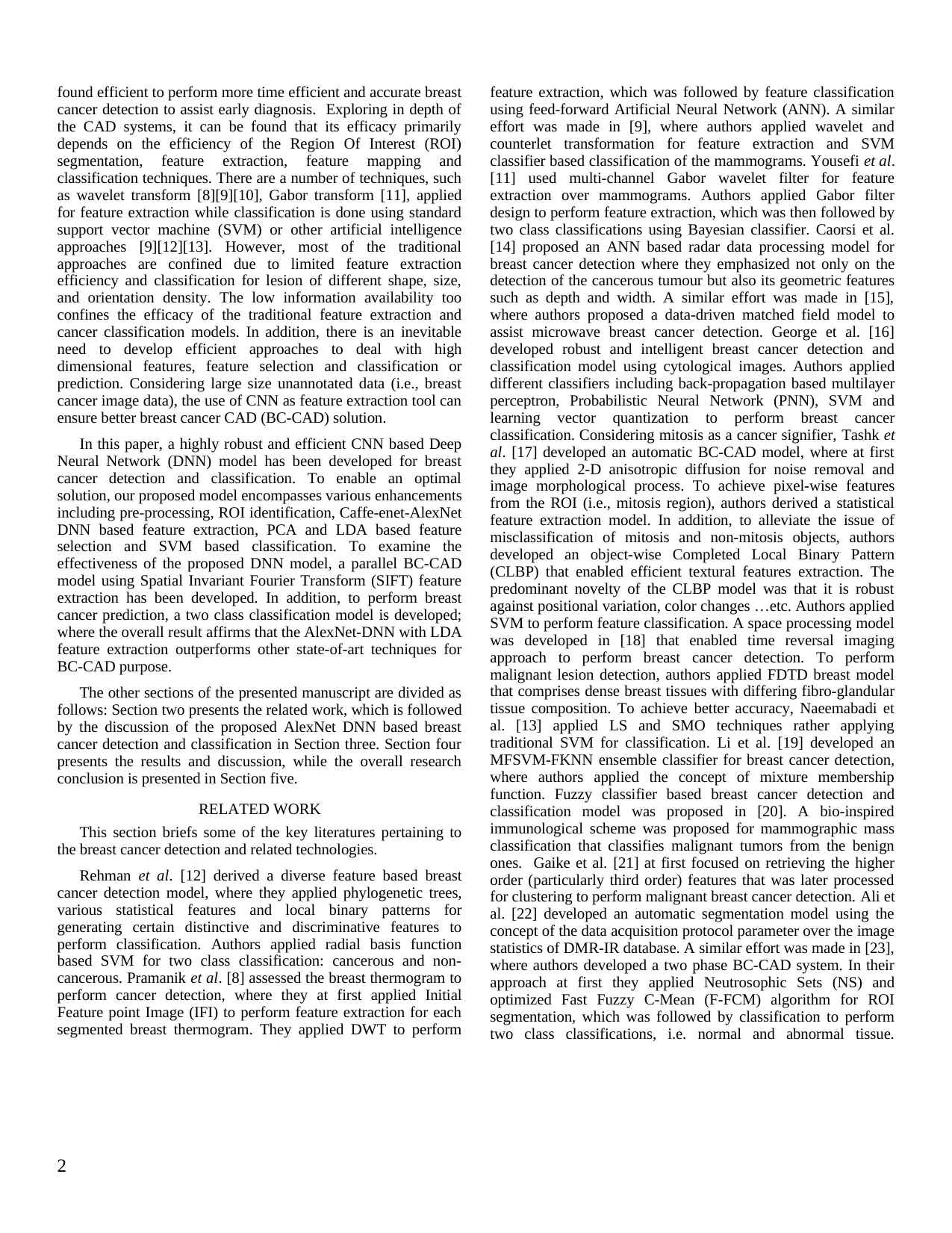
Breast Cancer Segmentation
Connected Components Analysis
ROI Localization
Alaxnet-DNN Based Feature
Extraction
Pre-processing and Anisotropic
Diffusion Based Noise Removal
Breast Cancer (Mass, Tumour, Calcification)
Detection
PCA/LDA based Feature Selection and
Dimensional Reduction
Feature Vector Preparation
10-Fold Cross Validation Based SVM
For BCD Diagnosis Prediction
Malignant
Breast Cancer Detection
and Segmentation
Breast Cancer
Prediction and
Classification
Breast Cancer Data
(BreakHis)
Benign
Performance Analysis
Fig 2. Proposed AlexNet-DNN based BC-CAD solution
The distribution of the data under study is given in Table II.
TABLE II. BREAKHIS 1.0 IS STRUCTURE
Magnification Benign Malignant Total
40X 652 1370 1995
100X 644 1437 2081
200X 623 1390 2013
400X 588 1232 1820
Total of Images 2480 5429 7909
Interestingly, both types of breast tumors can be sorted in
distinct categories on the basis of the way the tumoral cells look
under the microscope. Different types of the breast tumors can
have distinct prognoses and treatment implications. Our data sets
comprise the following distinct types of tumors. The III table
shown about different types of tumors such as benign and
5
shows the diversity of the data under consideration and the
suitability of BC-CAD model with such data can make it
applicable for different data types. BreakHis breast cancer dataset
contains a total of 2,480 benign and 5,429 malignant
Histopathological images with pixel-dimension 700X460, 3-
channel RGB, 8-bit depth in each channel. The applied BreakHis
datasets contains two distinct groups: benign tumors and
malignant tumors. Noticeably, histologically benign signifies
marked cellular atypia, mitosis…etc. In other words, it signifies
a lesion having no probability of malignancy. Usually, benign
tumors are stated to be “innocents”, which grows gradually and
often remains confined to a definite shape and size. On the other
hand, malignant tumor refers the cancer. It refers a lesion that
invades, destroys the neighboring structures and even spread
across distant sites causing eventual death. The applied BreaKHis
datasets has been obtained by means of partial mastectomy or
excisional biopsy [39].
Breast Cancer Segmentation
Connected Components Analysis
ROI Localization
Alaxnet-DNN Based Feature
Extraction
Pre-processing and Anisotropic
Diffusion Based Noise Removal
Breast Cancer (Mass, Tumour, Calcification)
Detection
PCA/LDA based Feature Selection and
Dimensional Reduction
Feature Vector Preparation
10-Fold Cross Validation Based SVM
For BCD Diagnosis Prediction
Malignant
Breast Cancer Detection
and Segmentation
Breast Cancer
Prediction and
Classification
Breast Cancer Data
(BreakHis)
Benign
Performance Analysis
Fig 2. Proposed AlexNet-DNN based BC-CAD solution
The distribution of the data under study is given in Table II.
TABLE II. BREAKHIS 1.0 IS STRUCTURE
Magnification Benign Malignant Total
40X 652 1370 1995
100X 644 1437 2081
200X 623 1390 2013
400X 588 1232 1820
Total of Images 2480 5429 7909
Interestingly, both types of breast tumors can be sorted in
distinct categories on the basis of the way the tumoral cells look
under the microscope. Different types of the breast tumors can
have distinct prognoses and treatment implications. Our data sets
comprise the following distinct types of tumors. The III table
shown about different types of tumors such as benign and
5
malignant along with their examples including overall breast
tumors which may or may not be cancer.
TABLE III. TUMOR CONTAINTS (DISTRIBUTION) IN BREAKHIS 1.0 IS
Benign Tumors: adenosis Malignant Tumors
Fibroadenoma carcinoma
phyllodes tumor lobular carcinoma
tubular adenona mucinous carcinoma
adenosis papillary carcinoma
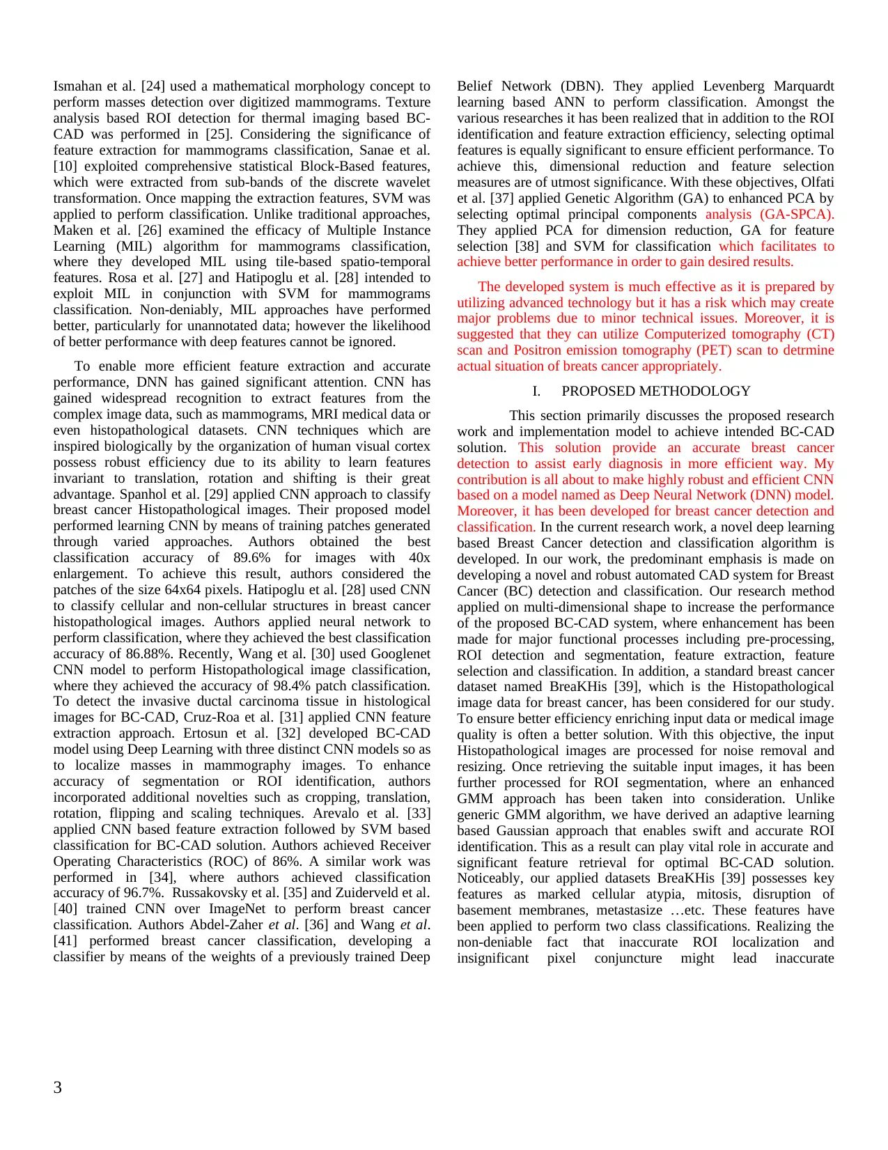
In the dataset, individual Histopathological image filename
stores significant image information containing biopsy method,
type of tumor, patient information (Patient ID), and
magnification factor. For illustration, for an image SOB_B_TA-
14-4659-40-001.png, as illustrated in Fig. 3, it signifies a benign
tumor of tubular adenoma type, obtained at the magnification
factor 40. The biopsy method used was SOB.
(a) (b)
(c) (d)
Fig 3. Snippet of the breast malignent cancer at different magnification
factors: (a) 40X, (b) 100X, (c) 200X, and (d) 400X [39].
.g Pre-processing
Non-deniably, the quality of data influences the final
classification accuracy and efficiency. To enable suitable data for
further processing, we have performed pre-processing. In our
research model, the pre-processing model is exceptionally
significant to ensure precise nuclei and allied vicinity
identification and characterization. In breast cancer detection, the
features, such as the shape and size of nuclei, glandular shape …
etc. is expected to be precisely identifiable. Performing pre-
processing assures that suppression of the original image
qualities required to estimate or identify texture representation
and other significant features, precisely [40]. Realizing
interchangeable nature of the pre-processing approaches, we have
performed pre-processing before performing candidate region
detection and feature extraction [40-41]. In our paper, we have
followed the pre-processing instructions provided in [35][41]
Once performing pre-processing, the concept region
identification or segmentation has been processed. In addition to
the above mentioned, in pre-processing, we have resized input
breast cancer Histopathological images to 224X224. A brief of
the proposed segmentation model or ROI identification approach
will be explained as follows.
.h Concept Region Identification
In this section, the process of breast cancer region detection
and segmentation is discussed. Unlike traditional approaches of
segmentation such as Otsu’s method [42], our model applies an
enhanced GMM technique. Here, we segment cancer tumor
region from the background so as to distinguish it from other
regions over Histopathological images. GMM has exhibited
better performance over other state-of-the-art techniques for
background subtraction and ROI identification because of b a
pixel-wise segmentation approach. In addition, GMM robustness
causes to be used for segmenting ROI from the background
region that can be further processed for feature extraction over
the detected ROI. A brief of the proposed GMM model for ROI
detection including several steps is given as follows:
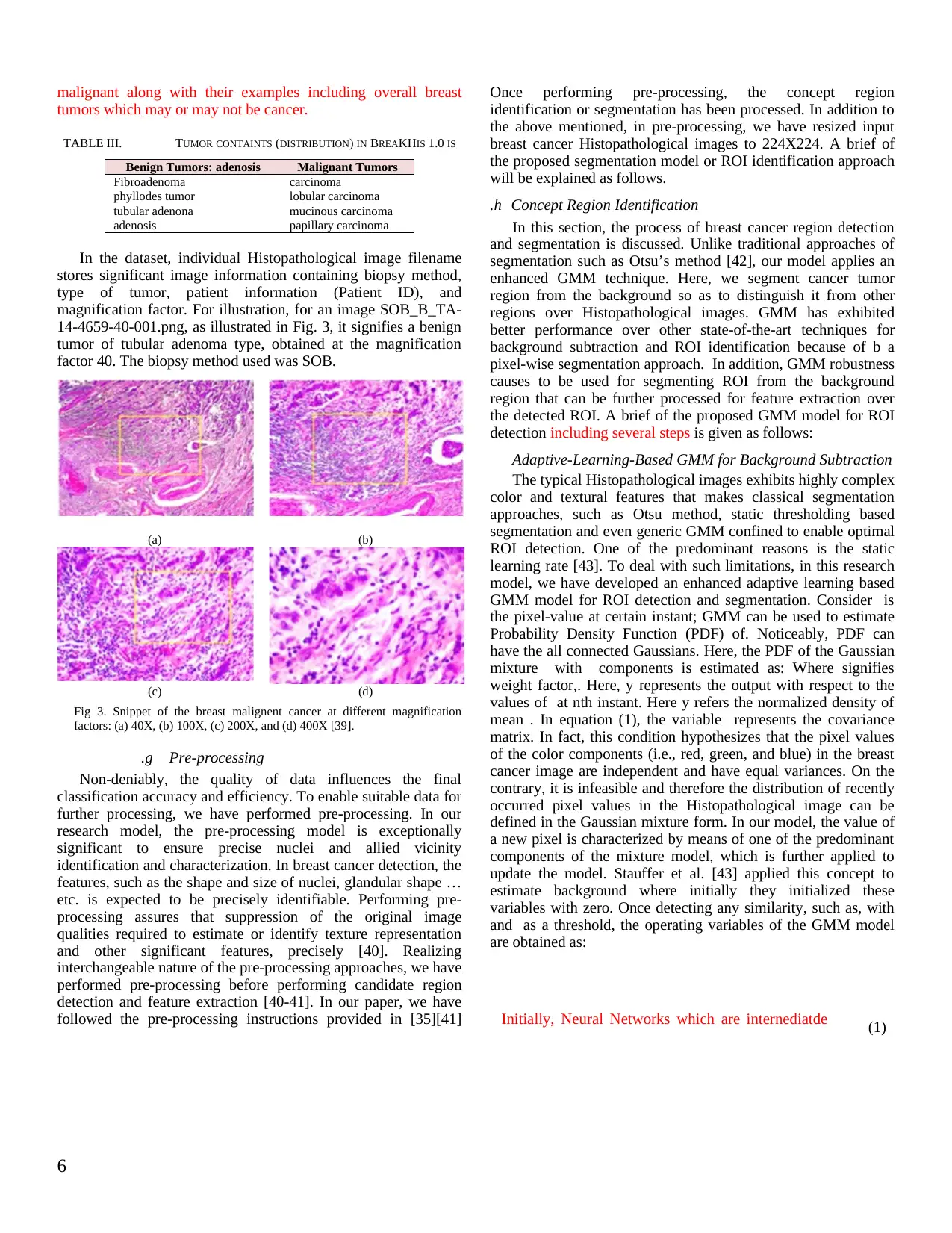
Adaptive-Learning-Based GMM for Background Subtraction
The typical Histopathological images exhibits highly complex
color and textural features that makes classical segmentation
approaches, such as Otsu method, static thresholding based
segmentation and even generic GMM confined to enable optimal
ROI detection. One of the predominant reasons is the static
learning rate [43]. To deal with such limitations, in this research
model, we have developed an enhanced adaptive learning based
GMM model for ROI detection and segmentation. Consider is
the pixel-value at certain instant; GMM can be used to estimate
Probability Density Function (PDF) of. Noticeably, PDF can
have the all connected Gaussians. Here, the PDF of the Gaussian
mixture with components is estimated as: Where signifies
weight factor,. Here, y represents the output with respect to the
values of at nth instant. Here y refers the normalized density of
mean . In equation (1), the variable represents the covariance
matrix. In fact, this condition hypothesizes that the pixel values
of the color components (i.e., red, green, and blue) in the breast
cancer image are independent and have equal variances. On the
contrary, it is infeasible and therefore the distribution of recently
occurred pixel values in the Histopathological image can be
defined in the Gaussian mixture form. In our model, the value of
a new pixel is characterized by means of one of the predominant
components of the mixture model, which is further applied to
update the model. Stauffer et al. [43] applied this concept to
estimate background where initially they initialized these
variables with zero. Once detecting any similarity, such as, with
and as a threshold, the operating variables of the GMM model
are obtained as:
Initially, Neural Networks which are internediatde (1)
6
tumors which may or may not be cancer.
TABLE III. TUMOR CONTAINTS (DISTRIBUTION) IN BREAKHIS 1.0 IS
Benign Tumors: adenosis Malignant Tumors
Fibroadenoma carcinoma
phyllodes tumor lobular carcinoma
tubular adenona mucinous carcinoma
adenosis papillary carcinoma
In the dataset, individual Histopathological image filename
stores significant image information containing biopsy method,
type of tumor, patient information (Patient ID), and
magnification factor. For illustration, for an image SOB_B_TA-
14-4659-40-001.png, as illustrated in Fig. 3, it signifies a benign
tumor of tubular adenoma type, obtained at the magnification
factor 40. The biopsy method used was SOB.
(a) (b)
(c) (d)
Fig 3. Snippet of the breast malignent cancer at different magnification
factors: (a) 40X, (b) 100X, (c) 200X, and (d) 400X [39].
.g Pre-processing
Non-deniably, the quality of data influences the final
classification accuracy and efficiency. To enable suitable data for
further processing, we have performed pre-processing. In our
research model, the pre-processing model is exceptionally
significant to ensure precise nuclei and allied vicinity
identification and characterization. In breast cancer detection, the
features, such as the shape and size of nuclei, glandular shape …
etc. is expected to be precisely identifiable. Performing pre-
processing assures that suppression of the original image
qualities required to estimate or identify texture representation
and other significant features, precisely [40]. Realizing
interchangeable nature of the pre-processing approaches, we have
performed pre-processing before performing candidate region
detection and feature extraction [40-41]. In our paper, we have
followed the pre-processing instructions provided in [35][41]
Once performing pre-processing, the concept region
identification or segmentation has been processed. In addition to
the above mentioned, in pre-processing, we have resized input
breast cancer Histopathological images to 224X224. A brief of
the proposed segmentation model or ROI identification approach
will be explained as follows.
.h Concept Region Identification
In this section, the process of breast cancer region detection
and segmentation is discussed. Unlike traditional approaches of
segmentation such as Otsu’s method [42], our model applies an
enhanced GMM technique. Here, we segment cancer tumor
region from the background so as to distinguish it from other
regions over Histopathological images. GMM has exhibited
better performance over other state-of-the-art techniques for
background subtraction and ROI identification because of b a
pixel-wise segmentation approach. In addition, GMM robustness
causes to be used for segmenting ROI from the background
region that can be further processed for feature extraction over
the detected ROI. A brief of the proposed GMM model for ROI
detection including several steps is given as follows:
Adaptive-Learning-Based GMM for Background Subtraction
The typical Histopathological images exhibits highly complex
color and textural features that makes classical segmentation
approaches, such as Otsu method, static thresholding based
segmentation and even generic GMM confined to enable optimal
ROI detection. One of the predominant reasons is the static
learning rate [43]. To deal with such limitations, in this research
model, we have developed an enhanced adaptive learning based
GMM model for ROI detection and segmentation. Consider is
the pixel-value at certain instant; GMM can be used to estimate
Probability Density Function (PDF) of. Noticeably, PDF can
have the all connected Gaussians. Here, the PDF of the Gaussian
mixture with components is estimated as: Where signifies
weight factor,. Here, y represents the output with respect to the
values of at nth instant. Here y refers the normalized density of
mean . In equation (1), the variable represents the covariance
matrix. In fact, this condition hypothesizes that the pixel values
of the color components (i.e., red, green, and blue) in the breast
cancer image are independent and have equal variances. On the
contrary, it is infeasible and therefore the distribution of recently
occurred pixel values in the Histopathological image can be
defined in the Gaussian mixture form. In our model, the value of
a new pixel is characterized by means of one of the predominant
components of the mixture model, which is further applied to
update the model. Stauffer et al. [43] applied this concept to
estimate background where initially they initialized these
variables with zero. Once detecting any similarity, such as, with
and as a threshold, the operating variables of the GMM model
are obtained as:
Initially, Neural Networks which are internediatde (1)
6
⊘ This is a preview!⊘
Do you want full access?
Subscribe today to unlock all pages.
Trusted by 1+ million students worldwide
varicables that are not observable known as hidden
units.
Support Vector Machine can be formulated in
context of binary classification to find decision
boudary for maximising margin between two
data classes.
(2)
Partial Least Square include principal
component analysis which has same concept as
PLS.
(3)
LDA technique which are combined in one
objective function.
(4)
where
If no element matches together then the element with the
minimal is re-initialized, and the variables (2-4) are updated as:
SVM and PLS (5)
In above expression (5), refers to the learning rate. Here, is
obtained as:
Support ventor machines is formulatde in order
to for dula optimization problem whihc ensures
unique global solution.
(6)
In our proposed GMM model, we normalize in a way that it
converges toward unit value (i.e., 1). To obtain background
region, authors [43] sorted Gaussians in decreasing order by
using threshold in conjunction with the sums of the weights. As
a result, this achieves a set, where is obtained as:
It summarise turning parameter of all models
which are inlcuded in proposed theory.
(7)
With decrease in variances, usually the Gaussian distribution
gains increases. This as a result makes it more evident to perform
background subtraction. Once estimating GMM parameters,
sorting is performed from the matched mixture distribution
toward most probable background. This is feasible only because
only the relative value of the matched model changes. In our
applied model, ordering is performed in a way that the most
feasible background distributions remains on the top, while the
low probability distributions move towards bottom. In such
manner, the initial value of (7) is considered as the background.
In (7), parameter signifies the result of the least section of the
data responsible for the background. With the minimal possible
value of , the background model is stated to be unimodal, where
using only the most probable distribution enhances
computational efficiency. On the contrary, the high signifies
multimodal distribution, is typically caused due to the iterative
background motion. In this paper, the breast cancer
Histopathological images with static features are taken as input
and hence the use of unimodal approach with low ensures
computationally efficient process. In our approach, the Gaussian
with the maximum and minimal refer to the background region.
Thus, applying aforementioned approach the background region
can be identified that consequently helps in segmenting the ROI.
In major cases, classical GMM updates with certain fixed
learning rate [43] are used. While, considering breast cancer
detection requirement, the static learning rate approach seems
questionable, particularly when there can be excessive texture
variation over the breast cancer Histopathological images. To
deal with such limitation, authors [44] developed an adaptive
learning rate based GMM for ROI segmentation over varying
surface or texture conditions. Non-deniably, there can be certain
texture or the surface conditions, where the pixel might neither be
the breast can tumor (i.e., ROI) nor the background; however, it
could be classified as either type Fibroadenoma and phyllodes
tumor. This as a result may influence accuracy of the proposed
BC-CAD solution. Increasing learning rate might cause high-
rate pixel feature changes [44] that consequently may make
breast cancer tumor or ROI detection susceptible. Though, the
adaptive learning based GMM model [43] can perform ROI
identification under varying background conditions. However,
the very minute and fine-connected texture or surface features
still make existing approach limited. Precisely for breast cancer
Histopathological images, textural or structural features, such as
cellular atypia, mitosis, disruption of basement membranes,
metastasize …etc. are highly intricate and minute feature that
requires more efficient ROI identification model. An enhanced
Gaussian mixture learning based GMM model was proposed in
[44]. However, authors could not address the saturation scenario
particularly during convergence. As an enhanced solution, in this
paper an adaptive learning based GMM model is developed that
detects precise ROI. In our proposed approach, initially, we have
decoupled learning rate from other components such as and. We
have applied a new learning rate variable called adaptive learning
rate that updates mean component using a probability
parameter. Here, states whether a pixel is a part of the nth
Gaussian distribution or not. In our model, the adaptive learning
rate parameter is estimated as follows:
Thresold (8)
Where, states the number of distributions.
Can provide fast and accurate Gaussian update that may
enhance the system to adapt with swift textural variations. As a
result, this can enable optimal ROI detection for the targeted BC-
CAD solution.
Now, substituting as in (3), it is observed that variance
updating might avoid Gaussian saturation. On the other hand, the
fast learning rate might cause degeneracy [44]. To deal with this
issue, we have developed a semi-parametric model to estimate
7
Paraphrase This Document
Need a fresh take? Get an instant paraphrase of this document with our AI Paraphraser
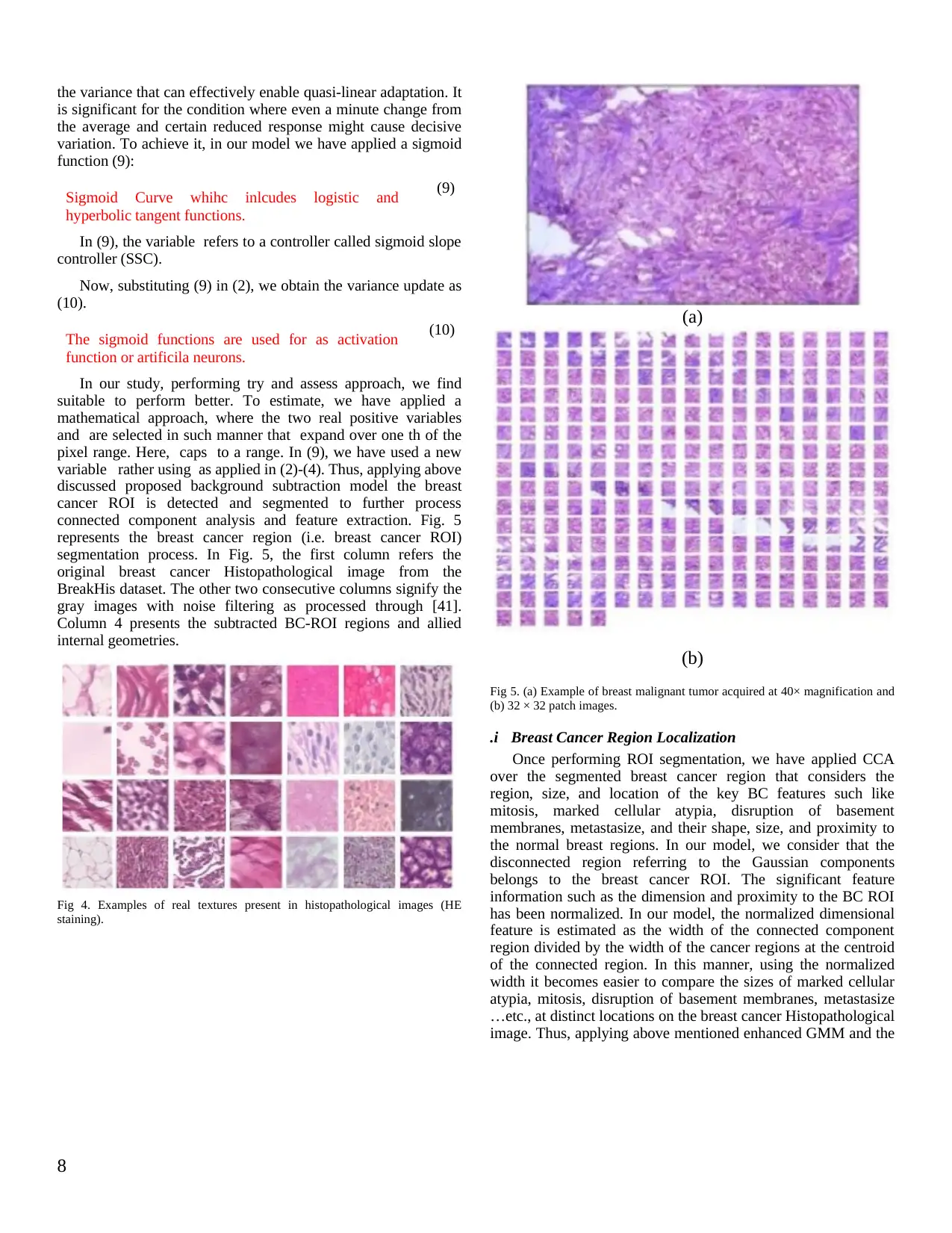
the variance that can effectively enable quasi-linear adaptation. It
is significant for the condition where even a minute change from
the average and certain reduced response might cause decisive
variation. To achieve it, in our model we have applied a sigmoid
function (9):
Sigmoid Curve whihc inlcudes logistic and
hyperbolic tangent functions.
(9)
In (9), the variable refers to a controller called sigmoid slope
controller (SSC).
Now, substituting (9) in (2), we obtain the variance update as
(10).
The sigmoid functions are used for as activation
function or artificila neurons.
(10)
In our study, performing try and assess approach, we find
suitable to perform better. To estimate, we have applied a
mathematical approach, where the two real positive variables
and are selected in such manner that expand over one th of the
pixel range. Here, caps to a range. In (9), we have used a new
variable rather using as applied in (2)-(4). Thus, applying above
discussed proposed background subtraction model the breast
cancer ROI is detected and segmented to further process
connected component analysis and feature extraction. Fig. 5
represents the breast cancer region (i.e. breast cancer ROI)
segmentation process. In Fig. 5, the first column refers the
original breast cancer Histopathological image from the
BreakHis dataset. The other two consecutive columns signify the
gray images with noise filtering as processed through [41].
Column 4 presents the subtracted BC-ROI regions and allied
internal geometries.
Fig 4. Examples of real textures present in histopathological images (HE
staining).
(a)
(b)
Fig 5. (a) Example of breast malignant tumor acquired at 40× magnification and
(b) 32 × 32 patch images.
.i Breast Cancer Region Localization
Once performing ROI segmentation, we have applied CCA
over the segmented breast cancer region that considers the
region, size, and location of the key BC features such like
mitosis, marked cellular atypia, disruption of basement
membranes, metastasize, and their shape, size, and proximity to
the normal breast regions. In our model, we consider that the
disconnected region referring to the Gaussian components
belongs to the breast cancer ROI. The significant feature
information such as the dimension and proximity to the BC ROI
has been normalized. In our model, the normalized dimensional
feature is estimated as the width of the connected component
region divided by the width of the cancer regions at the centroid
of the connected region. In this manner, using the normalized
width it becomes easier to compare the sizes of marked cellular
atypia, mitosis, disruption of basement membranes, metastasize
…etc., at distinct locations on the breast cancer Histopathological
image. Thus, applying above mentioned enhanced GMM and the
8
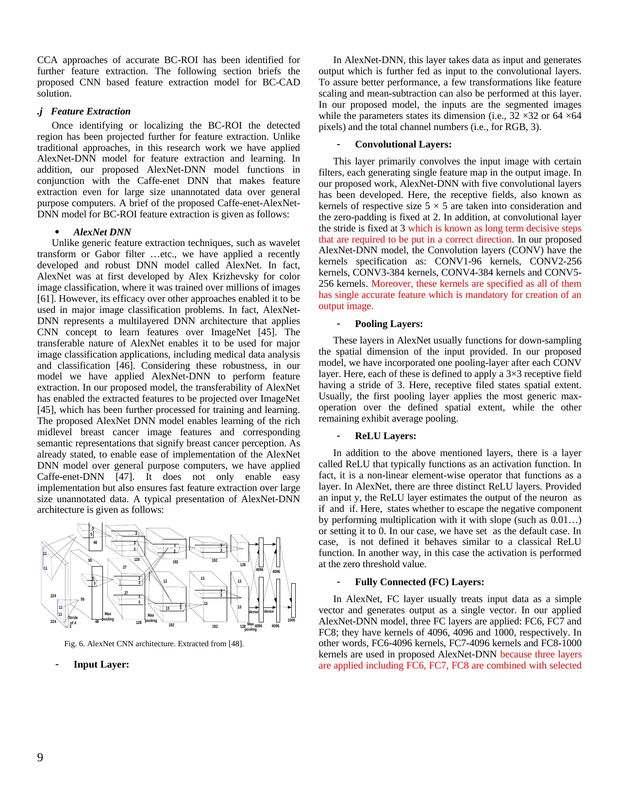
CCA approaches of accurate BC-ROI has been identified for
further feature extraction. The following section briefs the
proposed CNN based feature extraction model for BC-CAD
solution.
.j Feature Extraction
Once identifying or localizing the BC-ROI the detected
region has been projected further for feature extraction. Unlike
traditional approaches, in this research work we have applied
AlexNet-DNN model for feature extraction and learning. In
addition, our proposed AlexNet-DNN model functions in
conjunction with the Caffe-enet DNN that makes feature
extraction even for large size unannotated data over general
purpose computers. A brief of the proposed Caffe-enet-AlexNet-
DNN model for BC-ROI feature extraction is given as follows:
AlexNet DNN
Unlike generic feature extraction techniques, such as wavelet
transform or Gabor filter …etc., we have applied a recently
developed and robust DNN model called AlexNet. In fact,
AlexNet was at first developed by Alex Krizhevsky for color
image classification, where it was trained over millions of images
[61]. However, its efficacy over other approaches enabled it to be
used in major image classification problems. In fact, AlexNet-
DNN represents a multilayered DNN architecture that applies
CNN concept to learn features over ImageNet [45]. The
transferable nature of AlexNet enables it to be used for major
image classification applications, including medical data analysis
and classification [46]. Considering these robustness, in our
model we have applied AlexNet-DNN to perform feature
extraction. In our proposed model, the transferability of AlexNet
has enabled the extracted features to be projected over ImageNet
[45], which has been further processed for training and learning.
The proposed AlexNet DNN model enables learning of the rich
midlevel breast cancer image features and corresponding
semantic representations that signify breast cancer perception. As
already stated, to enable ease of implementation of the AlexNet
DNN model over general purpose computers, we have applied
Caffe-enet-DNN [47]. It does not only enable easy
implementation but also ensures fast feature extraction over large
size unannotated data. A typical presentation of AlexNet-DNN
architecture is given as follows:
Max
pooling
224
3
5
27
128 192
11
Stride
of 4
5
13
55
48
dense
4096
1000
11
11
11 13
13
13
13
13
3
3
3
3
3
3
3
3
3
3
3
3
3
3
3
27
55
48
Max
pooling
224
128
192
192192 128
128
dense
4096
4096
4096
5
5
Max
pooling
Fig. 6. AlexNet CNN architecture. Extracted from [48].
Input Layer:
In AlexNet-DNN, this layer takes data as input and generates
output which is further fed as input to the convolutional layers.
To assure better performance, a few transformations like feature
scaling and mean-subtraction can also be performed at this layer.
In our proposed model, the inputs are the segmented images
while the parameters states its dimension (i.e., 32 ×32 or 64 ×64
pixels) and the total channel numbers (i.e., for RGB, 3).
Convolutional Layers:
This layer primarily convolves the input image with certain
filters, each generating single feature map in the output image. In
our proposed work, AlexNet-DNN with five convolutional layers
has been developed. Here, the receptive fields, also known as
kernels of respective size 5 × 5 are taken into consideration and
the zero-padding is fixed at 2. In addition, at convolutional layer
the stride is fixed at 3 which is known as long term decisive steps
that are required to be put in a correct direction. In our proposed
AlexNet-DNN model, the Convolution layers (CONV) have the
kernels specification as: CONV1-96 kernels, CONV2-256
kernels, CONV3-384 kernels, CONV4-384 kernels and CONV5-
256 kernels. Moreover, these kernels are specified as all of them
has single accurate feature which is mandatory for creation of an
output image.
Pooling Layers:
These layers in AlexNet usually functions for down-sampling
the spatial dimension of the input provided. In our proposed
model, we have incorporated one pooling-layer after each CONV
layer. Here, each of these is defined to apply a 3×3 receptive field
having a stride of 3. Here, receptive filed states spatial extent.
Usually, the first pooling layer applies the most generic max-
operation over the defined spatial extent, while the other
remaining exhibit average pooling.
ReLU Layers:
In addition to the above mentioned layers, there is a layer
called ReLU that typically functions as an activation function. In
fact, it is a non-linear element-wise operator that functions as a
layer. In AlexNet, there are three distinct ReLU layers. Provided
an input y, the ReLU layer estimates the output of the neuron as
if and if. Here, states whether to escape the negative component
by performing multiplication with it with slope (such as 0.01…)
or setting it to 0. In our case, we have set as the default case. In
case, is not defined it behaves similar to a classical ReLU
function. In another way, in this case the activation is performed
at the zero threshold value.
Fully Connected (FC) Layers:
In AlexNet, FC layer usually treats input data as a simple
vector and generates output as a single vector. In our applied
AlexNet-DNN model, three FC layers are applied: FC6, FC7 and
FC8; they have kernels of 4096, 4096 and 1000, respectively. In
other words, FC6-4096 kernels, FC7-4096 kernels and FC8-1000
kernels are used in proposed AlexNet-DNN because three layers
are applied including FC6, FC7, FC8 are combined with selected
9
⊘ This is a preview!⊘
Do you want full access?
Subscribe today to unlock all pages.
Trusted by 1+ million students worldwide
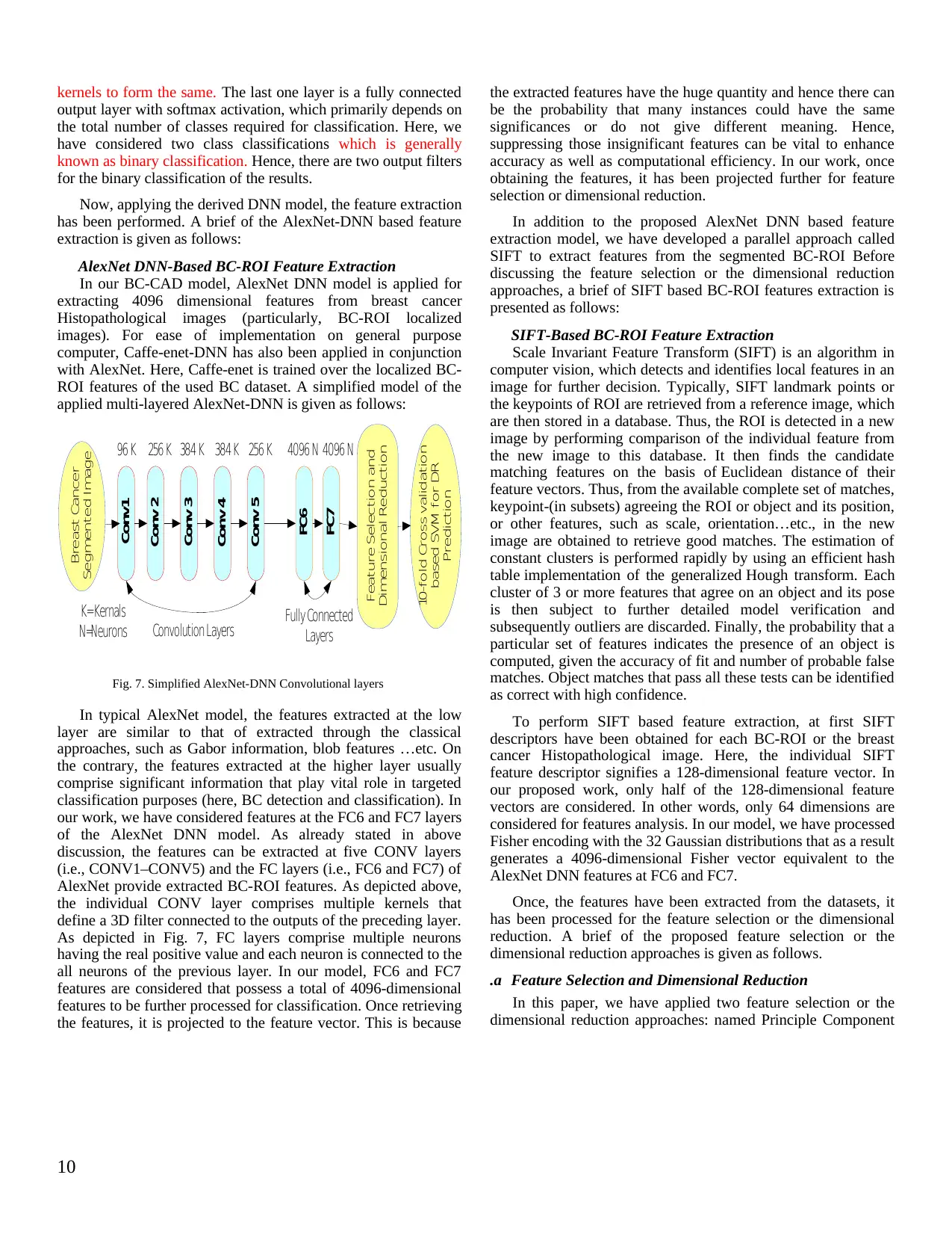
kernels to form the same. The last one layer is a fully connected
output layer with softmax activation, which primarily depends on
the total number of classes required for classification. Here, we
have considered two class classifications which is generally
known as binary classification. Hence, there are two output filters
for the binary classification of the results.
Now, applying the derived DNN model, the feature extraction
has been performed. A brief of the AlexNet-DNN based feature
extraction is given as follows:
AlexNet DNN-Based BC-ROI Feature Extraction
In our BC-CAD model, AlexNet DNN model is applied for
extracting 4096 dimensional features from breast cancer
Histopathological images (particularly, BC-ROI localized
images). For ease of implementation on general purpose
computer, Caffe-enet-DNN has also been applied in conjunction
with AlexNet. Here, Caffe-enet is trained over the localized BC-
ROI features of the used BC dataset. A simplified model of the
applied multi-layered AlexNet-DNN is given as follows:
Conv1
Conv 2
Conv 3
Conv 4
FC6
FC7
Feature Selection and
Dimensional Reduction
Breast Cancer
Segmented Image
10-fold Cross validation
based SVM for DR
Prediction
Conv 5
Convolution Layers Fully Connected
Layers
96 K 256 K 384 K384 K 4096 N256 K 4096 N
K=Kernals
N=Neurons
Fig. 7. Simplified AlexNet-DNN Convolutional layers
In typical AlexNet model, the features extracted at the low
layer are similar to that of extracted through the classical
approaches, such as Gabor information, blob features …etc. On
the contrary, the features extracted at the higher layer usually
comprise significant information that play vital role in targeted
classification purposes (here, BC detection and classification). In
our work, we have considered features at the FC6 and FC7 layers
of the AlexNet DNN model. As already stated in above
discussion, the features can be extracted at five CONV layers
(i.e., CONV1–CONV5) and the FC layers (i.e., FC6 and FC7) of
AlexNet provide extracted BC-ROI features. As depicted above,
the individual CONV layer comprises multiple kernels that
define a 3D filter connected to the outputs of the preceding layer.
As depicted in Fig. 7, FC layers comprise multiple neurons
having the real positive value and each neuron is connected to the
all neurons of the previous layer. In our model, FC6 and FC7
features are considered that possess a total of 4096-dimensional
features to be further processed for classification. Once retrieving
the features, it is projected to the feature vector. This is because
the extracted features have the huge quantity and hence there can
be the probability that many instances could have the same
significances or do not give different meaning. Hence,
suppressing those insignificant features can be vital to enhance
accuracy as well as computational efficiency. In our work, once
obtaining the features, it has been projected further for feature
selection or dimensional reduction.
In addition to the proposed AlexNet DNN based feature
extraction model, we have developed a parallel approach called
SIFT to extract features from the segmented BC-ROI Before
discussing the feature selection or the dimensional reduction
approaches, a brief of SIFT based BC-ROI features extraction is
presented as follows:
SIFT-Based BC-ROI Feature Extraction
Scale Invariant Feature Transform (SIFT) is an algorithm in
computer vision, which detects and identifies local features in an
image for further decision. Typically, SIFT landmark points or
the keypoints of ROI are retrieved from a reference image, which
are then stored in a database. Thus, the ROI is detected in a new
image by performing comparison of the individual feature from
the new image to this database. It then finds the candidate
matching features on the basis of Euclidean distance of their
feature vectors. Thus, from the available complete set of matches,
keypoint-(in subsets) agreeing the ROI or object and its position,
or other features, such as scale, orientation…etc., in the new
image are obtained to retrieve good matches. The estimation of
constant clusters is performed rapidly by using an efficient hash
table implementation of the generalized Hough transform. Each
cluster of 3 or more features that agree on an object and its pose
is then subject to further detailed model verification and
subsequently outliers are discarded. Finally, the probability that a
particular set of features indicates the presence of an object is
computed, given the accuracy of fit and number of probable false
matches. Object matches that pass all these tests can be identified
as correct with high confidence.
To perform SIFT based feature extraction, at first SIFT
descriptors have been obtained for each BC-ROI or the breast
cancer Histopathological image. Here, the individual SIFT
feature descriptor signifies a 128-dimensional feature vector. In
our proposed work, only half of the 128-dimensional feature
vectors are considered. In other words, only 64 dimensions are
considered for features analysis. In our model, we have processed
Fisher encoding with the 32 Gaussian distributions that as a result
generates a 4096-dimensional Fisher vector equivalent to the
AlexNet DNN features at FC6 and FC7.
Once, the features have been extracted from the datasets, it
has been processed for the feature selection or the dimensional
reduction. A brief of the proposed feature selection or the
dimensional reduction approaches is given as follows.
.a Feature Selection and Dimensional Reduction
In this paper, we have applied two feature selection or the
dimensional reduction approaches: named Principle Component
10
Paraphrase This Document
Need a fresh take? Get an instant paraphrase of this document with our AI Paraphraser
Analysis (PCA) and Linear Discriminate Analysis (LDA). In
major existing approaches, PCA based feature selection approach
has been applied; however, the efficacy of LDA towards most
discriminating features makes process more efficient.
Considering this fact, we have applied both algorithms distinctly
and respective performance has been assessed. A brief of these
techniques is given, as follows:
Principle Component Analysis (PCA)
It mainly projects feature space of a high dimension to the
lower dimension while ensuring that the obtained feature space
contains most significant features. In function, PCA rotates the p-
dimensional feature space’s axes to a new position called
principle axes in such way that the principle axis 1 has the
highest variance followed by axis 2, and so on. In practice among
extracted breast cancer features there can be the highly correlated
feature attributes of the elements. These highly correlated
features (sometimes called homogeneous feature) are known as
the most expressive features (MEF). In this case, PCA intends to
transfer the feature attributes or the elements to a new feature set
or feature vector having no correlation among the elements.
Thus, applying this approach PCA reduces a significant amount
of feature elements that makes computation more efficient. The
finally retrieved feature vector is then projected for classification.
Linear Discriminant Analysis (LDA)
Unlike PCA that uses MEF to perform feature selection or the
dimensional reduction, LDA employs the most discriminating
features (MDF) to perform dimensional reduction. LDA performs
dimensional reduction automatically. Executing PCA, the LDA
dimensional reduction approach projects extracted features to a
single principle component regardless of its class label. To
perform feature selection and the dimensional reduction, LDA
applied two distinct matrix called intra-class scatter matrix and
inter-class scatter matrix. These matrix values are estimated, as
follows:
(11)
(12)
In (12), refers the total number of classes, signifies average
vector of class , and presents the overall samples in class . We
estimate the as
(13)
In (13), refers the total number of classes.
LDA intends to increase the inter-class scatter and
decrease the intra-class scatter. In our model, it was
achieved by increasing a factor , given in equation
(14).
(14)
For a non-singular, is increased provided the column vectors
of the projection matrix are the eigenvectors of. Here, the
projection matrix with dimension projects the training data to
form Fisher vector (FV). In this way, the final Fischer Vector
obtained is given as input to the classifier. Noticeably, the overall
Fischer Vectors projected for classification be .
Once obtaining the final feature vectors, it is processed for
breast cancer classification. In our proposed work, a two class
classification has been done: BENIGN and MALIGNANT. To
further enhance performance, 10-fold cross validation has been
performed. A brief of the classification method used is presented
as follows:
.a Classification
Considering ease of implementation and better non-linear
pattern learning efficiency, in our model a Radial Basis Function
(RBF) kernel based SVM classifier is applied to perform two-
class BC-CAD classification. In our model, SVM has been
trained over extracted feature vectors that intend to achieve the
maximum margin by obtaining low as given in equation (15).
(15)
Where, ≥ 0 and signify the extent of error resiliency.
In our model, to perform two-class classification, FV is
grouped in the labeled pairs, say where presents the training
vector. The class label of the training vector is given by. In
classification, the hyper plane classifies the maximum possible
points of the same class on the same side of hyperplane. The use
of 10-fold cross-validation ensures higher accuracy and reliability
of the results. The overall performance of the proposed BC-CAD
model is examined in terms of classification accuracy and the
obtained results are compared with other state-of-art techniques.
The results obtained in this research work are discussed in the
next section.
V. RESULTS AND DISCUSSION
To assess the performance of our proposed BC-CAD
solution, we consider BreakHis dataset [39] that contains 9,109
Histopathological (say, microscopic) images of breast cancer
tissue with different tumor types or associated traits, such as
fibroadenoma, phyllodes tumor, tubular adenona, adenosis,
carcinoma, lobular carcinoma, mucinous carcinoma, and
papillary carcinoma. For ease of implementation, we consider
random 2000 Histopathological images for our study. The
original Histopathological images are in the size of 700X460,
which is further reduced using resampling approach (i.e., pixel
area relation) and finally the images with 350X230 dimensions
are obtained. It is then followed by the extraction of patches from
the considered Histopathological images. In our work, we apply a
sliding window approach with 50% overlapping. In addition, the
patches can be obtained randomly too without incorporating any
overlapping control between patches. In our simulation test, the
patches with 32X32 dimensions are obtained. As pre-processing,
11
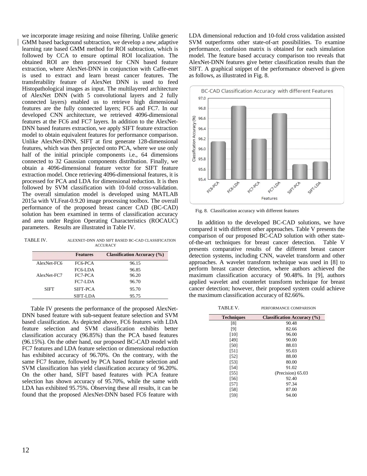
we incorporate image resizing and noise filtering. Unlike generic
GMM based background subtraction, we develop a new adaptive
learning rate based GMM method for ROI subtraction, which is
followed by CCA to ensure optimal ROI localization. The
obtained ROI are then processed for CNN based feature
extraction, where AlexNet-DNN in conjunction with Caffe-enet
is used to extract and learn breast cancer features. The
transferability feature of AlexNet DNN is used to feed
Histopathological images as input. The multilayered architecture
of AlexNet DNN (with 5 convolutional layers and 2 fully
connected layers) enabled us to retrieve high dimensional
features are the fully connected layers; FC6 and FC7. In our
developed CNN architecture, we retrieved 4096-dimensional
features at the FC6 and FC7 layers. In addition to the AlexNet-
DNN based features extraction, we apply SIFT feature extraction
model to obtain equivalent features for performance comparison.
Unlike AlexNet-DNN, SIFT at first generate 128-dimensional
features, which was then projected onto PCA, where we use only
half of the initial principle components i.e., 64 dimensions
connected to 32 Gaussian components distribution. Finally, we
obtain a 4096-dimensional feature vector for SIFT feature
extraction model. Once retrieving 4096-dimensional features, it is
processed for PCA and LDA for dimensional reduction. It is then
followed by SVM classification with 10-fold cross-validation.
The overall simulation model is developed using MATLAB
2015a with VLFeat-0.9.20 image processing toolbox. The overall
performance of the proposed breast cancer CAD (BC-CAD)
solution has been examined in terms of classification accuracy
and area under Region Operating Characteristics (ROCAUC)
parameters. Results are illustrated in Table IV.
TABLE IV. ALEXNET-DNN AND SIFT BASED BC-CAD CLASSIFICATION
ACCURACY
Features Classification Accuracy (%)
AlexNet-FC6 FC6-PCA 96.15
FC6-LDA 96.85
AlexNet-FC7 FC7-PCA 96.20
FC7-LDA 96.70
SIFT SIFT-PCA 95.70
SIFT-LDA 95.75
Table IV presents the performance of the proposed AlexNet-
DNN based feature with sub-sequent feature selection and SVM
based classification. As depicted above, FC6 features with LDA
feature selection and SVM classification exhibits better
classification accuracy (96.85%) than the PCA based features
(96.15%). On the other hand, our proposed BC-CAD model with
FC7 features and LDA feature selection or dimensional reduction
has exhibited accuracy of 96.70%. On the contrary, with the
same FC7 feature, followed by PCA based feature selection and
SVM classification has yield classification accuracy of 96.20%.
On the other hand, SIFT based features with PCA feature
selection has shown accuracy of 95.70%, while the same with
LDA has exhibited 95.75%. Observing these all results, it can be
found that the proposed AlexNet-DNN based FC6 feature with
LDA dimensional reduction and 10-fold cross validation assisted
SVM outperforms other state-of-art possibilities. To examine
performance, confusion matrix is obtained for each simulation
model. The feature based accuracy comparison too reveals that
AlexNet-DNN features give better classification results than the
SIFT. A graphical snippet of the performance observed is given
as follows, as illustrated in Fig. 8.
Fig. 8. Classification accuracy with different features
In addition to the developed BC-CAD solutions, we have
compared it with different other approaches. Table V presents the
comparison of our proposed BC-CAD solution with other state-
of-the-art techniques for breast cancer detection. Table V
presents comparative results of the different breast cancer
detection systems, including CNN, wavelet transform and other
approaches. A wavelet transform technique was used in [8] to
perform breast cancer detection, where authors achieved the
maximum classification accuracy of 90.48%. In [9], authors
applied wavelet and counterlet transform technique for breast
cancer detection; however, their proposed system could achieve
the maximum classification accuracy of 82.66%.
TABLE V. PERFORMANCE COMPARISON
Techniques Classification Accuracy (%)
[8] 90.48
[9] 82.66
[10] 96.00
[49] 90.00
[50] 88.03
[51] 95.03
[52] 88.00
[53] 80.00
[54] 91.02
[55] (Precision) 65.03
[56] 92.40
[57] 97.34
[58] 87.00
[59] 94.00
12
⊘ This is a preview!⊘
Do you want full access?
Subscribe today to unlock all pages.
Trusted by 1+ million students worldwide
1 out of 16

Your All-in-One AI-Powered Toolkit for Academic Success.
+13062052269
info@desklib.com
Available 24*7 on WhatsApp / Email
![[object Object]](/_next/static/media/star-bottom.7253800d.svg)
Unlock your academic potential
Copyright © 2020–2026 A2Z Services. All Rights Reserved. Developed and managed by ZUCOL.