University Report: Breast Cancer Pathophysiology and Imaging Findings

Verified

Added on  2022/10/17

|7
|1676
|15
Report
AI Summary
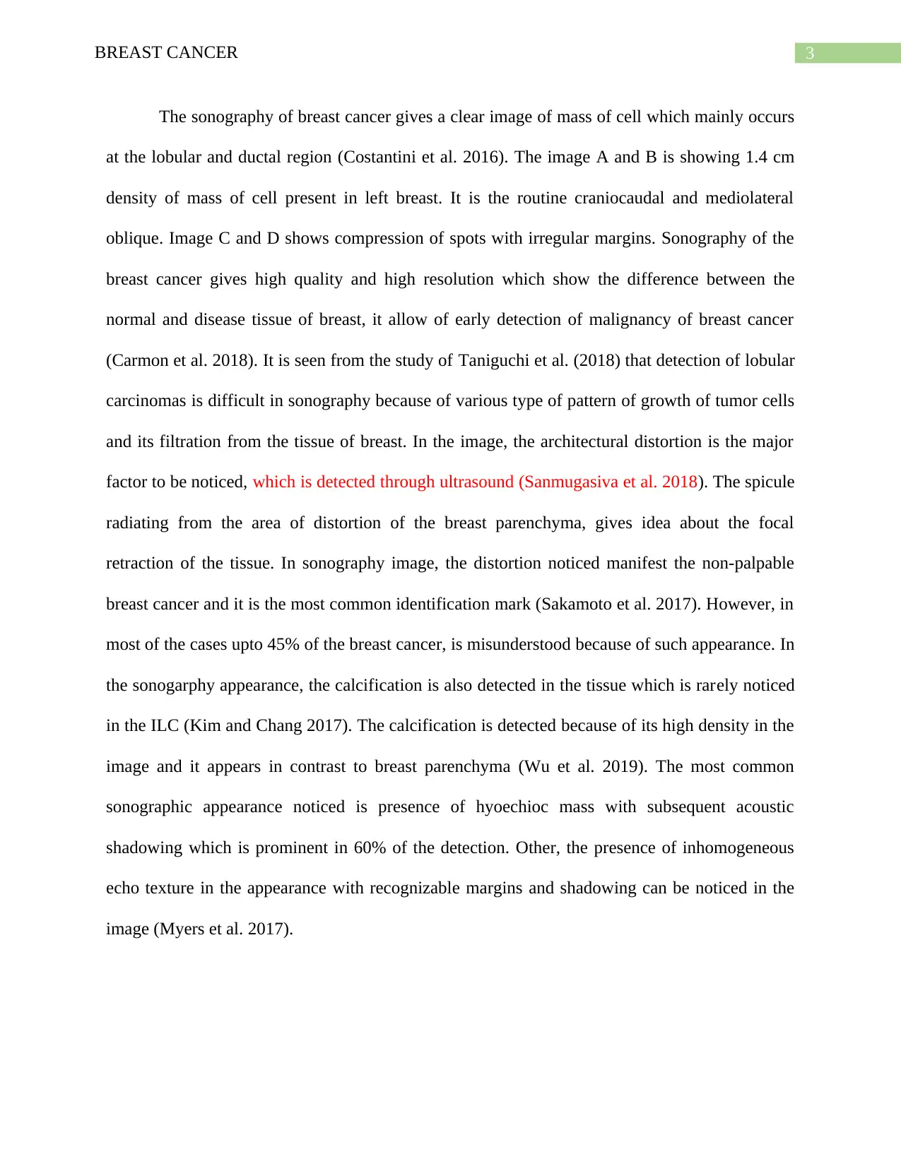
This report provides a comprehensive overview of breast cancer, beginning with its pathophysiology. It explains how hormonal and hereditary factors contribute to the development of breast carcinoma, including the roles of estrogen receptors, BRCA1 and BRCA2 genes, Human epidermal growth factor (EGFR), and the P53 protein in the progression of the disease. The report then transitions to the sonographic appearance of breast cancer, detailing how ultrasound imaging is used for early detection. It describes the typical sonographic features of breast cancer, such as the appearance of masses in the lobular and ductal regions, architectural distortions, and the presence of calcifications. The report also notes the challenges in sonographic detection, especially with lobular carcinomas. The report references several research studies to support its findings, providing a thorough examination of both the biological mechanisms and the diagnostic imaging techniques associated with breast cancer.
tabler-icon-diamond-filled.svg

Contribute Materials

Your contribution can guide someone’s learning journey. Share your documents today.
Document Page
Running head: BREAST CANCER
BREAST CANCER
Name of Student:
Name of University:
Author’s Note:
tabler-icon-diamond-filled.svg

Secure Best Marks with AI Grader

Need help grading? Try our AI Grader for instant feedback on your assignments.
Document Page
1BREAST CANCER
Introduction Breast -Breast carcinoma
Part A
Breast carcinoma is one of the type of neoplasm which arise from the breast tissue.
Among that the most common is the adenocarcinoma of those cells which underlines the
terminal duct lobular unit. Breast cancer is the common cancer among the women with 89%
death rate globally. The pathophysiology of the breast cancer can be understood as hormonal and
hereditary form of cancer. As per the study of Bland et al. (2017) it is understood that with breast
cancer requires hormone to grow and its incidence arise by the exposure of estrogen hormone.
78% of the breast cancer is hormone sensitive, which can be attributed by the fact that it carries
estrogen receptor and divide and grow in response of the hormone stimulation (Wefel et al.
2015). Estrogen receptor are basically of two type alpha and beta. The cells of breast express
these receptor mainly the ER alpha. When there is high amount of estrogen, it bind with the
receptor and the complex moved to the nucleus which causes formation of the transcription
protein which increase the cell division and result in imbalance of regulation of cell cycle. The
high degree of cell division result in breast carcinoma (Bloom et al. 2016).
In order to understand the pathophysiology of the breast cancer, it is important to
evaluate the gene expression of the BRCA1 and BRCA2 which is a type of tumour suppressor
gene. The major function of the gene is to code for the protein which is required in DNA repair
pathway which is necessary to protect against mutation (D.J et al. 2016) In breast cancer, there is
loss of function of these two genes which is linked with breast cancer in women. Such breast
cancer are ER (estrogen receptor) and PR (progesterone receptor) positive. There are many cause
Document Page
2BREAST CANCER
of the mutation in these gene which can be environmental, hormonal and genetic. According to
the study of Szabo (2016) it was seen that the mutated gene are inherited to next generation.
Jafari et al. (2018) has studied the role of Human epidermal growth factor which is also
associated with breast cancer. The Human epidermal growth factor receptor is proto-oncogene
which remain in inactive state. The main function of the gene is to code for growth factor that
increase the division of cell. The regulation of the cell is under control and in regulation. In
breast cancer, due to gain in mutation, EGFR are overexpressed and is associated with the
growth of malignant tumour in breast.
The guardian of cell cycle is P53 protein which is a tumour suppressor protein whose
major function is growth arrest, apoptosis and DNA repair. In breast cancer, the P53 is mutated
and there is loss of function of gene (Semenza 2016). Due to this, there is irregular division of
cell and DNA repair pathway is disrupted. The continue division of cell is not arrested and result
in breast cancer.
Part B. Sonographic appearance of breast cancer
Source: (Johnson and Hwang 2015).
Document Page
3BREAST CANCER
The sonography of breast cancer gives a clear image of mass of cell which mainly occurs
at the lobular and ductal region (Costantini et al. 2016). The image A and B is showing 1.4 cm
density of mass of cell present in left breast. It is the routine craniocaudal and mediolateral
oblique. Image C and D shows compression of spots with irregular margins. Sonography of the
breast cancer gives high quality and high resolution which show the difference between the
normal and disease tissue of breast, it allow of early detection of malignancy of breast cancer
(Carmon et al. 2018). It is seen from the study of Taniguchi et al. (2018) that detection of lobular
carcinomas is difficult in sonography because of various type of pattern of growth of tumor cells
and its filtration from the tissue of breast. In the image, the architectural distortion is the major
factor to be noticed, which is detected through ultrasound (Sanmugasiva et al. 2018). The spicule
radiating from the area of distortion of the breast parenchyma, gives idea about the focal
retraction of the tissue. In sonography image, the distortion noticed manifest the non-palpable
breast cancer and it is the most common identification mark (Sakamoto et al. 2017). However, in
most of the cases upto 45% of the breast cancer, is misunderstood because of such appearance. In
the sonogarphy appearance, the calcification is also detected in the tissue which is rarely noticed
in the ILC (Kim and Chang 2017). The calcification is detected because of its high density in the
image and it appears in contrast to breast parenchyma (Wu et al. 2019). The most common
sonographic appearance noticed is presence of hyoechioc mass with subsequent acoustic
shadowing which is prominent in 60% of the detection. Other, the presence of inhomogeneous
echo texture in the appearance with recognizable margins and shadowing can be noticed in the
image (Myers et al. 2017).
tabler-icon-diamond-filled.svg

Secure Best Marks with AI Grader

Need help grading? Try our AI Grader for instant feedback on your assignments.
Document Page
4BREAST CANCER
Reference
Bland, K.I., Copeland, E.M., Klimberg, V.S. and Gradishar, W.J., 2017. The breast:
comprehensive management of benign and malignant diseases. Elsevier Inc.
Bloom, M.W., Hamo, C.E., Cardinale, D., Ky, B., Nohria, A., Baer, L., Skopicki, H., Lenihan,
D.J., Gheorghiade, M., Lyon, A.R. and Butler, J., 2016. Cancer Therapy–Related Cardiac
Dysfunction and Heart Failure: Part 1: Definitions, Pathophysiology, Risk Factors, and
Imaging. Circulation: Heart Failure, 9(1), p.e002661.
Carmon, M., Zilber, S., Gekhtman, D., Olsha, O., Hadar, T. and Golomb, E., 2018. Hygroscopic
sonographically detectable clips form characteristic breast and lymph node pseudocysts. Modern
Pathology, 31(1), p.62.
Costantini, M., Belli, P., Bufi, E., Asunis, A. M., Ferra, E. and Bitti, G. T. (2016). Association
between sonographic appearances of breast cancers and their histopathologic features and
biomarkers. Journal of Clinical Ultrasound, 44(1), 26-33.
Jafari, S.H., Saadatpour, Z., Salmaninejad, A., Momeni, F., Mokhtari, M., Nahand, J.S.,
Rahmati, M., Mirzaei, H. and Kianmehr, M., 2018. Breast cancer diagnosis: Imaging techniques
and biochemical markers. Journal of cellular physiology, 233(7), pp.5200-5213.
Johnson, K., Sarma, D., & Hwang, E. S. (2015). Lobular breast cancer series: imaging. Breast
Cancer Research, 17(1), 94.
Kim, Y.S. and Chang, J.M., 2017. Sonographic appearance of a cholesterol granuloma
mimicking breast cancer. Journal of Clinical Ultrasound, 45(9), pp.608-611.
Document Page
5BREAST CANCER
Myers, K.S., Green, L.A., Lebron, L. and Morris, E.A., 2017. Imaging appearance and clinical
impact of preoperative breast MRI in pregnancy-associated breast cancer. American Journal of
Roentgenology, 209(3), pp.W177-W183.
Sakamoto, N., Ogawa, Y., Tsunoda, Y. and Fukuma, E., 2017. Evaluation of the sonographic
visibility and sonographic appearance of the breast biopsy marker (UltraClip®) placed in
phantoms and patients. Breast Cancer, 24(4), pp.585-592.
Sanmugasiva, V.V., Ramli, M.T., Fadzli, F., Kaur, S., Rahman, N.A. and Rahmat, K., 2018.
Myofibroblastoma of the breast. The Malaysian journal of pathology, 40(3), pp.349-353.
Semenza, G.L., 2016. The hypoxic tumor microenvironment: A driving force for breast cancer
progression. Biochimica et Biophysica Acta (BBA)-Molecular Cell Research, 1863(3), pp.382-
391.
Szabo, C., 2016. Gasotransmitters in cancer: from pathophysiology to experimental
therapy. Nature Reviews Drug Discovery, 15(3), p.185.
Taniguchi, C., Ohta, T., Inoue, M., Hirano, M., Nishioka, M., Nakata, N., Ojiri, H. And
Shirakawa, T., 2018. Four cases of mimic breast cancer as the location of a penetrating
artery. Choonpa Igaku, pp.JJMU-A.
Wefel, J.S., Kesler, S.R., Noll, K.R. and Schagen, S.B., 2015. Clinical characteristics,
pathophysiology, and management of noncentral nervous system cancer‐related cognitive
impairment in adults. CA: a cancer journal for clinicians, 65(2), pp.123-138.
Document Page
6BREAST CANCER
Wu, T., Li, J., Wang, D., Leng, X., Zhang, L., Li, Z., Jing, H., Kang, J. and Tian, J., 2019.
Identification of a correlation between the sonographic appearance and molecular subtype of
invasive breast cancer: A review of 311 cases. Clinical imaging, 53, pp.179-185.
chevron_up_icon
1 out of 7
circle_padding
hide_on_mobile
zoom_out_icon
logo.png

Your All-in-One AI-Powered Toolkit for Academic Success.

Available 24*7 on WhatsApp / Email

[object Object]