Comprehensive Case Study: C2 Fracture Diagnosis and Management
VerifiedAdded on 2020/02/05
|17
|3326
|125
Case Study
AI Summary
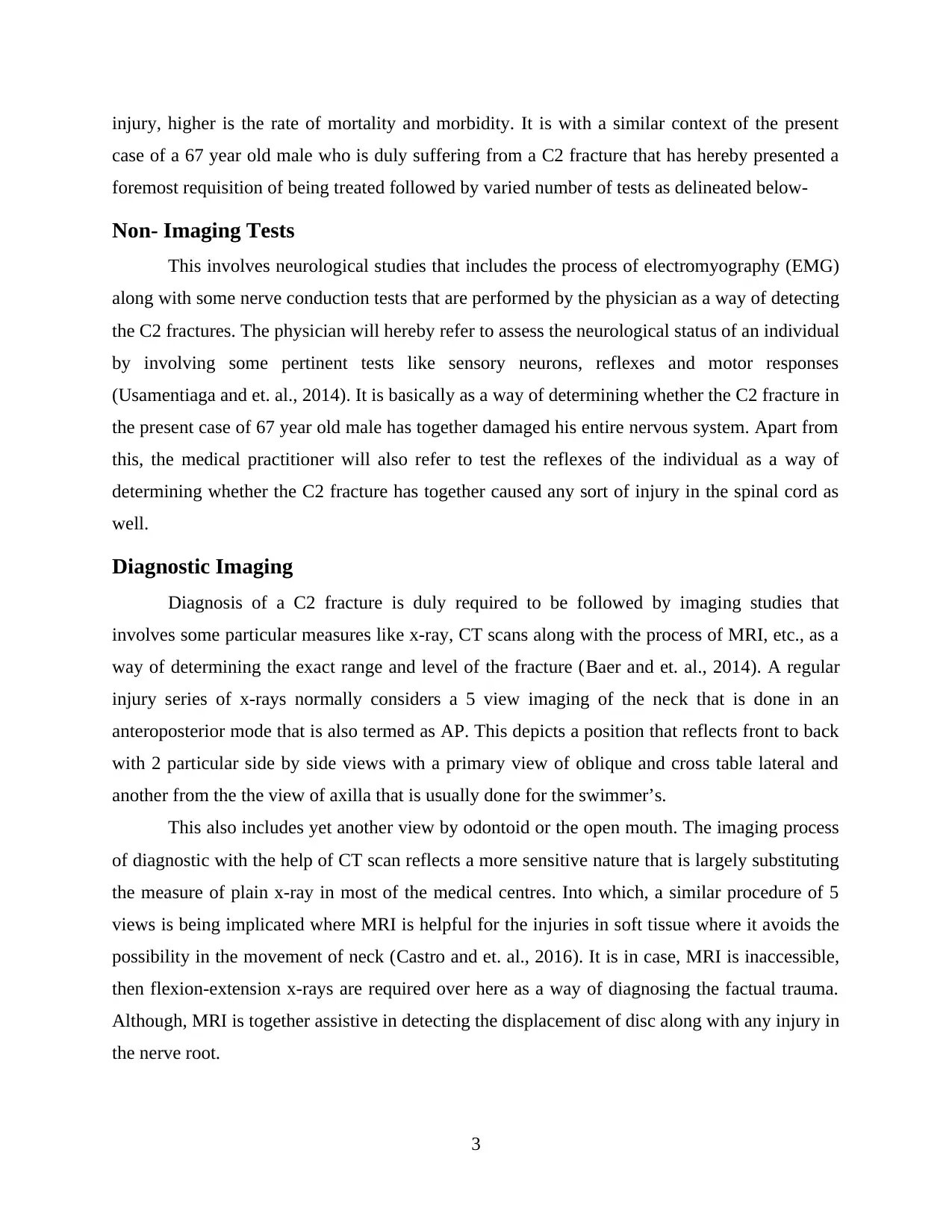
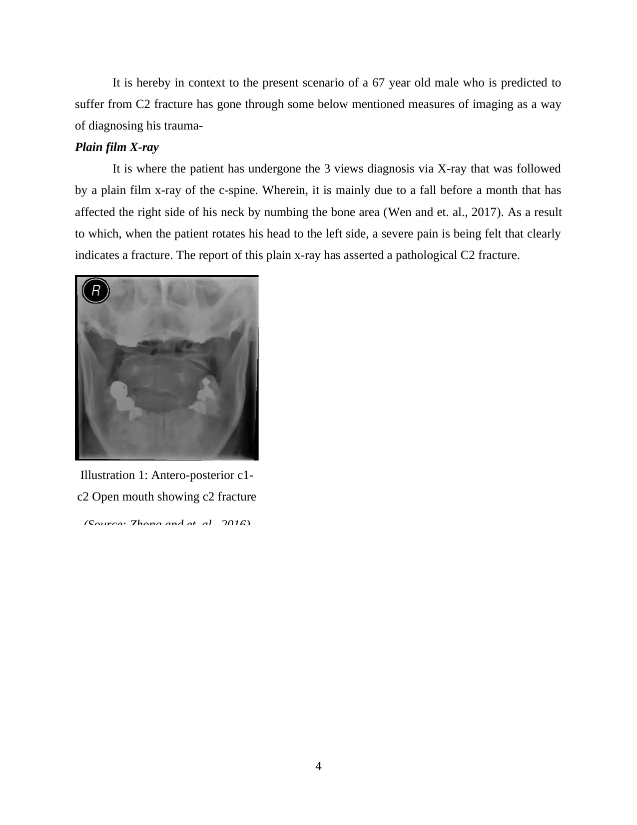
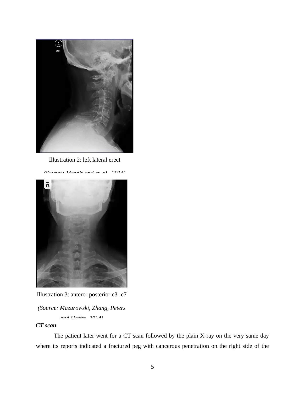
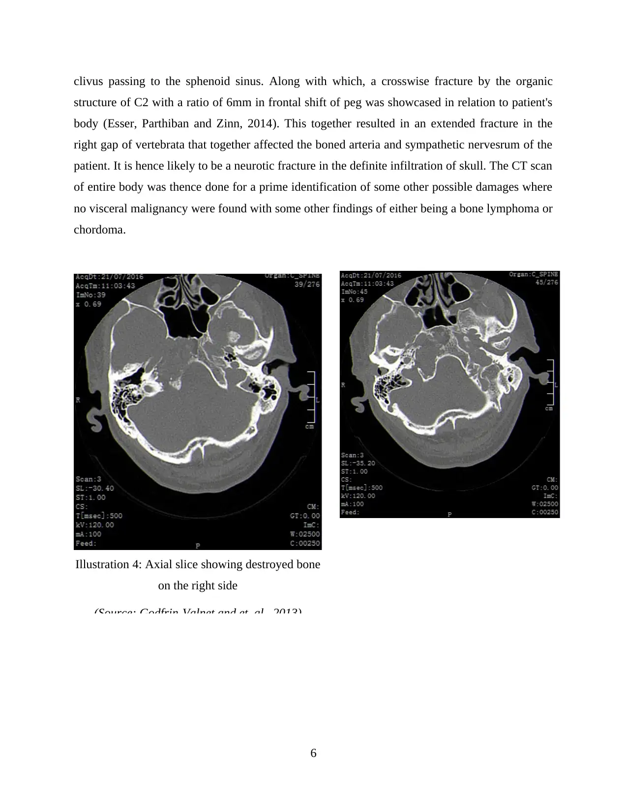
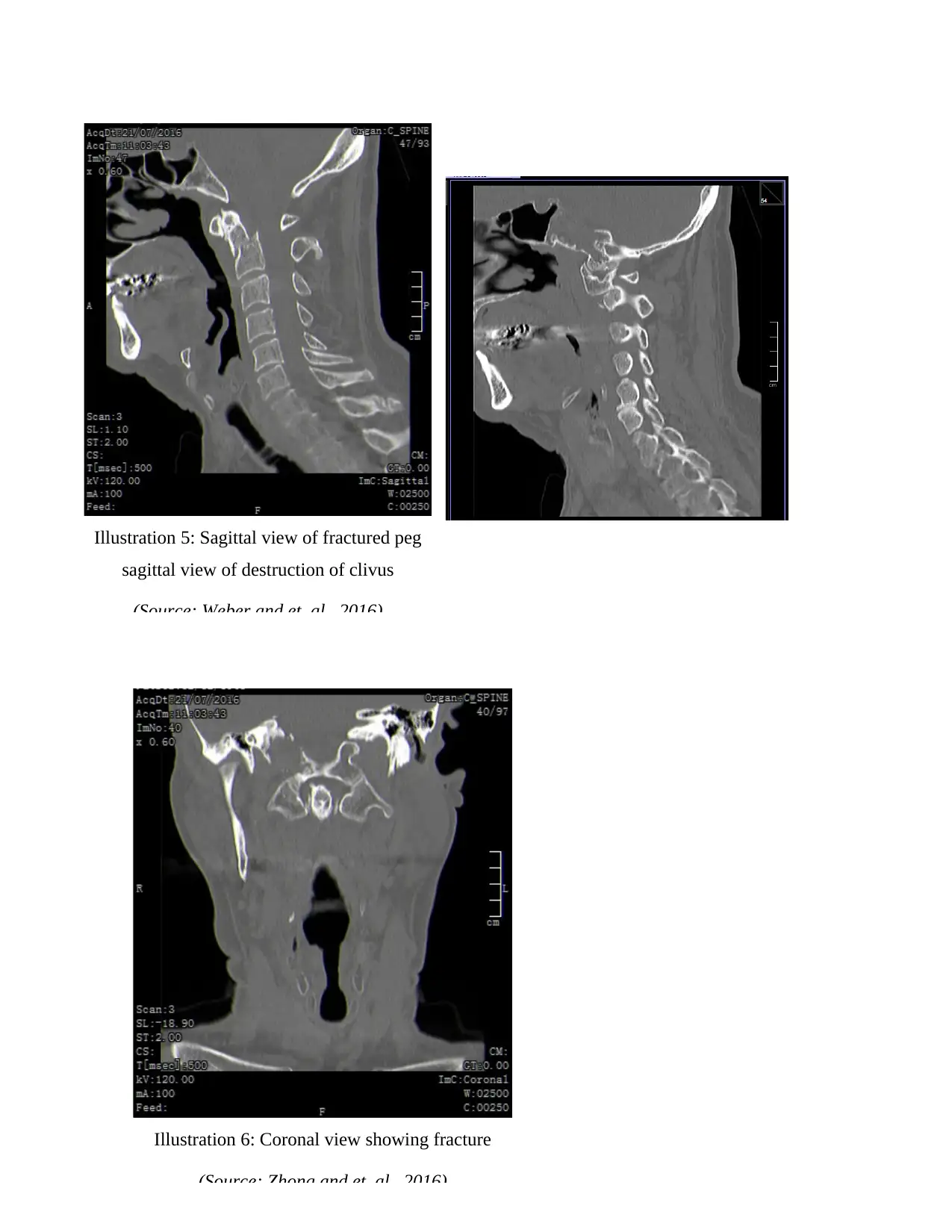
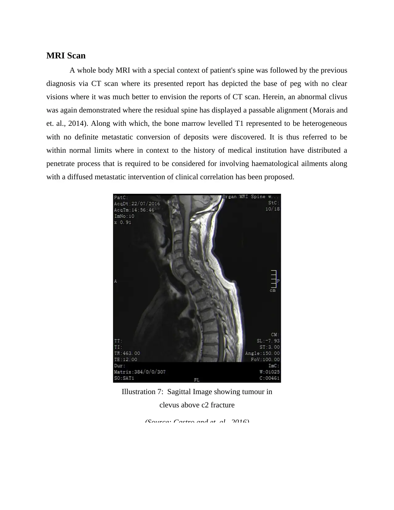
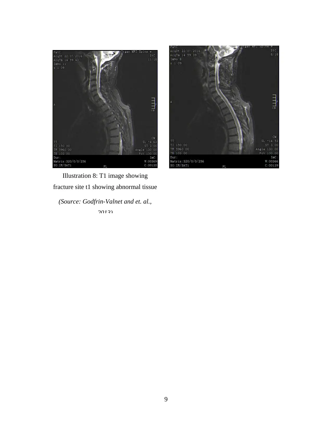
This case study examines the case of a 67-year-old male presenting with a C2 fracture, detailing the anatomy and physiology of the cervical spine. The assignment explores the aetiology of the injury, focusing on the patient's fall and subsequent symptoms like neck pain and numbness. It then delves into the diagnostic process, including non-imaging tests and detailed analysis of imaging results from X-rays, CT scans, and MRI scans, highlighting the findings of a pathological C2 fracture and cancerous penetration. The study further discusses treatment options, including palliative care, surgical methods (direct lateral mass screws), and follow-up procedures. Patient management pathways and relevant medical guidelines are also addressed to provide a comprehensive understanding of the case. The case study emphasizes the importance of prompt diagnosis and appropriate treatment for C2 fractures to prevent further complications and improve patient outcomes.

Case Study
(Medical)
(Medical)
Secure Best Marks with AI Grader
Need help grading? Try our AI Grader for instant feedback on your assignments.

TABLE OF CONTENT
Anatomy and Physiology.................................................................................................................1
Aetiology..........................................................................................................................................1
Disease Presentation........................................................................................................................2
Non- Imaging Tests..........................................................................................................................3
Diagnostic Imaging..........................................................................................................................3
MRI Scan.........................................................................................................................................9
Treatment.......................................................................................................................................11
Follow Up......................................................................................................................................11
Patient Management Pathways......................................................................................................11
Relevant Medical Guidelines.........................................................................................................12
REFERENCES..............................................................................................................................14


Anatomy and Physiology
Here, the patient used is a 67 year old male who has a c-spine injury because he fall a
month ago on the right side of the neck and had numbness in occipital area. However, when the
head is rotated to the left pain is felt and thus it needs to be identified that fracture is there in the
neck or not. Also, subsequent imaging and diagnostic tests needs to be carried out in order to
identify the C2 fracture. Cervical spine is also called C spine and thus injury in such area are
very tough to handle (Yue and et. al., 2016). It mainly occurs to the patients who had motor
vehicle accident. It is essential for care professionals to provide immediate treatment to the
patients with C2 injury so that they can overcome pain. Also, patient need to keep the head and
neck in a constant position so that fracture can be healed up fast. However, treatment for such
type of injury depends upon the individual circumstances and details of the injury itself. C2
injuries are more dangerous because it damages the spinal cord at any level and also it creates
issues to communicate signals to the rest of the body below that part. Thus, C2 fractures leaves
the patient paralyzed. There are around 10% of the unconscious patients who visit the emergency
department due to motor vehicle accident have a C2 spine fracture (Ertel, Robinson and Eckman,
2016).
Aetiology
C2 fracture is mainly caused due to abrupt impact or twisting the neck and thus occurs in
a short span of time during the trauma. It breaks the ligaments or crack the bone which needs
proper rest so that it can be recovered soon. It is also occurred through instability and cause
damage to the spinal cord and neurologic structures (Akella and Chendrasekhar, 2016). Cervical
C2 fracture is a break in the bone of the neck and affects the head and does not allow the head to
move on any of the side. Following are the main causes of C2 fracture such as-
Motor vehicle accident
Falls
Dive from height into deep water
Injuries from sports
Skating injuries
There are several other risk factors through which C2 injury occurs and thus care
professionals are required to provide proper medical treatment to patients in order to improve
1
Secure Best Marks with AI Grader
Need help grading? Try our AI Grader for instant feedback on your assignments.

their spinal cord and thus they are able to move their head in every location. All such causes
affects the individual and their health that results in C2 fracture. There is a strong evidence
available in order to suggest that some individuals possess a disability of not moving their head
in proper direction (Silva and et. al., 2016). Therefore, proper medical treatment needs to be
provided to patients suffering from C2 fracture so that proper medication could be provided to
patients.
Disease Presentation
C2 fracture is mainly caused due to fall or motor vehicle accident and thus it breaks the
ligaments or bone which affects the patients in the way that they are not able to move their head
in any direction. Signs or symptoms of spinal cord injury may present immediately or some
symptoms may be delayed as swelling and bleeding occur in or around the spinal cord (Garara
and et. al., 2016). Following are the symptoms that are occurred in C2 spinal cord injury-
Pain in the neck and numbness or burning sensation
Unable to move or walk
Muscle pain
Difficult in breathing
Unable to speak properly
Loss of feeling or sensation below the part of fracture
Paralysis in the part which is fractured
Very limited neck or head movement
Swallowing in the part of fracture
Lack of consciousness
Pain in the fractured part
Cervical C2 fracture- Damage in the spinal cord is considered as the most severe damage
because it can be like threatening. The neck is not able to move in any direction and it is still
which creates pain. It also affects the ability of patient to breathe properly and affects their health
(Haddad and et. al., 2016).
Injuries in the cervical spine also termed as C-spine are one of the most frightful traumas
out of all other type of spine injuries where it has the potential of significant hurtful abnormality.
It is where there together exists a correlation among the level of such injuries and the rate of
mortality or morbidity (Weber and et. al., 2016). It depicts that higher the level of a C-spine
2

injury, higher is the rate of mortality and morbidity. It is with a similar context of the present
case of a 67 year old male who is duly suffering from a C2 fracture that has hereby presented a
foremost requisition of being treated followed by varied number of tests as delineated below-
Non- Imaging Tests
This involves neurological studies that includes the process of electromyography (EMG)
along with some nerve conduction tests that are performed by the physician as a way of detecting
the C2 fractures. The physician will hereby refer to assess the neurological status of an individual
by involving some pertinent tests like sensory neurons, reflexes and motor responses
(Usamentiaga and et. al., 2014). It is basically as a way of determining whether the C2 fracture in
the present case of 67 year old male has together damaged his entire nervous system. Apart from
this, the medical practitioner will also refer to test the reflexes of the individual as a way of
determining whether the C2 fracture has together caused any sort of injury in the spinal cord as
well.
Diagnostic Imaging
Diagnosis of a C2 fracture is duly required to be followed by imaging studies that
involves some particular measures like x-ray, CT scans along with the process of MRI, etc., as a
way of determining the exact range and level of the fracture (Baer and et. al., 2014). A regular
injury series of x-rays normally considers a 5 view imaging of the neck that is done in an
anteroposterior mode that is also termed as AP. This depicts a position that reflects front to back
with 2 particular side by side views with a primary view of oblique and cross table lateral and
another from the the view of axilla that is usually done for the swimmer’s.
This also includes yet another view by odontoid or the open mouth. The imaging process
of diagnostic with the help of CT scan reflects a more sensitive nature that is largely substituting
the measure of plain x-ray in most of the medical centres. Into which, a similar procedure of 5
views is being implicated where MRI is helpful for the injuries in soft tissue where it avoids the
possibility in the movement of neck (Castro and et. al., 2016). It is in case, MRI is inaccessible,
then flexion-extension x-rays are required over here as a way of diagnosing the factual trauma.
Although, MRI is together assistive in detecting the displacement of disc along with any injury in
the nerve root.
3

It is hereby in context to the present scenario of a 67 year old male who is predicted to
suffer from C2 fracture has gone through some below mentioned measures of imaging as a way
of diagnosing his trauma-
Plain film X-ray
It is where the patient has undergone the 3 views diagnosis via X-ray that was followed
by a plain film x-ray of the c-spine. Wherein, it is mainly due to a fall before a month that has
affected the right side of his neck by numbing the bone area (Wen and et. al., 2017). As a result
to which, when the patient rotates his head to the left side, a severe pain is being felt that clearly
indicates a fracture. The report of this plain x-ray has asserted a pathological C2 fracture.
Illustration 1: Antero-posterior c1-
c2 Open mouth showing c2 fracture
(Source: Zhong and et. al., 2016)
4
Paraphrase This Document
Need a fresh take? Get an instant paraphrase of this document with our AI Paraphraser

Illustration 2: left lateral erect
(Source: Morais and et. al., 2014)
Illustration 3: antero- posterior c3- c7
(Source: Mazurowski, Zhang, Peters
and Hobbs, 2014)
CT scan
The patient later went for a CT scan followed by the plain X-ray on the very same day
where its reports indicated a fractured peg with cancerous penetration on the right side of the
5

clivus passing to the sphenoid sinus. Along with which, a crosswise fracture by the organic
structure of C2 with a ratio of 6mm in frontal shift of peg was showcased in relation to patient's
body (Esser, Parthiban and Zinn, 2014). This together resulted in an extended fracture in the
right gap of vertebrata that together affected the boned arteria and sympathetic nervesrum of the
patient. It is hence likely to be a neurotic fracture in the definite infiltration of skull. The CT scan
of entire body was thence done for a prime identification of some other possible damages where
no visceral malignancy were found with some other findings of either being a bone lymphoma or
chordoma.
6
Illustration 4: Axial slice showing destroyed bone
on the right side
(Source: Godfrin-Valnet and et. al., 2013)

7
Illustration 5: Sagittal view of fractured peg
sagittal view of destruction of clivus
(Source: Weber and et. al., 2016)
Illustration 6: Coronal view showing fracture
(Source: Zhong and et. al., 2016)
Illustration 5: Sagittal view of fractured peg
sagittal view of destruction of clivus
(Source: Weber and et. al., 2016)
Illustration 6: Coronal view showing fracture
(Source: Zhong and et. al., 2016)
Secure Best Marks with AI Grader
Need help grading? Try our AI Grader for instant feedback on your assignments.

MRI Scan
A whole body MRI with a special context of patient's spine was followed by the previous
diagnosis via CT scan where its presented report has depicted the base of peg with no clear
visions where it was much better to envision the reports of CT scan. Herein, an abnormal clivus
was again demonstrated where the residual spine has displayed a passable alignment (Morais and
et. al., 2014). Along with which, the bone marrow levelled T1 represented to be heterogeneous
with no definite metastatic conversion of deposits were discovered. It is thus referred to be
within normal limits where in context to the history of medical institution have distributed a
penetrate process that is required to be considered for involving haematological ailments along
with a diffused metastatic intervention of clinical correlation has been proposed.
Illustration 7: Sagittal Image showing tumour in
clevus above c2 fracture
(Source: Castro and et. al., 2016)

9
Illustration 8: T1 image showing
fracture site t1 showing abnormal tissue
(Source: Godfrin-Valnet and et. al.,
2013)
Illustration 8: T1 image showing
fracture site t1 showing abnormal tissue
(Source: Godfrin-Valnet and et. al.,
2013)

Treatment
According to the given case scenario, patient is reported for pathological C2 fracture. At
this juncture, palliative care can be provided to patient for the purpose of recovery. However,
under which soft tissue massage is used over cervical spine musculature in order to treat the pain
and muscle spasms. Not only this but gentle myofacila therapy can also be used for the purpose
of effective treatment. Though, many of the cases comes with results of quadriplegia and
subsequent death from respiratory distress. It is because rigid collar is used for the purpose of
stabilization. However, metastatic destruction remains progressed which might cause issue for
the this particular disease (Cervical spine, 2017).
Some of the regular treatment are done with the help of keeping the neck and head in
constant. However, in the current case study patient can be treated with the help of direct lateral
mass screws so as to ensure appropriate movement. Not only this, but surgical method can also
be used where complications remain minimum along with very low rate of morbidity. However,
appropriate method treatment is based on particular situation or circumstances of patient. Apart
from this, other options of treatment consists of anterior which might be without or with metal
plat and screws (Ying and et. al., 2008). However, initial treatment usually involve skeletal
traction and close reducing along with placement of mental pins. Moreover, the current case
reflect that Nuclear medicine treatment has been provided to patient.
Follow Up
Follow up is very important for patient suffering from any particular kind of disease
which make it possible to get the effective of treatment on a right time. However, it depends on
the extent of injury and accordingly longer length of hospitalization for surgical patient can be of
11.8 days whereas 4.4 days will be needed for nonsurgical patients. However, the follow up time
can be one month under which patient seek to doctor for light exercise or treatment etc. It would
be effective for healing because sufferer will get to know that how much time will be needed in
recovery (C2 Axis Spine Injury, 2017).
Patient Management Pathways
The patient management pathways in the current case consists of specific procedure
under which focus is laid on getting treatment effectively by communicating all related issues.
The clinical assessment of patient with C2 fracture consists of three steps such as history, general
10
Paraphrase This Document
Need a fresh take? Get an instant paraphrase of this document with our AI Paraphraser

examination with physician and evaluation of neurological status. Here, patients are generally
required the imaging in case they are unable to rotate their neck to 45 degrees. However, this
situation has been explained with the help of following image (Cervical Spine Injury, 2017).
(Source: Cervical spine, 2017)
Relevant Medical Guidelines
The guidelines of NICE reflected that the cervical spine immobilisation has become the
most common aspect behind the pre-hospital care. It also shed light on Nexus Low risk criteria
11

that it should be followed at the time of pre-hospital. However, the effective aspect of CCR
should be followed in-hospital assessment so as to treat the patient effectively. Apart from this, it
has not been recommended to use the MRI on routine basis for the purpose of cervical spine. In
addition to this, NICE guidelines show that 3 view plain radiographs are used for imaging the
cervical spine (Cervical Spine Injury. 2017).
12

REFERENCES
Books and Journal
Akella, K. and Chendrasekhar, A., 2016. Cervical Spine Injury Without Neurologic Deficit vs
Thoracic or Lumbar Spine Injury: Does Location Matter With Regard to
Complications?. CHEST Journal. 150(4_S). pp.336A-336A.
Baer, G. And et. al., 2014. Calibration of a non-null test interferometer for the measurement of
aspheres and free-form surfaces. Optics express. 22(25). pp.31200-31211.
Castro, M.P. And et. al., 2016. Construct validity of clinical spinal mobility tests in ankylosing
spondylitis: a systematic review and meta-analysis. Clinical rheumatology. 35(7).
pp.1777-1787.
Ertel, A.E., Robinson, B.R. and Eckman, M.H., 2016. Cost-effectiveness of cervical spine
clearance interventions with litigation and long-term-care implications in obtunded adult
patients following blunt injury. Journal of Trauma and Acute Care Surgery. 81(5).
pp.897-904.
Esser, C.M., Parthiban, C. and Zinn, M.R., 2014. Development of a parallel actuation approach
for MR-compatible robotics. IEEE/ASME Transactions on Mechatronics. 19(3). pp.904-
915.
Garara, B. and et. al., 2016. Intramuscular diaphragmatic stimulation for patients with traumatic
high cervical injuries and ventilator dependent respiratory failure: A systematic review of
safety and effectiveness. Injury. 47(3). pp.539-544.
Godfrin-Valnet, M. and et. al., 2013. Eighteen cases of crowned dens syndrome: presentation
and diagnosis. Neurochirurgie. 59(3). pp.115-120.
Haddad, S. and et. al., 2016. Diagnosis and neurologic status as predictors of surgical site
infection in primary cervical spinal surgery. The Spine Journal. 16(5). pp.632-642.
Mazurowski, M.A., Zhang, J., Peters, K.B. and Hobbs, H., 2014. Computer-extracted MR
imaging features are associated with survival in glioblastoma patients. Journal of neuro-
oncology. 120(3). pp.483-488.
Morais, D.F. And et. al., 2014. Clinical applicability of magnetic resonance imaging in acute
spinal cord trauma. European Spine Journal. 23(7). pp.1457-1463.
13
Secure Best Marks with AI Grader
Need help grading? Try our AI Grader for instant feedback on your assignments.

Silva, O.T.D. and et. al., 2016. Evaluation of the reliability and validity of the newer AOSpine
subaxial cervical injury classification (C-3 to C-7). Journal of Neurosurgery: Spine.
25(3). pp.303-308.
Usamentiaga, R. And et. al., 2014. Infrared thermography for temperature measurement and non-
destructive testing. Sensors. 14(7). pp.12305-12348.
Weber, K.A. And et. al., 2016. Lateralization of cervical spinal cord activity during an isometric
upper extremity motor task with functional magnetic resonance imaging. NeuroImage.
125. pp.233-243.
Wen, B.J. And et. al., 2017. Surface Properties of Nano-Film Type Patterning Electrode on
Flexible Substrate for Bending Test. Science of Advanced Materials. 9(1). pp.17-21.
Ying Z and et al. 2008. Anterior cervical discectomy and fusion for unstable traumatic
spondylolisthesis of the axis. Spine (Phila Pa 1976). 33(3). pp.255–258
Yue, J.K. and et. al., 2016. A review and update on the guidelines for the acute management of
cervical spinal cord injury-Part II. Journal of neurosurgical sciences. 60(3). pp.367-384.
Zhong, X. And et. al., 2016. A System-Theoretic Approach to Modeling and Analysis of
Mammography Testing Process. IEEE Transactions on Systems, Man, and Cybernetics:
Systems. 46(1). pp.126-138.
Online
C2 Axis Spine Injury. 2017. [Online]. Available through: <http://www.chop.edu/conditions-
diseases/c2-axis-spine-injury>. [Accessed on 2nd March 2017].
Cervical Spine Injury. 2017. [Online]. Available through:
<http://www.rcemlearning.co.uk/references/cervical-spine-injury/>. [Accessed on 2nd
March 2017].
Cervical spine. 2017. [Online]. Available through:
<http://www.racgp.org.au/afp/2012/april/cervical-spine/>. [Accessed on 2nd March 2017].
14
1 out of 17
Related Documents

Your All-in-One AI-Powered Toolkit for Academic Success.
+13062052269
info@desklib.com
Available 24*7 on WhatsApp / Email
Unlock your academic potential
© 2024 | Zucol Services PVT LTD | All rights reserved.