Case Study: Calcific Uremic Arteriolopathy, Haemodialysis & Treatment

Verified

Added on  2023/03/20

|5
|682
|59
Case Study
AI Summary
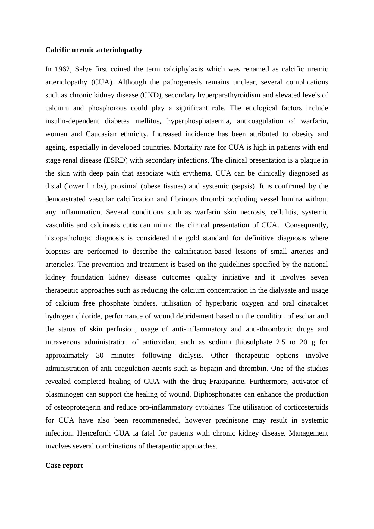
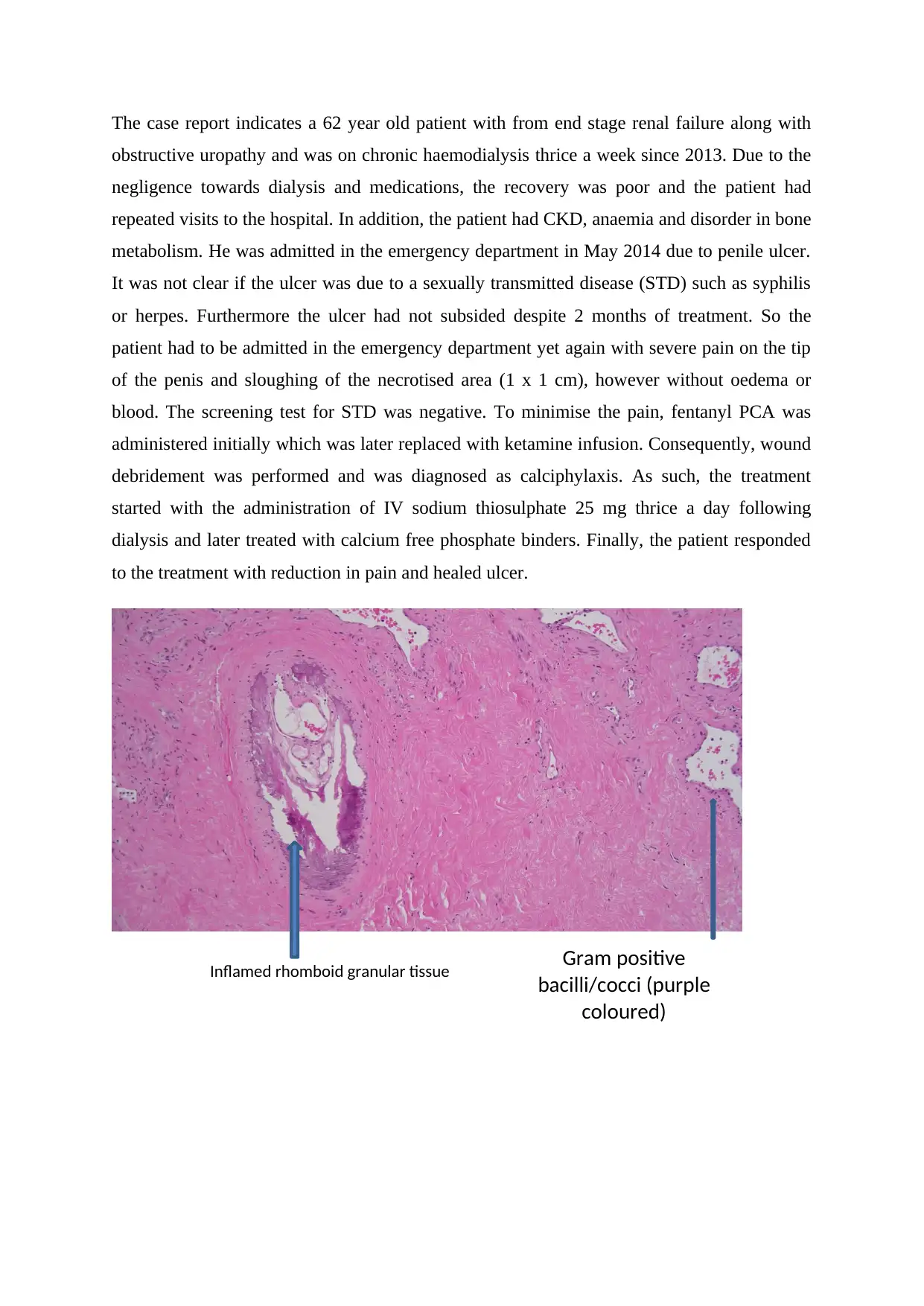
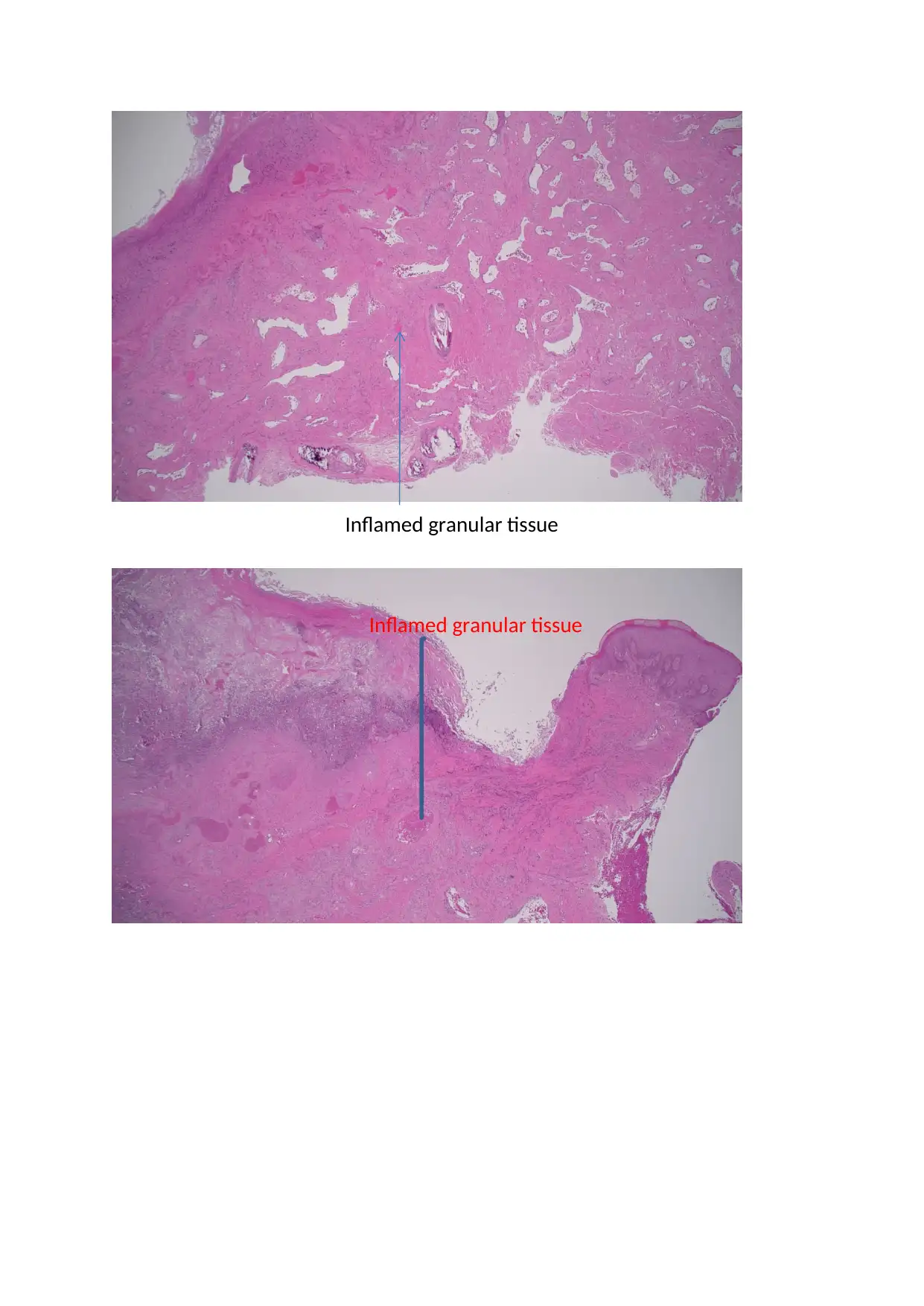
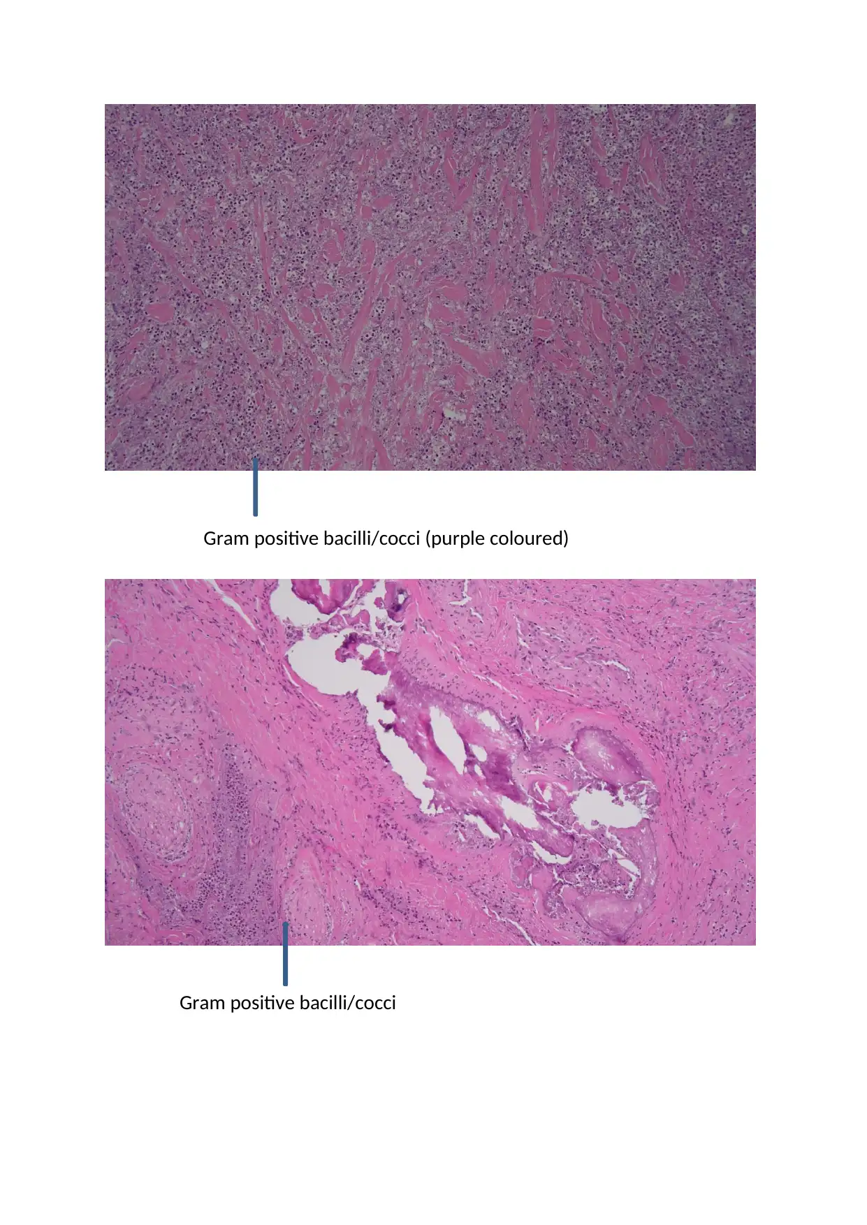
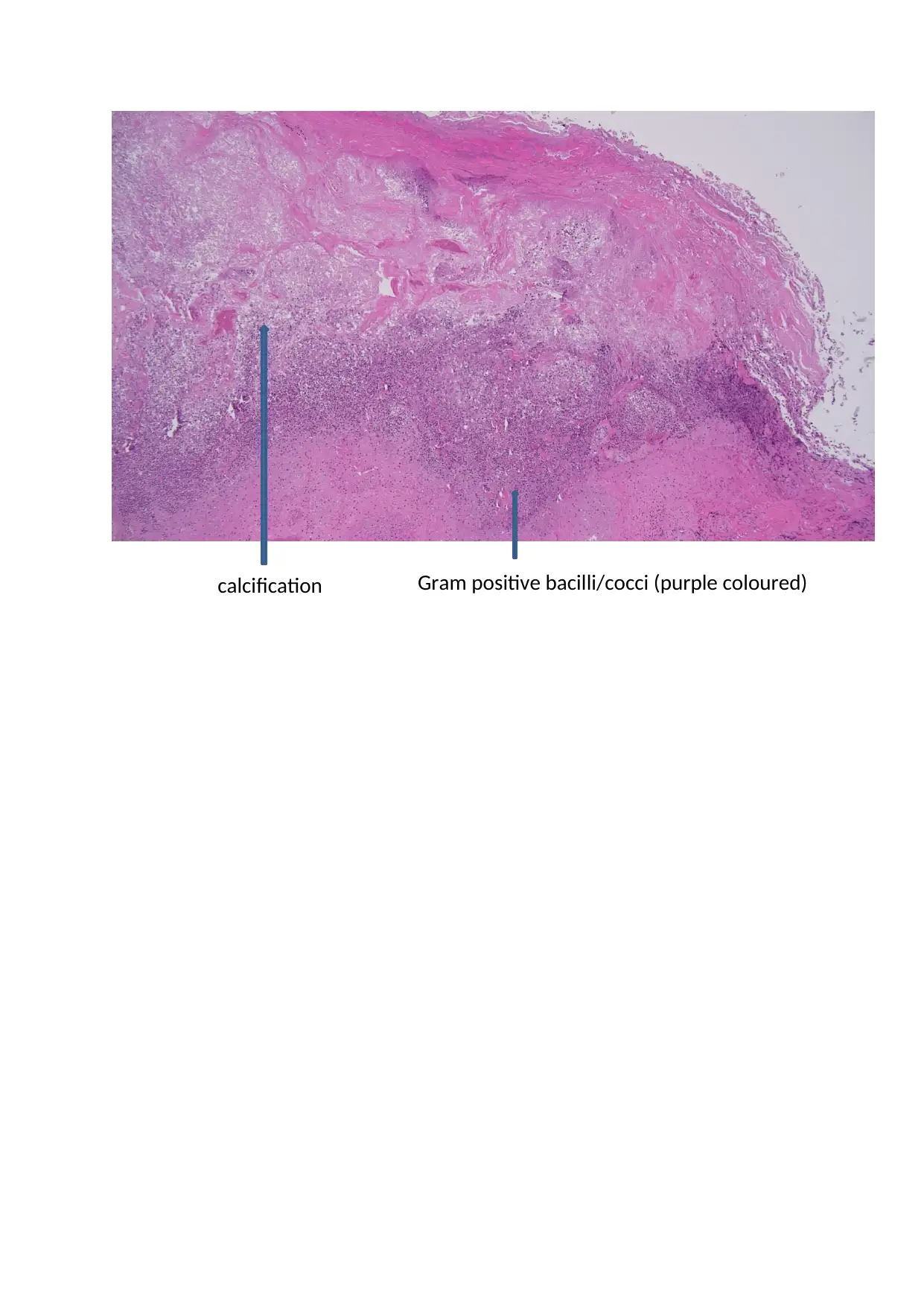
This case study examines a 62-year-old patient with end-stage renal failure (ESRF) on chronic haemodialysis who presented with a non-healing penile ulcer, ultimately diagnosed as calcific uremic arteriolopathy (CUA). The patient had a history of poor compliance with dialysis and medications, leading to multiple hospital visits. The presentation included severe pain and necrotic tissue, but the STD screen was negative. Treatment involved pain management with fentanyl and ketamine, wound debridement, and the administration of intravenous sodium thiosulphate and calcium-free phosphate binders. The patient responded positively to treatment, with pain reduction and ulcer healing. The case highlights the challenges of managing CUA, a condition associated with high mortality, and demonstrates the effectiveness of sodium thiosulphate in this context. The provided histology slides and the clinical presentation were consistent with the diagnosis of calciphylaxis.
Document Page
Calcific uremic arteriolopathy
In 1962, Selye first coined the term calciphylaxis which was renamed as calcific uremic
arteriolopathy (CUA). Although the pathogenesis remains unclear, several complications
such as chronic kidney disease (CKD), secondary hyperparathyroidism and elevated levels of
calcium and phosphorous could play a significant role. The etiological factors include
insulin-dependent diabetes mellitus, hyperphosphataemia, anticoagulation of warfarin,
women and Caucasian ethnicity. Increased incidence has been attributed to obesity and
ageing, especially in developed countries. Mortality rate for CUA is high in patients with end
stage renal disease (ESRD) with secondary infections. The clinical presentation is a plaque in
the skin with deep pain that associate with erythema. CUA can be clinically diagnosed as
distal (lower limbs), proximal (obese tissues) and systemic (sepsis). It is confirmed by the
demonstrated vascular calcification and fibrinous thrombi occluding vessel lumina without
any inflammation. Several conditions such as warfarin skin necrosis, cellulitis, systemic
vasculitis and calcinosis cutis can mimic the clinical presentation of CUA. Consequently,
histopathologic diagnosis is considered the gold standard for definitive diagnosis where
biopsies are performed to describe the calcification-based lesions of small arteries and
arterioles. The prevention and treatment is based on the guidelines specified by the national
kidney foundation kidney disease outcomes quality initiative and it involves seven
therapeutic approaches such as reducing the calcium concentration in the dialysate and usage
of calcium free phosphate binders, utilisation of hyperbaric oxygen and oral cinacalcet
hydrogen chloride, performance of wound debridement based on the condition of eschar and
the status of skin perfusion, usage of anti-inflammatory and anti-thrombotic drugs and
intravenous administration of antioxidant such as sodium thiosulphate 2.5 to 20 g for
approximately 30 minutes following dialysis. Other therapeutic options involve
administration of anti-coagulation agents such as heparin and thrombin. One of the studies
revealed completed healing of CUA with the drug Fraxiparine. Furthermore, activator of
plasminogen can support the healing of wound. Biphosphonates can enhance the production
of osteoprotegerin and reduce pro-inflammatory cytokines. The utilisation of corticosteroids
for CUA have also been recommeneded, however prednisone may result in systemic
infection. Henceforth CUA ia fatal for patients with chronic kidney disease. Management
involves several combinations of therapeutic approaches.
Case report
tabler-icon-diamond-filled.svg

Paraphrase This Document

Need a fresh take? Get an instant paraphrase of this document with our AI Paraphraser
Document Page
The case report indicates a 62 year old patient with from end stage renal failure along with
obstructive uropathy and was on chronic haemodialysis thrice a week since 2013. Due to the
negligence towards dialysis and medications, the recovery was poor and the patient had
repeated visits to the hospital. In addition, the patient had CKD, anaemia and disorder in bone
metabolism. He was admitted in the emergency department in May 2014 due to penile ulcer.
It was not clear if the ulcer was due to a sexually transmitted disease (STD) such as syphilis
or herpes. Furthermore the ulcer had not subsided despite 2 months of treatment. So the
patient had to be admitted in the emergency department yet again with severe pain on the tip
of the penis and sloughing of the necrotised area (1 x 1 cm), however without oedema or
blood. The screening test for STD was negative. To minimise the pain, fentanyl PCA was
administered initially which was later replaced with ketamine infusion. Consequently, wound
debridement was performed and was diagnosed as calciphylaxis. As such, the treatment
started with the administration of IV sodium thiosulphate 25 mg thrice a day following
dialysis and later treated with calcium free phosphate binders. Finally, the patient responded
to the treatment with reduction in pain and healed ulcer.
Gram positive
bacilli/cocci (purple
coloured)
Inflamed rhomboid granular tissue
Document Page
Inflamed granular tissue
Document Page
Gram positive bacilli/cocci (purple coloured)
Gram positive bacilli/cocci
tabler-icon-diamond-filled.svg

Paraphrase This Document

Need a fresh take? Get an instant paraphrase of this document with our AI Paraphraser
Document Page
Gram positive bacilli/cocci (purple coloured)calcification
chevron_up_icon
1 out of 5
circle_padding
hide_on_mobile
zoom_out_icon
[object Object]