Cell Biology Report: Nucleic Acids, Protein Synthesis, and Cancer
VerifiedAdded on 2022/10/04
|11
|2493
|377
Report
AI Summary
This report provides a detailed exploration of cell biology, beginning with the role of nucleic acids (DNA and RNA) in the nucleus and cytoplasm, emphasizing their importance in genetic inheritance and protein synthesis. The report explains the central dogma of biology, transcription, and translation. It then discusses embryonic stem cells, their differentiation, and the factors guiding this process. The importance of interphase in the cell cycle, including its phases (G1, S, and G2), and factors initiating cell division are analyzed. The process of mitosis and how identical genetic information is received by daughter cells is explained. Finally, the report compares and contrasts cancer cells with normal cells, highlighting key differences in growth, response to signals, programmed cell death, and metastasis. The report concludes by summarizing the key concepts discussed, emphasizing the interconnectedness of genetic mechanisms and cellular processes.
Running head: BIOLOGY
BIOLOGY
Name of the Student:
Name of the University:
Author Note:
BIOLOGY
Name of the Student:
Name of the University:
Author Note:
Paraphrase This Document
Need a fresh take? Get an instant paraphrase of this document with our AI Paraphraser
1BIOLOGY
The role of nucleic acids in the nucleus and cytoplasm.
Nucleic acids form the most integral macromolecules that are responsible for
maintaining the continuity of life. The nucleic acids contain the genetic blueprint of a cell and
are responsible for regulating the functional outcome of a cell. In this regard, it should be
noted that there are two types of nucleic acids that are known as the ribonucleic acid (RNA)
and the deoxyribonucleic acid (DNA). The deoxyribonucleic acid is the genetic substance
that is responsible for determining the heredity and is found in all organisms ranging from
unicellular prokaryotes to multicellular eukaryotes (Jun and Taheri-Araghi 2015). The DNA
is found within the nucleus and other cell organelles such as the chloroplasts, and
mitochondria in eukaryotic organisms. On the other hand, the DNA is randomly distributed
within the Prokaryotes and is not enclosed by a nuclear envelope. The DNA is primarily
responsible for regulating the cellular activities by the mechanism of switching the genes
‘On’ or ‘Off’. The RNA or the ribonucleic acid is primarily concerned with the process of
protein synthesis. It should however be noted that the DNA molecules do not traverse from
the nucleus but use an intermediary mechanism to communicate with the specific cells. The
messenger RNA or the mRNA assists with the intermediary mechanism of communication.
Also, other forms of ribonucleic acids such as the Rrna, tRna and the microRNA are
associated with the process of translation and protein synthesis (Jun and Taheri-Araghi 2015).
Within the cytoplasm the ribonucleic acid are linked with the ribosome which is responsible
for conducting the process of protein biosynthesis.
This section also requires you to discuss the synthesis of proteins.
The process of protein synthesis is responsible for maintain the functionality of all
important physiological process. In this regard, it is crucial to note that the central dogma
biology dictates the process of protein expression. The central dogma theory mentions that a
2BIOLOGY
gene that codes for a polypeptide is usually expressed in two subsequent steps, transcription
and translation. The process of transcription marks the flow of information from a DNA to a
RNA which then through a cascade of biochemical reactions regulate the synthesis of a
protein. The first step of protein expression includes genetic coding (Heim and Mitelman
2015). This process is marked by copying a nucleotide sequence from the DNA to RNA
which is followed by alignment of the amino acids in the subsequent step. It is integral to
note in this context that the process in which the amino acids are joined with one another
determine the properties, function as well as shape of a protein. The 4 nucleotide bases
Adenine, Cytosine, Guanine and Uracil are read as three bases which form a codon. Each
distinct codon codes for a single amino acid or initiates the start and stop of a sequence.
The process of transcription is marked by the process of transcribing the DNA into a
similar RNA sequence. The RNA within eukaryotic organisms undergo processing in order to
transform into mRNA or the messenger RNA. The mRNA comprises of a specific coding
sequence which is then decoded and translated to a functional amino acid. However, it is
important to note that there might be instances where on account of faulty copying of the
genetic information, mutations are induced. Mutations result in faulty protein expression,
however in certain cases mutations can be irrelevant and cause no significant alternation in
the level of protein expression.
The generation of specialised tissues from embryonic stem cells.
The embryonic stem cells abbreviated as (ESCs) are the undifferentiated cellular mass
that are present within the human embryo. The ESCs are pluripotent in nature which means
that are able to differentiate further into any of the three primary germ layers that include the
ectoderm, mesoderm as well as the endoderm. Research studies mention that the embryonic
stem cells can potentially divide and develop into more than two hundred types of cells in the
⊘ This is a preview!⊘
Do you want full access?
Subscribe today to unlock all pages.
Trusted by 1+ million students worldwide
3BIOLOGY
adult body (Bretones et al. 2015). The broad category of differentiating embryonic stem cells
is based on two characteristic properties that include the ability to differentiate indefinitely
and the characteristic of pleuripotency. The development of the embryo causes the cells to
grow and proliferate and then migrate in specialised patterns to different regions of the body
to form an elaborate body. In order to enhance the impact of differentiation it is essential to
ensure that the body comprises of a defined head and a tail. In addition to this, the
differentiation of the cells also require the collection of multicellular organs and other
structure that are specifically positioned at the right spots adjacent to the aces and are
interconnected to one another in a proper manner. The differentiation and the migration of the
embryonic stem cells is guided by the two important processes of intrinsic lineage and
extrinsic processing (Bretones et al. 2015). The intrinsic lineage refers to the set of instruction
that is inherited from the parent cell by virtue of the process of cell division. For instance, a
cell might inherit a set of instruction that specifically mentions that the cell is a part of the
neurological lineage of the body.
The extrinsic information on the other hand refers to the information that is received
by the surroundings of the cell. For instance, a cell might receive chemical signals from the
neighbouring cells that instruct it to become a specific type of photoreceptor. During the
process of development, the cells make use of both the extrinsic as well as intrinsic
information to undertake decisions about the behaviour and identity of the cells. The
embryonic cells eventually differentiate into three germ layers known as the ectoderm,
endoderm and the mesoderm. Under specific conditions, each of the layers give rise to a
specific set of cells or tissues.
The importance of interphase and factors that initiate cell division.
Paraphrase This Document
Need a fresh take? Get an instant paraphrase of this document with our AI Paraphraser
4BIOLOGY
Research studies mention that on an average each cell spends almost 90% of its life
cycle in the interphase stage (Nagano et al. 2017; Heim and Mitelman 2015). The interphase
state is one of the most integral stages of the cell cycle and without this stage, there is no
possibility that a cell would properly divide. The interphase stage is typically marked the
point in the cell cycle that ensures growth of the cells and at the same time creates essential
proteins and duplicates the set of chromosomes, It should be crucially noted in this regard
that if the DNA does not replicate, the cell would be deprived of the optimal quantity of the
genetic material that is required for the process of cell division. In addition to this, it should
also be noted that a broad range of factors are responsible for the process of cell division. A
number of factors are associated with the improvement of health and development whereas
some of the factors are responsible for physiological problems such as congenital birth
defects, cancer and a variety of other physiological abnormalities. The interphase broadly
comprises of three independent phases that include the G1 phase, the S phase and the G2
phase. The G1 phase marks the first gap phase that dictates the cells to grow larger in size and
replicate genetic content and give rise to integral proteins that is required in the subsequent
cell division states. The S phase on the other hand synthesises a complete copy of the DNA
within the nucleus and replicates an organelle which is known as the centrosome. The
centrosome is primarily concerned with the separation of the DNA in the M phase. The G2
phase forms the second gap phase and gives rise to proteins and organelles that start to
reorganize during the preparatory phase of mitosis (Nagano et al. 2017).
How the same genetic information is received by each daughter cell.
The process of mitosis gives rise to daughter cells that are genetically similar to the
parent cells. The process of mitosis can be briefly explained as the procedure where the cell
replicates the chromosomes and then divides the replicated chromosomes into each daughter
cells in a manner that each of the daughter cell comprises of a complete set of chromosome.
5BIOLOGY
The process of mitosis gives rise to cell proliferation and basically generates identical cells
that comprise of the similar genetic material as that of the parent cells. The process of mitosis
also helps to replace and regenerate the worn out cells or renew damaged cells. The process
involves the differentiation of a cell into two identical daughter cells. The daughter cells
comprise a copy of each of the chromosomes because the process includes replication of the
chromosome first and then segregating or separation of the copy to give rise to a new set
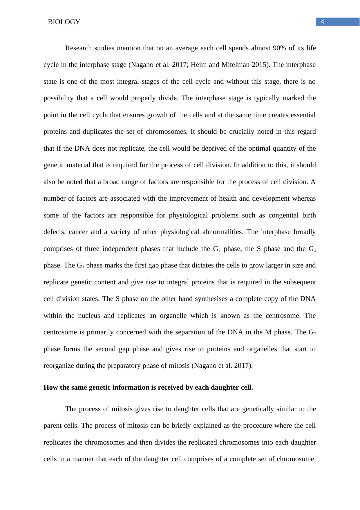
(Karimian et al. 2016). Before the initiation of the process of mitosis, the chromosomes are
replicated. Post replication the chromosomes are coiled and condensed and resemble the
share of the letter ‘X’ inside the nucleus of the cell. The chromosomes at this point comprise
of two sister chromatids which is separated in a manner that each of the new cells contain a
replicated copy of each of the chromosomes.
(Source: Genome.gov 2019)
The process of mitosis gives rise to cell proliferation and basically generates identical cells
that comprise of the similar genetic material as that of the parent cells. The process of mitosis
also helps to replace and regenerate the worn out cells or renew damaged cells. The process
involves the differentiation of a cell into two identical daughter cells. The daughter cells
comprise a copy of each of the chromosomes because the process includes replication of the
chromosome first and then segregating or separation of the copy to give rise to a new set
(Karimian et al. 2016). Before the initiation of the process of mitosis, the chromosomes are
replicated. Post replication the chromosomes are coiled and condensed and resemble the
share of the letter ‘X’ inside the nucleus of the cell. The chromosomes at this point comprise
of two sister chromatids which is separated in a manner that each of the new cells contain a
replicated copy of each of the chromosomes.
(Source: Genome.gov 2019)
⊘ This is a preview!⊘
Do you want full access?
Subscribe today to unlock all pages.
Trusted by 1+ million students worldwide
6BIOLOGY
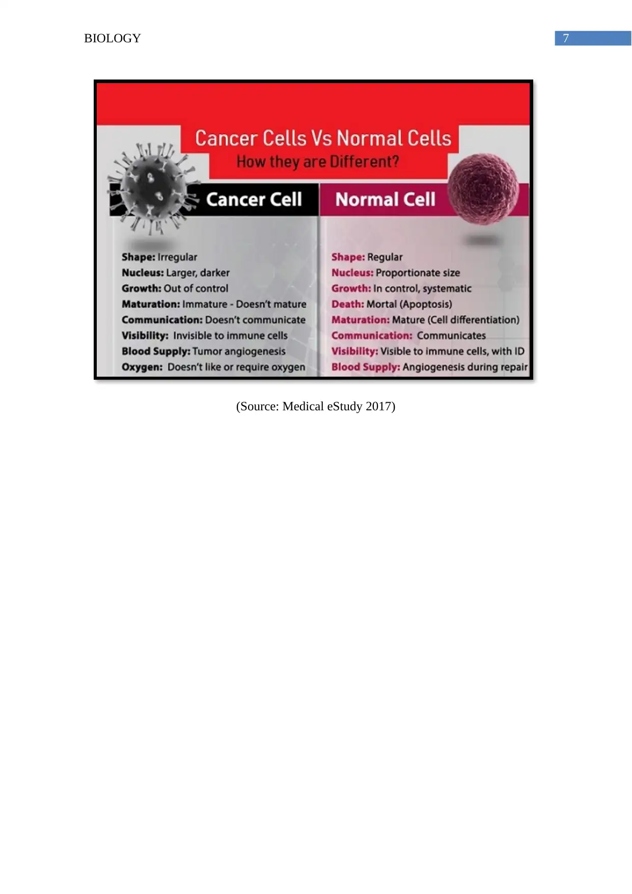
This section also requires you to compare and contrast cancer cells with normal cells.
A number of differences exist between cancer cells and normal cells. Normal cells
typically cease to grow after a certain point of time when sufficient number of cells are
present (Mjelle et al. 2015). For instance, in case of an injury or a wear and tear of the skin,
once a substantial amount of cells are produced for the repair mechanism, the proliferation
process stops, on the other hand, growth and proliferation in cancerous cells do not stop. On
account of the uninhibited growth and proliferation, a tumour is formed which acquires the
characteristic of malignancy and results in cancer. Further normal cells actively respond to
signals acquired from adjacent cells and undertake appropriate actions. However, cancerous
cells do not respond to cell signalling pathway and continue to proliferate which result in
metastasis. Further normal cells undergo an appropriate programmed cell death, on the other
hand, cancerous cells do not undergo apoptosis (Fischer et al. 2015). It should be noted here
that it is the normal cells that turn cancerous due to induced mutation within the tumour
suppressor gene which causes loss of function and on account of induced mutation within the
oncogene, the cancerous cells acquire the property to grow uninterruptedly due to gain of
function mutation (Chen 2016). Also, cancerous cells are able to metastasize and traverse to
different parts of the body. On the other hand, normal cells do not possess the ability to
metastasise and it is on account of these properties that the cancerous cells drastically impact
the normal physiology of the body.
Paraphrase This Document
Need a fresh take? Get an instant paraphrase of this document with our AI Paraphraser
7BIOLOGY
(Source: Medical eStudy 2017)
(Source: Medical eStudy 2017)
8BIOLOGY
Conclusion:
Therefore, to conclude it should be mentioned that assessment provided a clear and
articulate overview about cell biology and the involved genetic mechanism. Typically the
process of cell division is tightly concerned with the process of genetic inheritance as well as
repair mechanism. Further, the central dogma concept provides an overview about how the
genes are transcribed to messenger RNA which ultimately dictates the process of protein
synthesis. In addition to this, the paper also provides a broad overview about embryonic stem
cells and specifically defines the existing differences between cancerous cells and normal
cells. The basic knowledge is integral to understand the mechanism of normal physiological
activities.
⊘ This is a preview!⊘
Do you want full access?
Subscribe today to unlock all pages.
Trusted by 1+ million students worldwide
9BIOLOGY
References:
Bretones, G., Delgado, M.D. and León, J., 2015. Myc and cell cycle control. Biochimica et
Biophysica Acta (BBA)-Gene Regulatory Mechanisms, 1849(5), pp.506-516.
Chen, J., 2016. The cell-cycle arrest and apoptotic functions of p53 in tumor initiation and
progression. Cold Spring Harbor perspectives in medicine, 6(3), p.a026104.
Fischer, M., Quaas, M., Steiner, L. and Engeland, K., 2015. The
p53-p21-DREAM-CDE/CHR pathway regulates G2/M cell cycle genes. Nucleic acids
research, 44(1), pp.164-174.
Genome.gov (2019). National Human Genome Research Institute Home | NHGRI. [online]
Genome.gov. Available at: https://www.genome.gov/ [Accessed 12 Aug. 2019].
Heim, S. and Mitelman, F. eds., 2015. Cancer cytogenetics: chromosomal and molecular
genetic aberrations of tumor cells. John Wiley & Sons.
Jun, S. and Taheri-Araghi, S., 2015. Cell-size maintenance: universal strategy
revealed. Trends in microbiology, 23(1), pp.4-6.
Karimian, A., Ahmadi, Y. and Yousefi, B., 2016. Multiple functions of p21 in cell cycle,
apoptosis and transcriptional regulation after DNA damage. DNA repair, 42, pp.63-71.
Medical eStudy (2019). Cancer Cells Vs Normal Cells: How they are Different - Medical
eStudy. [online] Medical eStudy. Available at: http://www.medicalestudy.com/cancer-cells-
vs-normal-cells-different/ [Accessed 12 Aug. 2019].
Mjelle, R., Hegre, S.A., Aas, P.A., Slupphaug, G., Drabløs, F., Sætrom, P. and Krokan, H.E.,
2015. Cell cycle regulation of human DNA repair and chromatin remodeling genes. DNA
repair, 30, pp.53-67.
Paraphrase This Document
Need a fresh take? Get an instant paraphrase of this document with our AI Paraphraser
10BIOLOGY
Nagano, T., Lubling, Y., Várnai, C., Dudley, C., Leung, W., Baran, Y., Cohen, N.M.,
Wingett, S., Fraser, P. and Tanay, A., 2017. Cell-cycle dynamics of chromosomal
organization at single-cell resolution. Nature, 547(7661), p.61.
Nagano, T., Lubling, Y., Várnai, C., Dudley, C., Leung, W., Baran, Y., Cohen, N.M.,
Wingett, S., Fraser, P. and Tanay, A., 2017. Cell-cycle dynamics of chromosomal
organization at single-cell resolution. Nature, 547(7661), p.61.
1 out of 11
Related Documents


Your All-in-One AI-Powered Toolkit for Academic Success.
+13062052269
info@desklib.com
Available 24*7 on WhatsApp / Email
![[object Object]](/_next/static/media/star-bottom.7253800d.svg)
Unlock your academic potential
Copyright © 2020–2026 A2Z Services. All Rights Reserved. Developed and managed by ZUCOL.