Cardiac Conduction System: Physiology, Disease, and Homeostasis Report

Verified

Added on  2022/08/22

|6
|1130
|28
Report
AI Summary
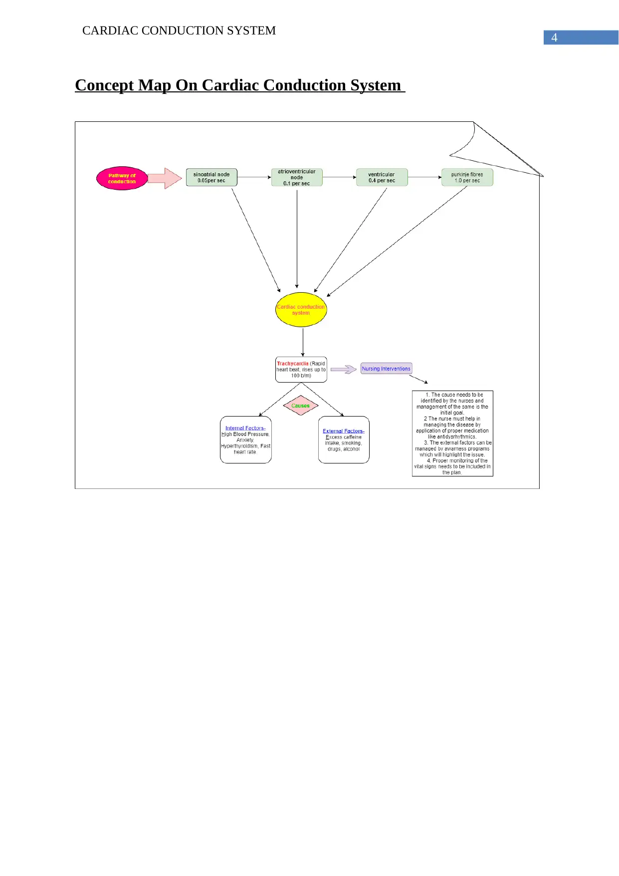
This report provides a comprehensive overview of the cardiac conduction system, detailing its components, including the sinoatrial node, atrioventricular node, bundle of His, Purkinje fibers, and bundle branches, and their roles in initiating and conducting electrical impulses throughout the heart. It explains the process of electrical conduction, the role of transmembrane potential, and the factors that can disrupt normal heart rhythm. The report discusses atrial flutter and tachycardia as examples of arrhythmias, outlining their causes and effects. A concept map is included to visually represent the system. The report references key literature on cardiac electrophysiology and conduction mechanisms, offering insights into the maintenance of homeostasis and the impact of disease. This report is a great resource to understand the physiology of the heart, its functions, and potential diseases.
Document Page
Running head: CARDIAC CONDUCTION SYSTEM
Cardiac conduction system
Name of the Student
Name of the University
Author Note
tabler-icon-diamond-filled.svg

Paraphrase This Document

Need a fresh take? Get an instant paraphrase of this document with our AI Paraphraser
Document Page
1
CARDIAC CONDUCTION SYSTEM
Introduction
The conducting system of heart consists of mainly conducting fibres and cardiac
muscle cells, which are involved in commencing impulses, and carrying these impulses
quickly through the heart (Veeraraghavan, Gourdie & Poelzing, 2014). The process mainly
controls the heart rate. It creates electrical impulses and conducts them all the way through
the muscle of the heart, invigorating the heart to contract and pump blood for maintaining
normal function of the body. The components of this system are sinoatrial node,
atrioventricular node, bundle of his, Purkinje fibers and bundle branches. Disruption of the
electrical conduction leads to defective heartbeat and rhythmicity. The factors that mainly
disrupts the pathway are high blood pressure and blockages in the arteries.
Discussion
Electrical conduction of the heart
The electrical conduction system of heart transmits signals, which is initiated by the
sinoatrial node (SA), which causes tightening of the heart muscle. The signal that is
originated in the SA node travels through the right atrium of the heart and propagates to the
atrioventricular node(AV) along with the Bundle of His with the help of bundle branches to
which ultimately leads to the contraction of the heart muscles, this process helps in the
pumping of the blood throughout the body (Park & Fishman, 2017). The conduction system
comprises of heart muscle cells which are located within the myocardium. The skeleton tissue
is recognised by the electrocardiogram technique. The disruption of this function leads to the
fast, irregular and slow heart rhythms.
Document Page
2
CARDIAC CONDUCTION SYSTEM
The rhythmicity of sinoatrial node is 70 to 80 beats per minutes and that of
atrioventricular node is 40 to 60 beats per minutes. The SA node has the highest rhythmicity
capacity and it maintains the AV node maintains the normal activity of the muscle, if the SA
node fails to regulate the normal function then the normal beat (Boyette & Manna, 2019).
This conduction process leads to the alteration of the membrane potential known as
Transmembrane potential. The galvanometer measures the potential directly which ranges
from -80 to -90mV from outside the cell. The resting potential in the SV note is -80mV and
those of other parts are -90mV. The resting potential is disrupted and resting state is altered
due to the onset of excitation which drops the potential at -20mV, the potential remains in
this state for few minutes. This is known as depolarising state which alters the membrane
permeability to sodium, in this phase the an influx of sodium takes places till the potential
reaches to +20mV (Klabunde, 2017). After this, the potassium efflux out of the cell, which is,
knows as repolarisation. The pacemaker phase also known as diastolic depolarization, which
controls the cardiac chronotropism. The rhythmic is also altered by the stimulation of
sympathetic nerve connected to the heart. The slow heart rate is due to the depolarisation
state of the heart.
Contractility and excitability is another important property of the heart. It is achieved
by the cardiac muscle unit known as myofibrils composed of myosin and actin. This phase is
governed by the generation of ATP which slows or fasts the rate according to the production.
Extra calcium keeps the muscle in contracting state known are calcium rigor. The contraction
happens when the ATP binds to the myosin ultimately pulling the actin to the centre of the
sarcomere.
The all or none law is an important factor in conduction system. It states that the
muscles are only excited if a proper threshold is stimulated. The heart muscle has another
phase known are refractory period where the muscle fails to response in second stimulus if
Document Page
3
CARDIAC CONDUCTION SYSTEM
two stimulus is applied consecutively. These process collectively helps in the propagation of
electrical stimulus which helps in contraction of heart muscle.
Atrial flutter
Atrial flutter or arrhythmia is a condition of heart, which happens due to the defective
rhythmicity. The condition happens when the atria beats very rapidly, which results in a rapid
heartbeat. This can lead to stroke or heart failure (Cosio, 2017).
Tachycardia
Tachycardia is a type of arrhythmia, which leads to the rapid heartbeats compared to
the normal rate, originates from both lower and upper chambers of the heart. It happens due
to deformity in the electrical signals of the heart, which elevates the rate up to 100beats per
minute. It may lead to serious complication such as stroke, cardiac arrest and heart failure.
Atrial flutter is type of tachycardia, which results in the defective high heart rate. It also
caused due to defective electric activity of the heart and is accompanied by the atrial
fibrillation. The factors that may lead to this condition are injury in heart tissue, stress, and
difference in electrolytes, hyperthyroidism and many more (Gopinathannair & Olshansky,
2015).
A concept map is attached to provide a better understanding of the system.
tabler-icon-diamond-filled.svg

Paraphrase This Document

Need a fresh take? Get an instant paraphrase of this document with our AI Paraphraser
Document Page
4
CARDIAC CONDUCTION SYSTEM
Concept Map On Cardiac Conduction System
Document Page
5
CARDIAC CONDUCTION SYSTEM
References
Boyette, L. C., & Manna, B. (2019). Physiology, Myocardial Oxygen Demand. In StatPearls
[Internet]. StatPearls Publishing.
Cosío F. G. (2017). Atrial Flutter, Typical and Atypical: A Review. Arrhythmia &
electrophysiology review, 6(2), 55–62. https://doi.org/10.15420/aer.2017.5.2
Gopinathannair, R., & Olshansky, B. (2015). Management of tachycardia. F1000prime
reports, 7, 60. https://doi.org/10.12703/P7-60
Klabunde, R. E. (2017). Cardiac electrophysiology: normal and ischemic ionic currents and
the ECG. Advances in physiology education, 41(1), 29-37.
Park, D. S., & Fishman, G. I. (2017). Development and Function of the Cardiac Conduction
System in Health and Disease. Journal of cardiovascular development and
disease, 4(2), 7. https://doi.org/10.3390/jcdd4020007
Veeraraghavan, R., Gourdie, R. G., & Poelzing, S. (2014). Mechanisms of cardiac
conduction: a history of revisions. American journal of physiology. Heart and
circulatory physiology, 306(5), H619–H627. https://doi.org/10.1152/ajpheart.00760.2013
chevron_up_icon
1 out of 6
circle_padding
hide_on_mobile
zoom_out_icon
[object Object]