Case Study Analysis: Cardiovascular and Respiratory System Disorders
VerifiedAdded on 2020/11/02
|12
|3491
|77
Case Study
AI Summary
This case study analyzes two medical scenarios: cardiovascular disease (CVD) and asthma. The CVD scenario covers coronary artery disease, including atherosclerosis, angina (stable and variant), acute coronary syndrome, and myocardial infarction (STEMI). It details the pathophysiology, clinical manifestations, diagnostic methods (ECG, serum markers), and treatments such as statins, anti-platelets, beta-blockers, and angioplasty. The asthma scenario describes the obstructive lung disease, its causes (environmental factors, drugs), pathophysiology (atopic and nonatopic attacks), and clinical manifestations. The case study explains the mechanisms of asthma attacks, including bronchospasm, airway inflammation, and mucus production, along with the importance of managing asthma to prevent chronic airway damage. The study also includes a brief overview of COPD, its causes, and effects of smoking on respiratory health.
Scenario 1/ cardio:
CVD lecture:
Coronary arteries:
Left CA – branch directly from the aorta
Right CA – branch directly from the aorta
Right marginal branch of RCA
Left auricle (reflected)
Circumflex branch of LCA
Perfusion of the heart (blood going to the coronary arteries) cannot happen during systole
and only occurs during diastole. The aortic valve cusps close off the coronary arteries during
systole.
Intramyocardial coronary vessels – blood also doesn’t flow during systole.
Autoregulatory mechanisms tell vessels to contract or constrict, usually metabolic factors. If
a cell has a higher level of activity more metabolic factors are released which tells the artery
supplying that factor to dilate to get more blood and increase oxygen and nutrient
absorption.
When blood flows through the coronary arteries the muscle tissue extracts 60-80% of
oxygen in that blood, skeletal only 20-40%. Meaning, the cells will die quickly if blood supply
stops.
Coronary Heart/Artery Disease:
Involves narrowing of the lumen of one or more coronary arteries enough that the
downstream myocardial perfusion is reduced.
Clinical manifestations depend on:
- Which artery
- Transient or permanent obstruction (sometimes or always blocked)
- Partial or complete obstruction (some blood flow or no blood flow)
- Development of collateral blood supply (stimulate the growth of new blood vessels
(anastomosis) to supply blood to cells, takes long time to occur)
Ischemia: reduced blood flow to/in an area
Hypoxia: low oxygenation in an area
Cause of CHD:
Hypertension and hyperlipidaemia, increases risk of atherosclerosis which increases risk of
CAD.
Atherosclerosis:
Common site is proximal coronary arteries, carotid arteries, vertebral, basilic and cerebral
arteries and abdominal, iliac, femoral and popliteal arteries.
Paraphrase This Document
Need a fresh take? Get an instant paraphrase of this document with our AI Paraphraser
It is the process of plaque building up in vessels with can obstruct the pathway of blood,
causing primarily by hyperlipidaemia.
The negative affecting cholesterol is a high level of low density lipoprotien.
The process happens between the epithelium and the elastic layer of the blood vessel
creating an overgrowth of muscle that is bumpy instead of smooth.
Plaque results in a reduced luminal diameter and a reduced capacity to vasodilate.
Chronic ischemic heart disease: Recurrent episodes of transient (happens every now and
again) myocardial ischemia
= partial obstruction to coronary blood flow causing imbalance between O2 supply and O2
demand.
Heart doesn’t have a big stored source of energy in anerobic metabolism (60 seconds before
it starts losing function)
Types of CIHD:
- Stable angina: occurs predictably with exertion or known stressors (temperature
regulation), caused by atherosclerotic plaques in main coronary arteries. Meaning at
rest the myocardium has adequate oxygen supply but with the known stressors the
increased oxygen cannot be met.
The pain is caused by biomechanical fatigue, switching to aerobic to anaerobic
metabolism producing things like: adenosine, lactic acid, hydrogen ions, etc. Glyceryl
trinitrate relieves it.
- Variant angina: occurs unpredictably and can occur at rest. Caused by episodes of
coronary arteries having vasospasms (contracting for no reason)
- Silent myocardial angina: recurrent episodes of myocardial ischemia but no angina
pectoris. Can be causes by autonomic sensory neuron loss like due to diabetes. Can
be diagnosed by exercise stress test.
Usually results in characteristic pain, angina pectoris (symptom) this is the pain associated
with heart attack. Crushing pain in the left side of the chest radiating down the arm and into
the jaw.
This can cause arrhythmias: AF, VT
Treatments:
Statins are a medication that decrease the progression of atherosclerosis.
Anti-platelets: When plaque ruptures platelets adhere to plaques and generate thrombus
which blocks artery.
Beta blockers:
Glyceryl trinitrate: dilates veins, = decreased venous return, decreased energy and oxygen
for heart to contract (decrease pre load). And dialates arterioles = decreased afterload.
Organic nitrates decreased the work of the heart.
Acute Coronary Syndrome:
Chronic ischemic heart disease can develop into/cause acute coronary syndrome.
Classification of ACS:
Distinguishing features:
Onset: can occur when resting
Duration: pain does not go away at rest, can go on for much longer.
Intensity: Pain is worse
Myocardial ischemia, resulting from acute coronary occlusion causing pain and or
discomfort that is more severe, prolonged and or frequent or is not relieved by glyceryl
trinitrate.
Other clinical features:
Diaphoresis – sympathetic nervous system gone into overdrive
Dyspnoea – alterations to breathing rate
Nausea/ and or vomiting –
Feeling of impending doom – emotional response, fight or flight response into overdrive
Angina pectoris: symptom, chest pain
Angina: atherosclerotic plaque
- Unstable angina: this is commonly a progression of stable (plaque ruptures and
platelets aggregate) or variant angina OR secondary to a non-cardiac condition such
as severe anaemia or infection OR post MI.
Pain is more persistent and severe than stable angina.
To determine whether it is this or a non-STEMI is the absence of serum markers for
myocardial damage.
- No ST-segment elevation: no change on ECG, needs cardiac blood work to determine
- ST-segment elevation: MI STEMI
- Acute myocardial infarction
ST segment can be depressed but is usually elevated in ACS. Either depression or elevation
indicate ischemia within the ventricle wall and injury of the cells.
Myocardial infarction type 1:
Thrombus forms on atherosclerotic plaque blocking off blood flow causing ischemia and cell
death.
Process:
Plaque ruptures
Thrombus is over the plaque
Occlusion of coronary artery (either all or partial blockage)
Prolonged ischemia of the cells past the blockage
Hypoxic injury of myocardial cells
Cell necrosis (infarction = tissue death)
Cell membrane breaks down and the content is released into the blood
Troponin I and Troponin T are higher and indicate myocardial infarction, creatine kinases-
MB and myoglobin can be tested also but aren’t as relevant.
However, there are other caused of MI not atherosclerosis related.
⊘ This is a preview!⊘
Do you want full access?
Subscribe today to unlock all pages.

Trusted by 1+ million students worldwide
Scenario:
Stable angina – unstable angina – transient ischemia – sustained ischemia – myocardial
infarction (STEMI) – myocardial inflammation and necrosis (can cause remodelling of the
heart and lead to heart failure.
Where in the patient’s heart will the ischemia be: because in the left anterior descending
artery, the anterior (front) section of the intraventricular septum will become ischemic.
Functional significance:
Once cells start dying cells stop contracting, reduces stroke volume and leads to decreases
in cardiac output or complete stoppages of cardiac output. Can also lead to arrhythmias.
If the patients present to hospital with chest pain and persistent ST elevation on the ECG the
diagnosis is ST elevation myocardial infarction (stemi). No biomarkers required.
STEMI: Within less than 120 minutes reperfusion (return of blood flow) of the heart is the
aim.
Immediate management for chest pain admission as a nurse: Aim to re-establish to maintain
myocardial perfusion
- ECG, Serum markers
- IV Opioid to reduce pain and anxiety reduce sympathetic nervous system activation
- O2 therapy if SaO2 below 93%
- Nitrates such as glyceryl trinitrate, dilate blood vessels reduce workload of heart.
- Aspirin to prevent thrombus formation
- Angioplasty if STEMI is evident
Angioplasty:
Catheter is threaded up through femoral artery through abdominal aorta to the heart and
into the coronary artery where the blockage is found. A balloon is placed to reopen the
coronary artery that is blocked.
Coronary artery bypass graft: CABG
Join the aorta with a vein usually the saphenous vein from the leg and join the aorta to the
coronary artery.
What might happen to the patient if the treatment was prolonged:
Heart Failure:
Symptoms typically occur upon exertion but can also occur at rest. It is secondary to an
abnormality of cardiac structure or function that impairs the ability of the heart to fill with
blood at normal pressure to eject blood sufficiently to fulfil the needs of metabolising
organs.
Paraphrase This Document
Need a fresh take? Get an instant paraphrase of this document with our AI Paraphraser
Scenario 2:
Asthma:
Is an obstructive lung disease
Characterised by reversible obstruction to outflow of air during expiration, meaning
asthmatic episodes are reversible with treatment.
Chronic uncontrolled asthma can lead to permanent remodelling of airways and progressive
airway damage.
Asthma must be managed well to avoid life threatening episodes and progressive airway
damage.
10-15% in children and 5-10% in adults.
Serious cases can lead to respiratory failure and death. Education and upkeep on asthma
treatment is critical in management.
Common causes:
- Environmental factors: tobacco, temperature, thunderstorms, air pollution, perfume
- Drugs: aspirin, beta-blockers
- Immunological challenges: dust mites, pollens, wood dust
- Psychological – emotional stress – sympathetic nervous system activation
- Exercise – tissues need more oxygen & sympathetic nervous system activation
- Respiratory infection
Demographic factors/risk factors:
- Age (children more likely)
- Ethnicity
- genetics
- Co-morbidities
Pathophysiology:
There are 2 types of asthma attacks
Atopic: Heightened immune response
- Onset usually childhood, related to family history
- Brought on by extrinsic antigen such as pollen or allergen
- Response has 2 phases: early or acute which occurs in 10 minutes and late-phase
occurring 4-8 hours post exposure.
Nonatopic:
- Infections
- Exercise
- Inhaled irritants (tobacco smoke)
Underlying mechanisms:
Inflammatory changes through bronchial walls
- Stimulates parasympathetic nervous system nerves in lung tissue
This causes contraction or spasm of bronchiolar smooth muscle
Leads to constriction of bronchioles
Reduced ventilation
- Histamine release causes vasodilation
Leads to oedema of interstitum surrounding bronchioles (obstruction of small
airways)
Increased mucus production (plugs) can block the airways
All leads to ventilation and perfusion mismatch and reduced gas exchange.
Phases of an atopic asthma attack: (early asthmatic response)
Antigen enter the airway (e.g. pollen)
Antigen binds to mast cells
Mast cell degranulation and release of mediators (contents), this activates the
parasympathetic nervous system and caused bronchospasm/bronchioles constricting.
Degranulation causes the lining of the air way to form gaps where antigens get into
Antigen binds to mast cells to the mucosa
Mucus secretion increased leading to mucus plugs
Vascular leak of fluid leading to oedema
Airway smooth muscle constriction (due to parasympathetic nervous system activation
Phases of an Atopic asthma attack: (late phase)
Damage to lining of airway more severe
Lots of mucus produced blocking airways
Immune cells (t cells) arrived to site
Lead to increases in oedema
3 main things:
- Bronchospasm/ bronchiolar constriction caused by the sympathetic nervous system
- Airway inflammation causes oedema
- Production and accumulation of mucus blocking airways
Expiration is difficult in expiratory diseases.
Untreated chronic asthmatic damage:
- Smooth muscle constriction
- Degranulation of cell tissue
- Mucus accumulation and plugs the airway
- Hyperinflation of alveoli
Asthma manifestations:
Presentation:
- Expiratory wheeze caused by constriction making it harder for air to leave the lungs
- Reduced FEV1
- Dyspnoea
- Non-productive cough
⊘ This is a preview!⊘
Do you want full access?
Subscribe today to unlock all pages.

Trusted by 1+ million students worldwide
- Accessory muscle use
- Anxiety and fear – increased SNS causes tachycardia
- Hypoxia in later stages
The patient in the scenario is likely having a moderate exacerbation of asthma due to his
FEV1 being within 50 – 75%.
COPD:
COPD is characterised by airflow limitation in the lungs which can lead to milk or severe
shortness of breath that is not fully reversible by medication/treatment. It is a serious
chronic condition affecting mainly the older population. It includes: bronchitis and
emphysema.
CO2 levels are monitored by peripheral chemoreceptors.
FEV1/FVC is lower with obstruction.
Hypoxia: low oxygen at a tissue level
Hypoxemia: low oxygen in the blood.
Causes of respiratory failure:
- Inadequate ventilation
- Ventilation and perfusion not matched
- Impaired gas diffusion from changes to the respiratory membrane
Chronic respiratory disorders - obstructive lung diseases – COPD – emphysema and
bronchitis
Females present different symptoms to male pts, more severe dyspnoea and are also at
higher risk to more significant harm from tobacco.
Effects of smoking on respiratory health:
FEV naturally declines
- Smokers can lose double their forced expiratory volume in comparison to non -
smokers.
- Peak is at age 25 and healthy individuals can usually expire 80% of their lung
capacity.
- Young smokers might not reach peak capacity.
Tobacco is a constant irritant to lining of the lung.
- Abnormal growth of bronchial epithelium
- Loss of mucocillary escalator
- Chronic inflammation
- Progression to neoplasia – lung cancer
Mucocillary escalator; brushes to push mucus and pathogens up.
Paraphrase This Document
Need a fresh take? Get an instant paraphrase of this document with our AI Paraphraser
Mucus is a great home for bacteria.
Obstructive lung disease - airflow limitation that is not reversible
- Chronic inflammatory response impacts on ventilation of lungs and diffusion of
gases.
- Pathological lung changes
Pathophysiology: bronchitis
Tobacco smoke and/or air pollution (causes)
inflamed airway epithelium (leads to)
infiltration of inflammatory cells + release of cytokines. Causes systemic effects
(muscle weakness, weight loss) (leads to)
continuous bronchial inflammation (results in)
chronic bronchitis, mucus production and hypersecretion, bronchial oedema,
bacterial colonisation of airways
Chronic bronchitis:
- bronchial oedema
- overproduction of mucus
- bacterial infections,
- inflammation in the larger bronchi and then eventually the entire airway
- chronic productive cough
Pathophysiology: emphysema
Tobacco smoke and/or air pollution (causes)
inflamed airway epithelium (leads to)
infiltration of inflammatory cells + release of cytokines. Causes systemic effects
(muscle weakness, weight loss) (leads to)
breakdown of lung elastic tissue (stretch and recoil of the lungs) (causes)
difficult expiration (leads to)
Destruction of alveolar septa – the wall between alveoli.
Emphysema:
- Destruction of connective tissue in alveolar walls leads to reduction of surface area
for gas exchange
- Loss of elastic in walls of alveoli and bronchioles leads to expiratory limitation
because of obstruction and airway collapse.
Centriacinar: the centre of the airway, more prominent with smoking due to smoke only
reaching the bronchi
Panacinar: can still occur with copd but not as common, more commonly genetic cause.
During expiration air movement is trapped via the bronchial walls collapsing and the mucus
plug, causing airway obstruction. Air becomes trapped in the lungs and causes
hyperinflation of the chest.
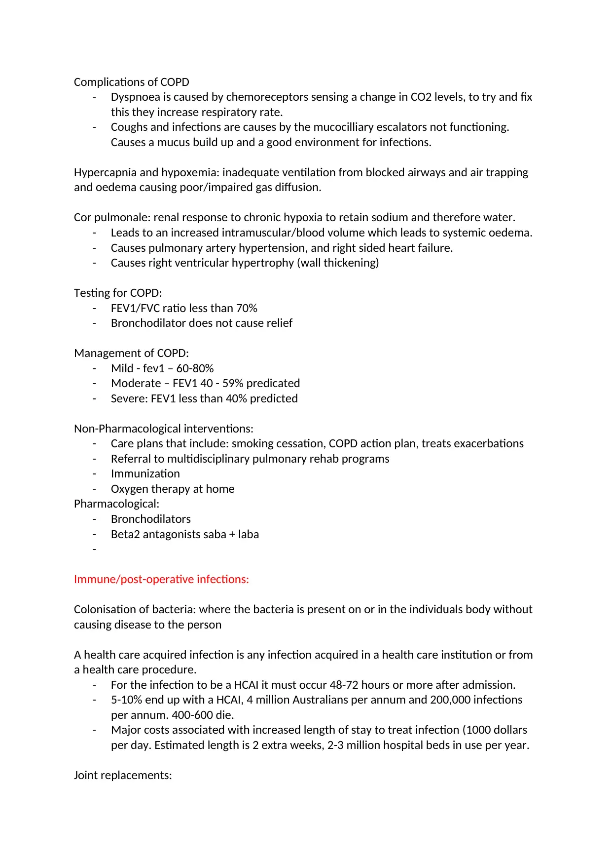
Complications of COPD
- Dyspnoea is caused by chemoreceptors sensing a change in CO2 levels, to try and fix
this they increase respiratory rate.
- Coughs and infections are causes by the mucocilliary escalators not functioning.
Causes a mucus build up and a good environment for infections.
Hypercapnia and hypoxemia: inadequate ventilation from blocked airways and air trapping
and oedema causing poor/impaired gas diffusion.
Cor pulmonale: renal response to chronic hypoxia to retain sodium and therefore water.
- Leads to an increased intramuscular/blood volume which leads to systemic oedema.
- Causes pulmonary artery hypertension, and right sided heart failure.
- Causes right ventricular hypertrophy (wall thickening)
Testing for COPD:
- FEV1/FVC ratio less than 70%
- Bronchodilator does not cause relief
Management of COPD:
- Mild - fev1 – 60-80%
- Moderate – FEV1 40 - 59% predicated
- Severe: FEV1 less than 40% predicted
Non-Pharmacological interventions:
- Care plans that include: smoking cessation, COPD action plan, treats exacerbations
- Referral to multidisciplinary pulmonary rehab programs
- Immunization
- Oxygen therapy at home
Pharmacological:
- Bronchodilators
- Beta2 antagonists saba + laba
-
Immune/post-operative infections:
Colonisation of bacteria: where the bacteria is present on or in the individuals body without
causing disease to the person
A health care acquired infection is any infection acquired in a health care institution or from
a health care procedure.
- For the infection to be a HCAI it must occur 48-72 hours or more after admission.
- 5-10% end up with a HCAI, 4 million Australians per annum and 200,000 infections
per annum. 400-600 die.
- Major costs associated with increased length of stay to treat infection (1000 dollars
per day. Estimated length is 2 extra weeks, 2-3 million hospital beds in use per year.
Joint replacements:
⊘ This is a preview!⊘
Do you want full access?
Subscribe today to unlock all pages.

Trusted by 1+ million students worldwide
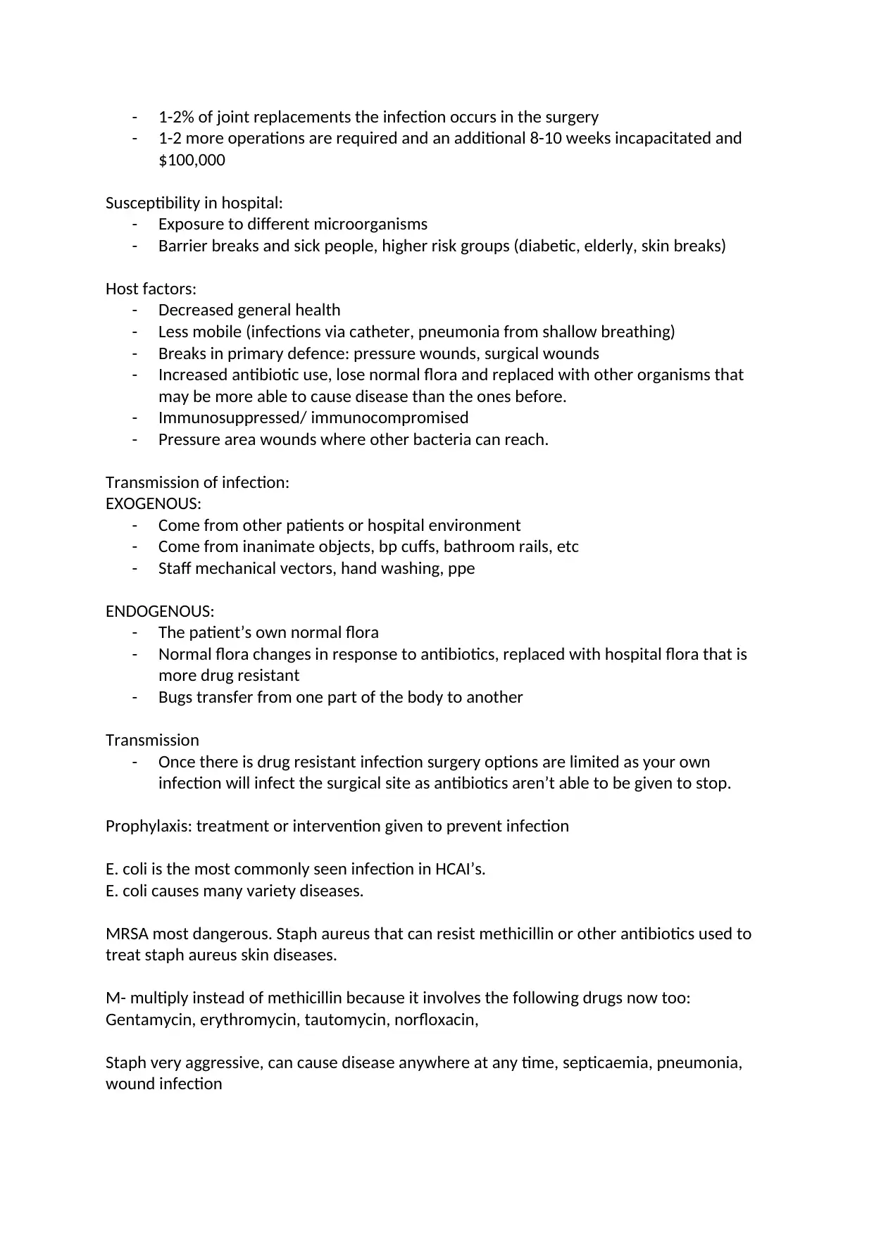
- 1-2% of joint replacements the infection occurs in the surgery
- 1-2 more operations are required and an additional 8-10 weeks incapacitated and
$100,000
Susceptibility in hospital:
- Exposure to different microorganisms
- Barrier breaks and sick people, higher risk groups (diabetic, elderly, skin breaks)
Host factors:
- Decreased general health
- Less mobile (infections via catheter, pneumonia from shallow breathing)
- Breaks in primary defence: pressure wounds, surgical wounds
- Increased antibiotic use, lose normal flora and replaced with other organisms that
may be more able to cause disease than the ones before.
- Immunosuppressed/ immunocompromised
- Pressure area wounds where other bacteria can reach.
Transmission of infection:
EXOGENOUS:
- Come from other patients or hospital environment
- Come from inanimate objects, bp cuffs, bathroom rails, etc
- Staff mechanical vectors, hand washing, ppe
ENDOGENOUS:
- The patient’s own normal flora
- Normal flora changes in response to antibiotics, replaced with hospital flora that is
more drug resistant
- Bugs transfer from one part of the body to another
Transmission
- Once there is drug resistant infection surgery options are limited as your own
infection will infect the surgical site as antibiotics aren’t able to be given to stop.
Prophylaxis: treatment or intervention given to prevent infection
E. coli is the most commonly seen infection in HCAI’s.
E. coli causes many variety diseases.
MRSA most dangerous. Staph aureus that can resist methicillin or other antibiotics used to
treat staph aureus skin diseases.
M- multiply instead of methicillin because it involves the following drugs now too:
Gentamycin, erythromycin, tautomycin, norfloxacin,
Staph very aggressive, can cause disease anywhere at any time, septicaemia, pneumonia,
wound infection
Paraphrase This Document
Need a fresh take? Get an instant paraphrase of this document with our AI Paraphraser
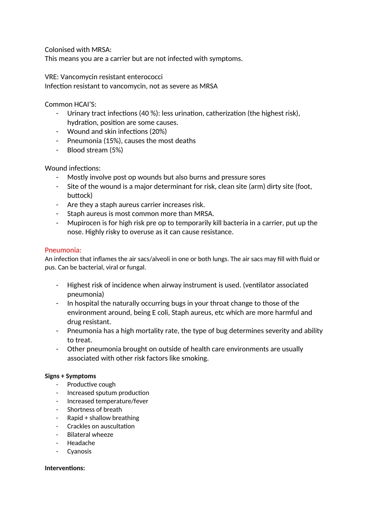
Colonised with MRSA:
This means you are a carrier but are not infected with symptoms.
VRE: Vancomycin resistant enterococci
Infection resistant to vancomycin, not as severe as MRSA
Common HCAI’S:
- Urinary tract infections (40 %): less urination, catherization (the highest risk),
hydration, position are some causes.
- Wound and skin infections (20%)
- Pneumonia (15%), causes the most deaths
- Blood stream (5%)
Wound infections:
- Mostly involve post op wounds but also burns and pressure sores
- Site of the wound is a major determinant for risk, clean site (arm) dirty site (foot,
buttock)
- Are they a staph aureus carrier increases risk.
- Staph aureus is most common more than MRSA.
- Mupirocen is for high risk pre op to temporarily kill bacteria in a carrier, put up the
nose. Highly risky to overuse as it can cause resistance.
Pneumonia:
An infection that inflames the air sacs/alveoli in one or both lungs. The air sacs may fill with fluid or
pus. Can be bacterial, viral or fungal.
- Highest risk of incidence when airway instrument is used. (ventilator associated
pneumonia)
- In hospital the naturally occurring bugs in your throat change to those of the
environment around, being E coli, Staph aureus, etc which are more harmful and
drug resistant.
- Pneumonia has a high mortality rate, the type of bug determines severity and ability
to treat.
- Other pneumonia brought on outside of health care environments are usually
associated with other risk factors like smoking.
Signs + Symptoms
- Productive cough
- Increased sputum production
- Increased temperature/fever
- Shortness of breath
- Rapid + shallow breathing
- Crackles on auscultation
- Bilateral wheeze
- Headache
- Cyanosis
Interventions:

- Oxygen therapy
- IV Antibiotics
- Positioning
- Pain assessment
-
Why do patients with COPD have increased risk of pneumonia?
- They already have mucus in air sacs, impaired gas exchange, perfect condition for
colonisation of bacteria: warm environment, prescence of bacteria.
Why is pneumonia dangerous for patients with COPD?
- Further obstruction of the airway, decreased level of gas exchange, full body effect as less
oxygen to the body and cells.
⊘ This is a preview!⊘
Do you want full access?
Subscribe today to unlock all pages.

Trusted by 1+ million students worldwide
1 out of 12
Related Documents
Your All-in-One AI-Powered Toolkit for Academic Success.
+13062052269
info@desklib.com
Available 24*7 on WhatsApp / Email
![[object Object]](/_next/static/media/star-bottom.7253800d.svg)
Unlock your academic potential
Copyright © 2020–2026 A2Z Services. All Rights Reserved. Developed and managed by ZUCOL.


