Critical Analysis: CBCT's Influence on Endodontic Treatment Planning
VerifiedAdded on 2023/05/29
|6
|1364
|191
Literature Review
AI Summary
This literature review explores the effectiveness of Cone Beam Computed Tomography (CBCT) in endodontics, focusing on its impact on clinical decision-making. It begins by establishing the rationale for using CBCT over traditional 2D radiographs due to limitations like anatomical noise and geometric distortion. The review analyzes existing literature based on the Fryback and Thornbury hierarchical model of efficacy, specifically levels 3 to 6, which assess CBCT's influence on clinicians' diagnostic estimations, treatment planning, and patient outcomes. The methodology involves a comprehensive literature search across PubMed, Google Scholar, and Open Grey, resulting in the selection of 12 articles that meet the inclusion criteria. The review aims to determine whether CBCT improves diagnostic and prognostic assessments and alters treatment plans compared to conventional radiography, ultimately contributing to better patient care in endodontics. Desklib provides access to this review along with other solved assignments.

Running head: BACKGROUND OF THE STUDY
BACKGROUND OF THE STUDY
Name of the Student
Name of the university
Author’s note
BACKGROUND OF THE STUDY
Name of the Student
Name of the university
Author’s note
Paraphrase This Document
Need a fresh take? Get an instant paraphrase of this document with our AI Paraphraser
1BACKGROUND OF THE STUDY
Rationale for the study
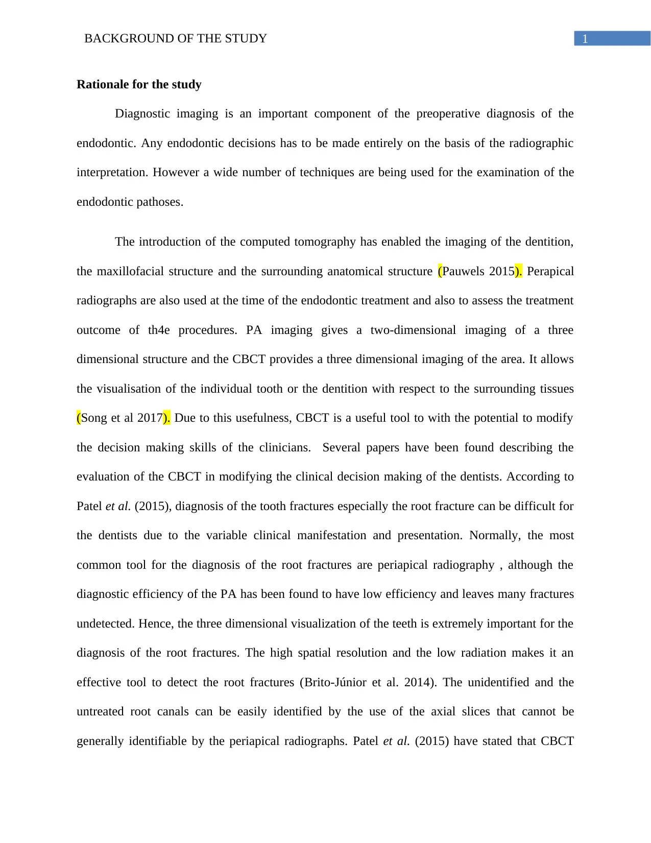
Diagnostic imaging is an important component of the preoperative diagnosis of the
endodontic. Any endodontic decisions has to be made entirely on the basis of the radiographic
interpretation. However a wide number of techniques are being used for the examination of the
endodontic pathoses.
The introduction of the computed tomography has enabled the imaging of the dentition,
the maxillofacial structure and the surrounding anatomical structure (Pauwels 2015). Perapical
radiographs are also used at the time of the endodontic treatment and also to assess the treatment
outcome of th4e procedures. PA imaging gives a two-dimensional imaging of a three
dimensional structure and the CBCT provides a three dimensional imaging of the area. It allows
the visualisation of the individual tooth or the dentition with respect to the surrounding tissues
(Song et al 2017). Due to this usefulness, CBCT is a useful tool to with the potential to modify
the decision making skills of the clinicians. Several papers have been found describing the
evaluation of the CBCT in modifying the clinical decision making of the dentists. According to
Patel et al. (2015), diagnosis of the tooth fractures especially the root fracture can be difficult for
the dentists due to the variable clinical manifestation and presentation. Normally, the most
common tool for the diagnosis of the root fractures are periapical radiography , although the
diagnostic efficiency of the PA has been found to have low efficiency and leaves many fractures
undetected. Hence, the three dimensional visualization of the teeth is extremely important for the
diagnosis of the root fractures. The high spatial resolution and the low radiation makes it an
effective tool to detect the root fractures (Brito‐Júnior et al. 2014). The unidentified and the
untreated root canals can be easily identified by the use of the axial slices that cannot be
generally identifiable by the periapical radiographs. Patel et al. (2015) have stated that CBCT
2BACKGROUND OF THE STUDY
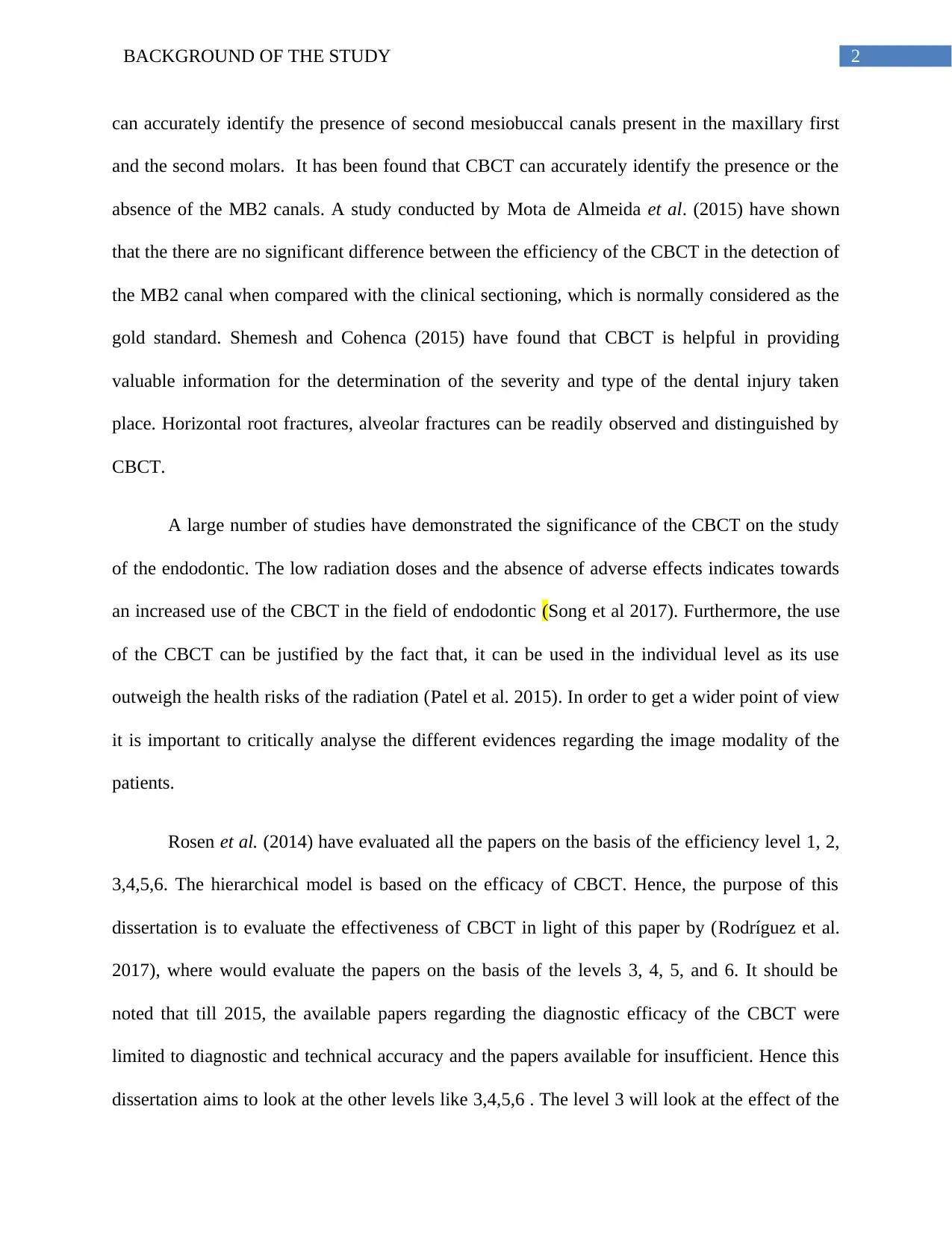
can accurately identify the presence of second mesiobuccal canals present in the maxillary first
and the second molars. It has been found that CBCT can accurately identify the presence or the
absence of the MB2 canals. A study conducted by Mota de Almeida et al. (2015) have shown
that the there are no significant difference between the efficiency of the CBCT in the detection of
the MB2 canal when compared with the clinical sectioning, which is normally considered as the
gold standard. Shemesh and Cohenca (2015) have found that CBCT is helpful in providing
valuable information for the determination of the severity and type of the dental injury taken
place. Horizontal root fractures, alveolar fractures can be readily observed and distinguished by
CBCT.
A large number of studies have demonstrated the significance of the CBCT on the study
of the endodontic. The low radiation doses and the absence of adverse effects indicates towards
an increased use of the CBCT in the field of endodontic (Song et al 2017). Furthermore, the use
of the CBCT can be justified by the fact that, it can be used in the individual level as its use
outweigh the health risks of the radiation (Patel et al. 2015). In order to get a wider point of view
it is important to critically analyse the different evidences regarding the image modality of the
patients.
Rosen et al. (2014) have evaluated all the papers on the basis of the efficiency level 1, 2,
3,4,5,6. The hierarchical model is based on the efficacy of CBCT. Hence, the purpose of this
dissertation is to evaluate the effectiveness of CBCT in light of this paper by (Rodríguez et al.
2017), where would evaluate the papers on the basis of the levels 3, 4, 5, and 6. It should be
noted that till 2015, the available papers regarding the diagnostic efficacy of the CBCT were
limited to diagnostic and technical accuracy and the papers available for insufficient. Hence this
dissertation aims to look at the other levels like 3,4,5,6 . The level 3 will look at the effect of the
⊘ This is a preview!⊘
Do you want full access?
Subscribe today to unlock all pages.

Trusted by 1+ million students worldwide
3BACKGROUND OF THE STUDY
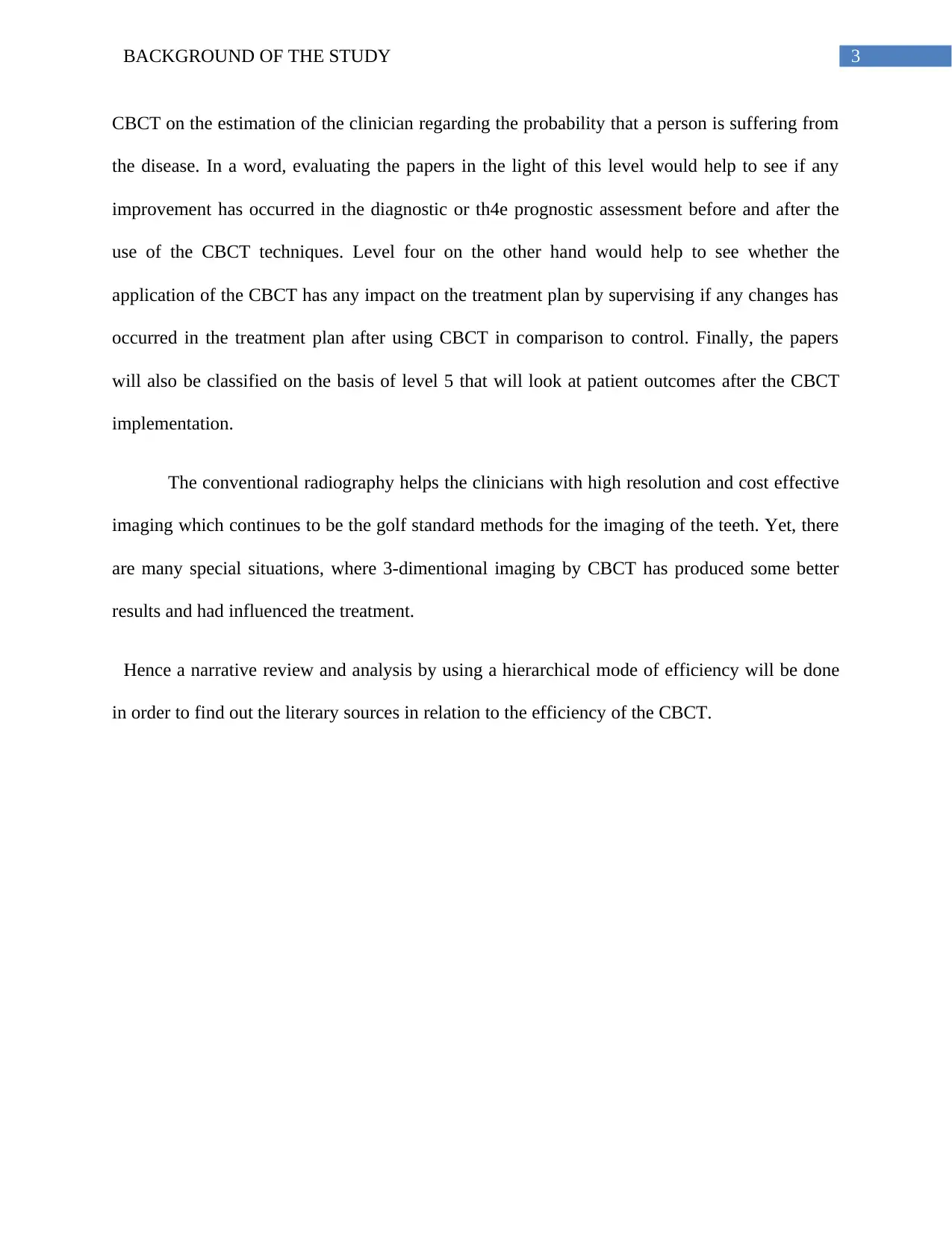
CBCT on the estimation of the clinician regarding the probability that a person is suffering from
the disease. In a word, evaluating the papers in the light of this level would help to see if any
improvement has occurred in the diagnostic or th4e prognostic assessment before and after the
use of the CBCT techniques. Level four on the other hand would help to see whether the
application of the CBCT has any impact on the treatment plan by supervising if any changes has
occurred in the treatment plan after using CBCT in comparison to control. Finally, the papers
will also be classified on the basis of level 5 that will look at patient outcomes after the CBCT
implementation.
The conventional radiography helps the clinicians with high resolution and cost effective
imaging which continues to be the golf standard methods for the imaging of the teeth. Yet, there
are many special situations, where 3-dimentional imaging by CBCT has produced some better
results and had influenced the treatment.
Hence a narrative review and analysis by using a hierarchical mode of efficiency will be done
in order to find out the literary sources in relation to the efficiency of the CBCT.
Paraphrase This Document
Need a fresh take? Get an instant paraphrase of this document with our AI Paraphraser
4BACKGROUND OF THE STUDY
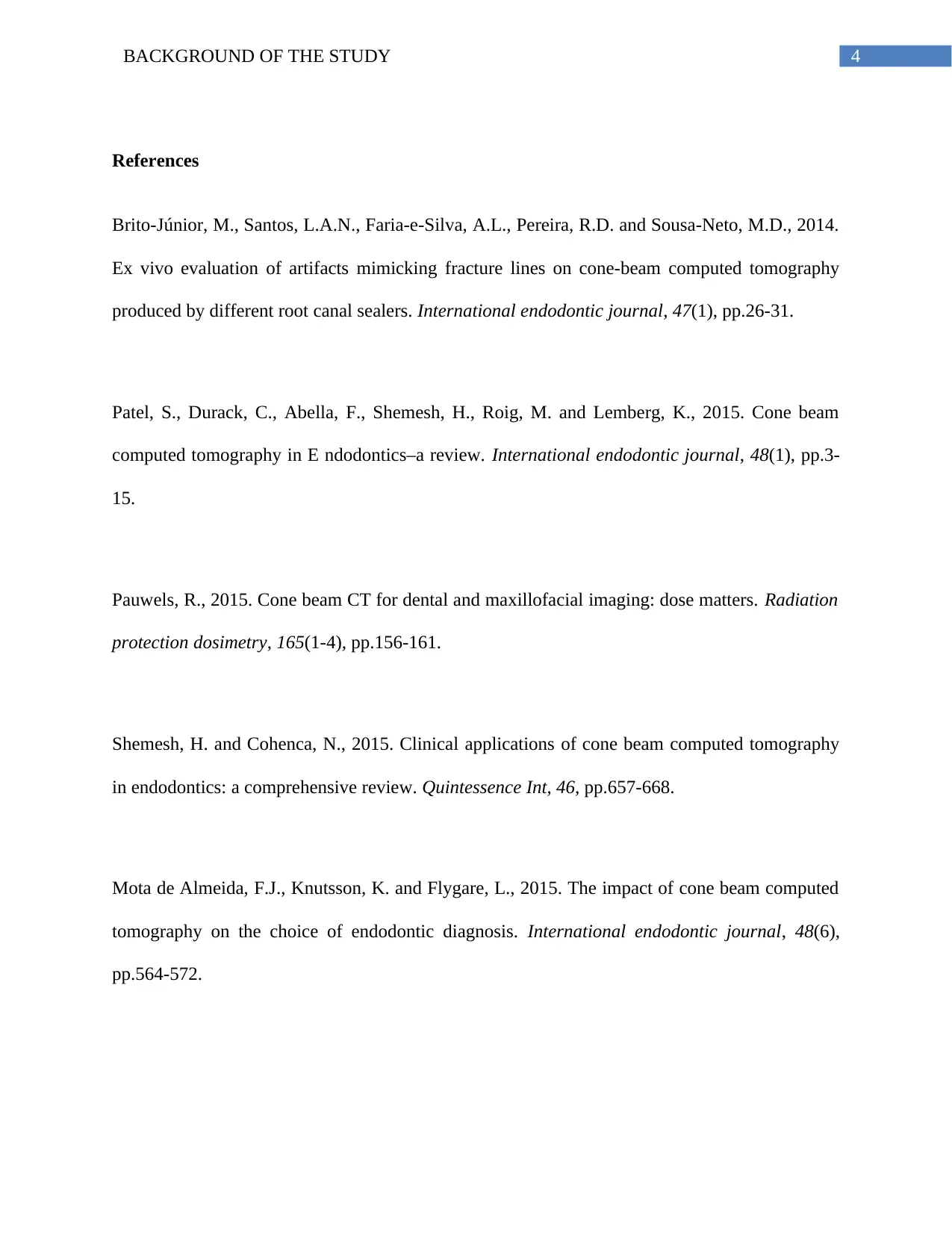
References
Brito‐Júnior, M., Santos, L.A.N., Faria‐e‐Silva, A.L., Pereira, R.D. and Sousa‐Neto, M.D., 2014.
Ex vivo evaluation of artifacts mimicking fracture lines on cone‐beam computed tomography
produced by different root canal sealers. International endodontic journal, 47(1), pp.26-31.
Patel, S., Durack, C., Abella, F., Shemesh, H., Roig, M. and Lemberg, K., 2015. Cone beam
computed tomography in E ndodontics–a review. International endodontic journal, 48(1), pp.3-
15.
Pauwels, R., 2015. Cone beam CT for dental and maxillofacial imaging: dose matters. Radiation
protection dosimetry, 165(1-4), pp.156-161.
Shemesh, H. and Cohenca, N., 2015. Clinical applications of cone beam computed tomography
in endodontics: a comprehensive review. Quintessence Int, 46, pp.657-668.
Mota de Almeida, F.J., Knutsson, K. and Flygare, L., 2015. The impact of cone beam computed
tomography on the choice of endodontic diagnosis. International endodontic journal, 48(6),
pp.564-572.
5BACKGROUND OF THE STUDY
Rodríguez, G., Patel, S., Durán-Sindreu, F., Roig, M. and Abella, F., 2017. Influence of Cone-
beam Computed Tomography on Endodontic Retreatment Strategies among General Dental
Practitioners and Endodontists. Journal of endodontics, 43(9), pp.1433-1437.
Song, D., Zhang, L., Zhou, W., Zheng, Q., Duan, X., Zhou, X. and Huang, D., 2017. Comparing
cone-beam computed tomography with periapical radiography for assessing root canal obturation
in vivo using microsurgical findings as validation. Dentomaxillofacial Radiology, 46(5),
p.20160463.
Rosen, E., Taschieri, S., Del Fabbro, M., Beitlitum, I. and Tsesis, I., 2015. The diagnostic
efficacy of cone-beam computed tomography in endodontics: a systematic review and analysis
by a hierarchical model of efficacy. Journal of endodontics, 41(7), pp.1008-1014.
⊘ This is a preview!⊘
Do you want full access?
Subscribe today to unlock all pages.

Trusted by 1+ million students worldwide
1 out of 6
Your All-in-One AI-Powered Toolkit for Academic Success.
+13062052269
info@desklib.com
Available 24*7 on WhatsApp / Email
![[object Object]](/_next/static/media/star-bottom.7253800d.svg)
Unlock your academic potential
Copyright © 2020–2026 A2Z Services. All Rights Reserved. Developed and managed by ZUCOL.


