Cell Structure, Metabolism, and Growth: A Detailed Biological Report
VerifiedAdded on 2022/08/30
|35
|6788
|35
Report
AI Summary
This report provides a detailed analysis of cell structure, metabolism, and growth. It begins with an overview of cell characteristics, comparing and contrasting prokaryotic and eukaryotic cells, and discussing the impact of viruses. The report then delves into eukaryotic sub-cellular structures and organelles. Section 2 focuses on cellular metabolism, examining the role of the cell membrane in nutrient regulation and waste removal, the use of nutrients for energy in animal cells, and the roles of nucleic acids in the nucleus and cytoplasm, including protein synthesis. Section 3 explores cell growth and division, covering the generation of specialized tissues from embryonic stem cells, the importance of interphase, factors initiating cell division, and how genetic information is maintained during cell division.
Running head: UNDERSTANDING THE CELL STRUCTURE, METABOLISM AND CELL GROWTH
UNDERSTANDING THE CELL STRUCTURE, METABOLISM AND
CELL GROWTH
Name of the Student:
Name of the University:
Author Note:
UNDERSTANDING THE CELL STRUCTURE, METABOLISM AND
CELL GROWTH
Name of the Student:
Name of the University:
Author Note:
Paraphrase This Document
Need a fresh take? Get an instant paraphrase of this document with our AI Paraphraser
1CELL STRUCTURE, METABOLISM AND CELL GROWTH
Introduction
The cell is the membrane-bound unit that contains the main functional molecules of life.
Living organelles are present in the cell, and it helps in the building blocks of molecular
organisms like humans and other animals. The animal body or the plant body is composed of
fundamental functional units known as microscopic cells. A number of organisms are made
of single-cell; on the other hand, multicellular organisms are composed of multiple cells. A
single-cell organism is known as unicellular or acellular such as amoeba, Chlamydomonas,
and Acetabidaria. Typically, cells are classified into three main types: stem cells, post-mitotic
cells, and dedifferentiated cells (Anderson, 2015, p. 1723). Differentiated cells are
specialized, which return to the undifferentiated zone to take over the work of the division.
Dedifferentiation helps in healing the wounds and vegetative propagation in cells. Eukaryotes
are multicellular, have a cell of size between 5-100 μm. Among all cells, muscle and nerve
cells are comparatively larger. In the case of plants, algae contain large cells as compared to
others. Typically eggs are large size cells that provide food for the development of the
embryo. The human egg is 0.1mm in diameter. Plant cell contains a cell wall and protoplast,
whereas no cell wall is present in the animal cell. It is differentiated into the plasma
membrane, cytoplasm, nucleus, and vacuoles. The cytoplasm is divided into a cytoplasmic
matrix and endoplasmic reticulum (Threadgold, 2017). Cells are generally classified into two
types: Prokaryotic and eukaryotic. Prokaryotes are found in bacteria, and eukaryotes are
observed in plants and animals. A eukaryotic cell contains a nucleus, mitochondria, Golgi
body, endoplasmic reticulum, vacuoles, centriole, vesicle, lysosome, and ribosome. The cell
membrane is present in the eukaryotic cell, which comprises of phospholipids and proteins.
The phospholipid bilayer is found to work as the fluid mosaic model (Turlier, 2018, p. 114).
ATP is the primary energy source of the cell and is produced in the mitochondria. Cell
2CELL STRUCTURE, METABOLISM AND CELL GROWTH
metabolism refers to the different set of chemical reactions, which occurs in the cell. Cell
gain nutrients and energy from the food that is consumed by the animal. Foods are digested in
the animal body through different organelles, which provide nutrients to the cell. Cell
division is significant, and it is fragmented into some phases: G1 phase, S phase, G2 phase,
and M phase (mitosis and cytokinesis).
Discussion
Section 1
Living cells have eight important characteristics. First, ingestion is a system to absorb
through cell boundaries or chemical release in the regions. Two, assimilation, is a system in
which a series of conversions of ingested food into the chemical substances takes place,
which is referred to become a biochemical pathway. The release of energy converted into
ATP is an example of assimilation. Third, growth, which is the result of assimilation that
occurs through repairing the parts of the body (Shrimal, 2019, p. 288). Fourth, reproduction
means the production of more individual cells like self. Fifth, elimination refers to the release
of the unused materials or toxic substances, which are converted into molecules by the cell.
Sixth, Responsiveness means to respond to the environment that is a type of reaction.
Seventh, coordination is a complex metabolic reaction that happens with the inhibitors.
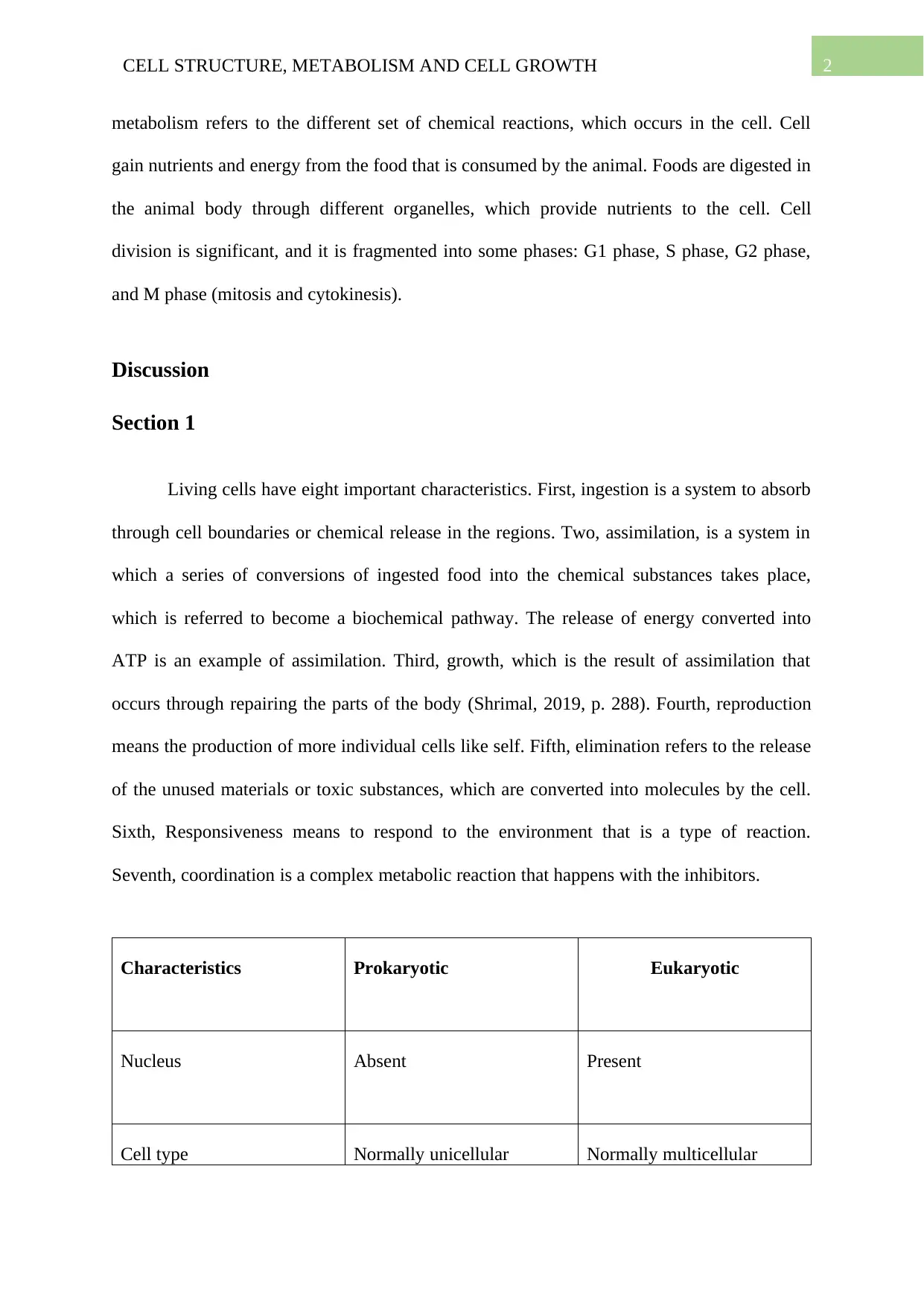
Characteristics Prokaryotic Eukaryotic
Nucleus Absent Present
Cell type Normally unicellular Normally multicellular
metabolism refers to the different set of chemical reactions, which occurs in the cell. Cell
gain nutrients and energy from the food that is consumed by the animal. Foods are digested in
the animal body through different organelles, which provide nutrients to the cell. Cell
division is significant, and it is fragmented into some phases: G1 phase, S phase, G2 phase,
and M phase (mitosis and cytokinesis).
Discussion
Section 1
Living cells have eight important characteristics. First, ingestion is a system to absorb
through cell boundaries or chemical release in the regions. Two, assimilation, is a system in
which a series of conversions of ingested food into the chemical substances takes place,
which is referred to become a biochemical pathway. The release of energy converted into
ATP is an example of assimilation. Third, growth, which is the result of assimilation that
occurs through repairing the parts of the body (Shrimal, 2019, p. 288). Fourth, reproduction
means the production of more individual cells like self. Fifth, elimination refers to the release
of the unused materials or toxic substances, which are converted into molecules by the cell.
Sixth, Responsiveness means to respond to the environment that is a type of reaction.
Seventh, coordination is a complex metabolic reaction that happens with the inhibitors.
Characteristics Prokaryotic Eukaryotic
Nucleus Absent Present
Cell type Normally unicellular Normally multicellular
⊘ This is a preview!⊘
Do you want full access?
Subscribe today to unlock all pages.
Trusted by 1+ million students worldwide
3CELL STRUCTURE, METABOLISM AND CELL GROWTH
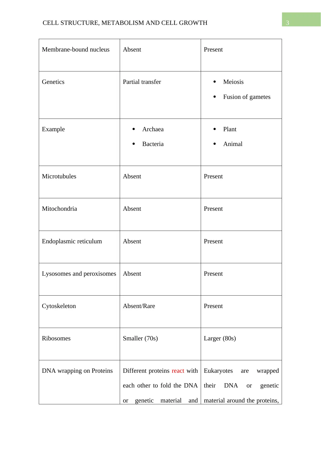
Membrane-bound nucleus Absent Present
Genetics Partial transfer Meiosis
Fusion of gametes
Example Archaea
Bacteria
Plant
Animal
Microtubules Absent Present
Mitochondria Absent Present
Endoplasmic reticulum Absent Present
Lysosomes and peroxisomes Absent Present
Cytoskeleton Absent/Rare Present
Ribosomes Smaller (70s) Larger (80s)
DNA wrapping on Proteins Different proteins react with
each other to fold the DNA
or genetic material and
Eukaryotes are wrapped
their DNA or genetic
material around the proteins,
Paraphrase This Document
Need a fresh take? Get an instant paraphrase of this document with our AI Paraphraser
4CELL STRUCTURE, METABOLISM AND CELL GROWTH
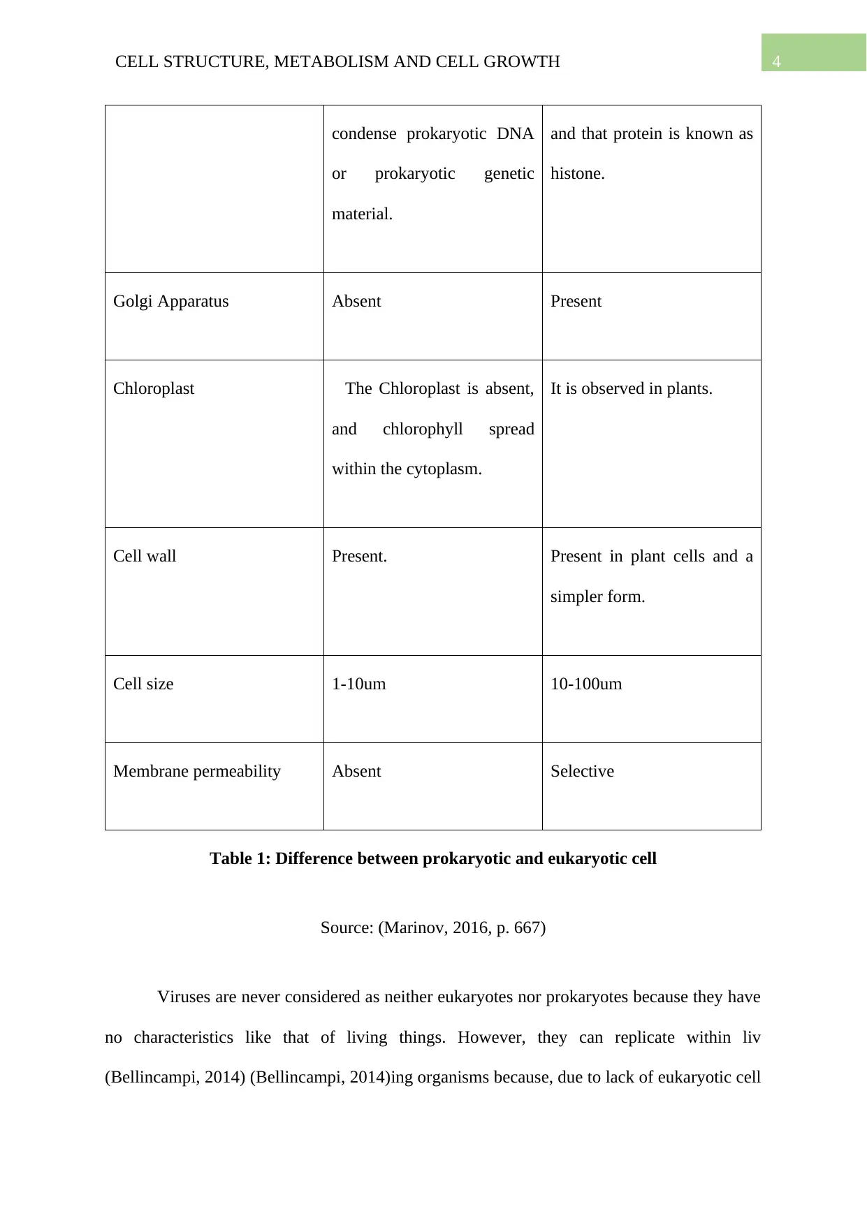
condense prokaryotic DNA
or prokaryotic genetic
material.
and that protein is known as
histone.
Golgi Apparatus Absent Present
Chloroplast The Chloroplast is absent,
and chlorophyll spread
within the cytoplasm.
It is observed in plants.
Cell wall Present. Present in plant cells and a
simpler form.
Cell size 1-10um 10-100um
Membrane permeability Absent Selective
Table 1: Difference between prokaryotic and eukaryotic cell
Source: (Marinov, 2016, p. 667)
Viruses are never considered as neither eukaryotes nor prokaryotes because they have
no characteristics like that of living things. However, they can replicate within liv
(Bellincampi, 2014) (Bellincampi, 2014)ing organisms because, due to lack of eukaryotic cell
5CELL STRUCTURE, METABOLISM AND CELL GROWTH
or prokaryotic cell, they cannot replicate and reproduce outside of the living cell. Two
microbiologists, Schleiden and Schwann, who demonstrated the structure and characteristics
of cells, described cell theory in 1839. The cell wall is a rigid layer, which is also a non-living
being found outside of the cell membrane. Cell walls are observed in plants, fungi, and
bacteria (Bellincampi, 2014, p. 228). In the case of a plant, cellulose is the primary
component of the cell wall. There are three layers present which supports and helps the plant
as a structural framework. The cell wall gives plant cells an equal shape and offers support to
the plant body.
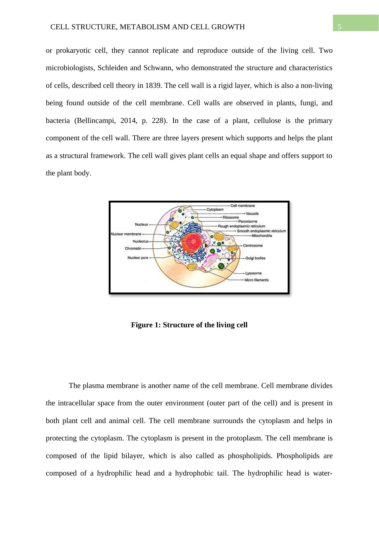
Figure 1: Structure of the living cell
The plasma membrane is another name of the cell membrane. Cell membrane divides
the intracellular space from the outer environment (outer part of the cell) and is present in
both plant cell and animal cell. The cell membrane surrounds the cytoplasm and helps in
protecting the cytoplasm. The cytoplasm is present in the protoplasm. The cell membrane is
composed of the lipid bilayer, which is also called as phospholipids. Phospholipids are
composed of a hydrophilic head and a hydrophobic tail. The hydrophilic head is water-
or prokaryotic cell, they cannot replicate and reproduce outside of the living cell. Two
microbiologists, Schleiden and Schwann, who demonstrated the structure and characteristics
of cells, described cell theory in 1839. The cell wall is a rigid layer, which is also a non-living
being found outside of the cell membrane. Cell walls are observed in plants, fungi, and
bacteria (Bellincampi, 2014, p. 228). In the case of a plant, cellulose is the primary
component of the cell wall. There are three layers present which supports and helps the plant
as a structural framework. The cell wall gives plant cells an equal shape and offers support to
the plant body.
Figure 1: Structure of the living cell
The plasma membrane is another name of the cell membrane. Cell membrane divides
the intracellular space from the outer environment (outer part of the cell) and is present in
both plant cell and animal cell. The cell membrane surrounds the cytoplasm and helps in
protecting the cytoplasm. The cytoplasm is present in the protoplasm. The cell membrane is
composed of the lipid bilayer, which is also called as phospholipids. Phospholipids are
composed of a hydrophilic head and a hydrophobic tail. The hydrophilic head is water-
⊘ This is a preview!⊘
Do you want full access?
Subscribe today to unlock all pages.
Trusted by 1+ million students worldwide
6CELL STRUCTURE, METABOLISM AND CELL GROWTH
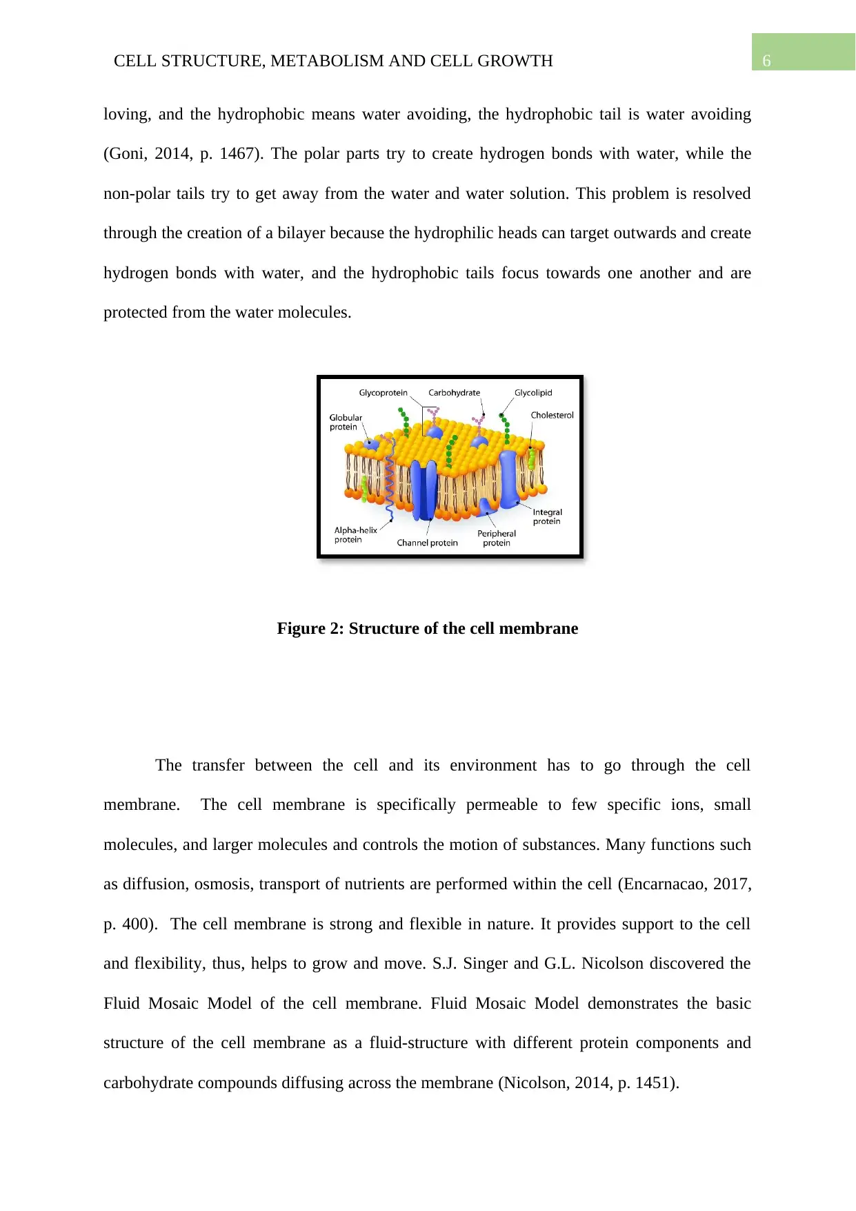
loving, and the hydrophobic means water avoiding, the hydrophobic tail is water avoiding
(Goni, 2014, p. 1467). The polar parts try to create hydrogen bonds with water, while the
non-polar tails try to get away from the water and water solution. This problem is resolved
through the creation of a bilayer because the hydrophilic heads can target outwards and create
hydrogen bonds with water, and the hydrophobic tails focus towards one another and are
protected from the water molecules.
Figure 2: Structure of the cell membrane
The transfer between the cell and its environment has to go through the cell
membrane. The cell membrane is specifically permeable to few specific ions, small
molecules, and larger molecules and controls the motion of substances. Many functions such
as diffusion, osmosis, transport of nutrients are performed within the cell (Encarnacao, 2017,
p. 400). The cell membrane is strong and flexible in nature. It provides support to the cell
and flexibility, thus, helps to grow and move. S.J. Singer and G.L. Nicolson discovered the
Fluid Mosaic Model of the cell membrane. Fluid Mosaic Model demonstrates the basic
structure of the cell membrane as a fluid-structure with different protein components and
carbohydrate compounds diffusing across the membrane (Nicolson, 2014, p. 1451).
Paraphrase This Document
Need a fresh take? Get an instant paraphrase of this document with our AI Paraphraser

7CELL STRUCTURE, METABOLISM AND CELL GROWTH
Figure 3: Phospholipid bilayer
Cell membrane proteins are found crossing the membrane from the inner division to
the outer division of the cell. The membrane proteins consist of a hydrophilic and
hydrophobic area, which allow proteins to pass suitable into the cell membrane. Membrane
protein works as a carrier protein and restricts the motion of the ion and molecule over the
cell membrane. Glycoproteins are observed on the extracellular space of the cell membrane,
which is also linked to polypeptide chains. It the case of a cell-to-cell recognition,
glycoproteins are used and is most) suitable (Anderson, 2015, p. 1723) Glycolipids are
attached to phospholipids on the outer surface of the cell membrane. It works as the
recognition site for specified chemicals and is important in cell-to-cell linkage to form
tissues. The motion of substances over cell membranes is hugely beneficial as it allows cells
to obtain oxygen and nutrients, remove waste products, and control the number of required
substances in the cell. The significant processes by which such motion and transportation take
place include diffusion, osmosis, facilitated diffusion, and active transport (Ayala, 2014, p.
3).
8CELL STRUCTURE, METABOLISM AND CELL GROWTH
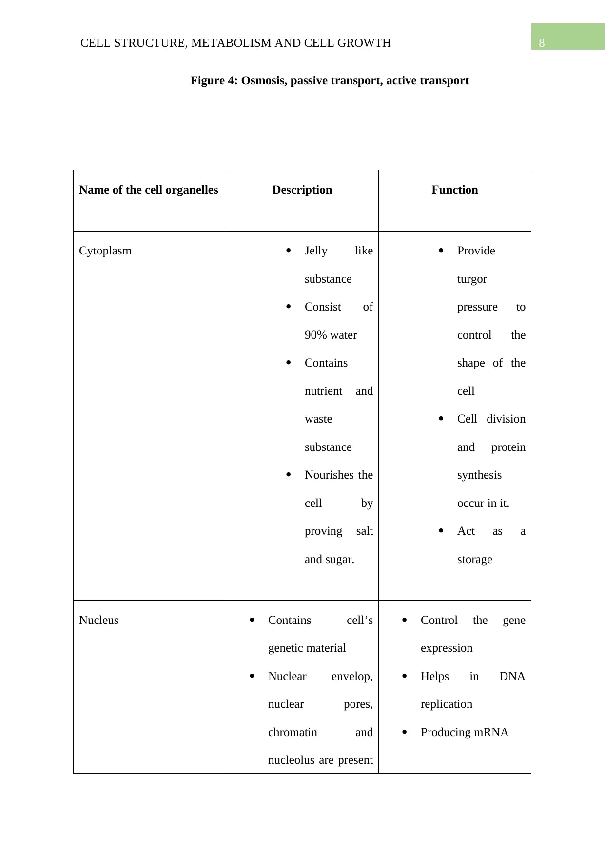
Figure 4: Osmosis, passive transport, active transport
Name of the cell organelles Description Function
Cytoplasm Jelly like
substance
Consist of
90% water
Contains
nutrient and
waste
substance
Nourishes the
cell by
proving salt
and sugar.
Provide
turgor
pressure to
control the
shape of the
cell
Cell division
and protein
synthesis
occur in it.
Act as a
storage
Nucleus Contains cell’s
genetic material
Nuclear envelop,
nuclear pores,
chromatin and
nucleolus are present
Control the gene
expression
Helps in DNA
replication
Producing mRNA
⊘ This is a preview!⊘
Do you want full access?
Subscribe today to unlock all pages.
Trusted by 1+ million students worldwide
9CELL STRUCTURE, METABOLISM AND CELL GROWTH
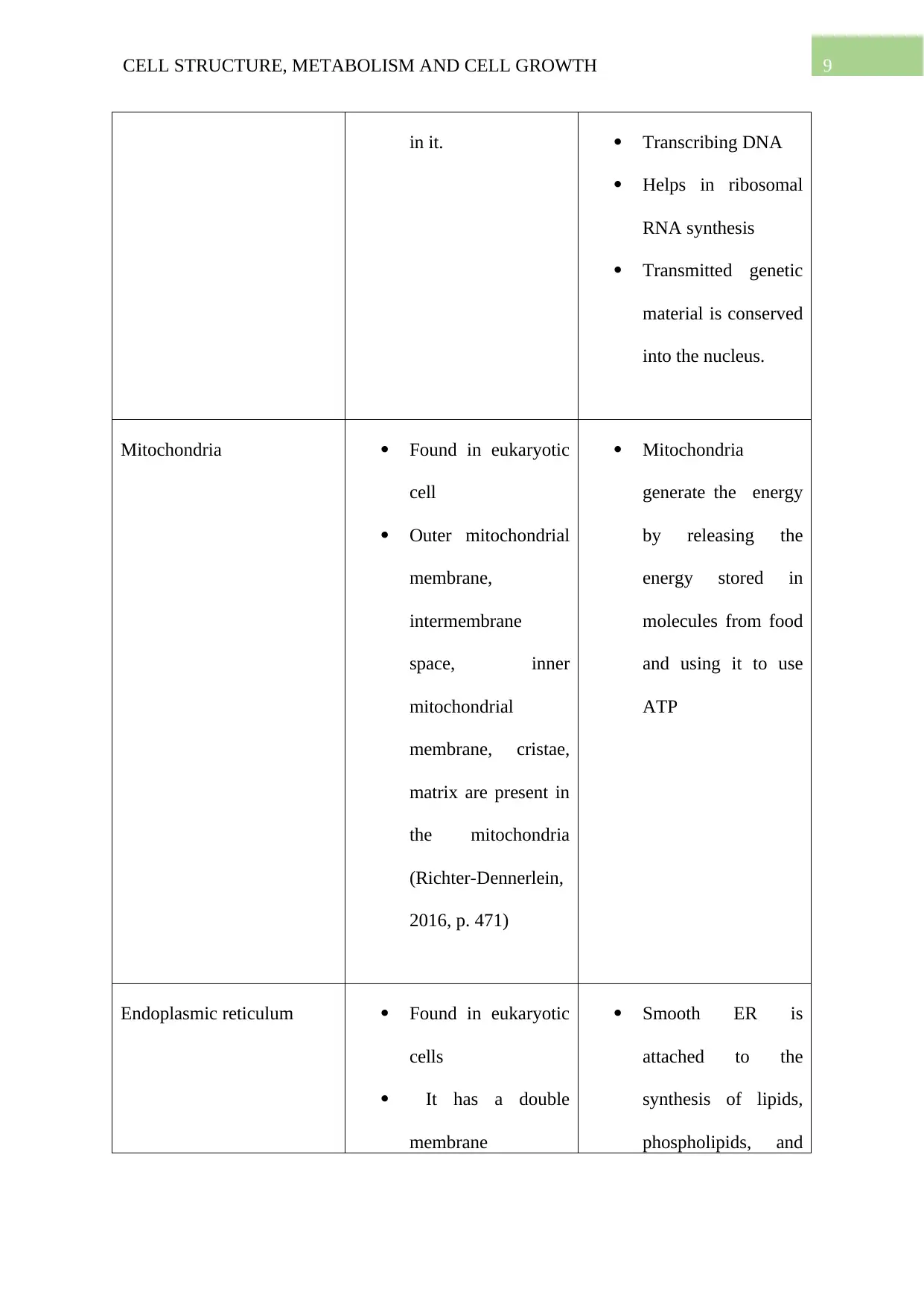
in it. Transcribing DNA
Helps in ribosomal
RNA synthesis
Transmitted genetic
material is conserved
into the nucleus.
Mitochondria Found in eukaryotic
cell
Outer mitochondrial
membrane,
intermembrane
space, inner
mitochondrial
membrane, cristae,
matrix are present in
the mitochondria
(Richter-Dennerlein,
2016, p. 471)
Mitochondria
generate the energy
by releasing the
energy stored in
molecules from food
and using it to use
ATP
Endoplasmic reticulum Found in eukaryotic
cells
It has a double
membrane
Smooth ER is
attached to the
synthesis of lipids,
phospholipids, and
Paraphrase This Document
Need a fresh take? Get an instant paraphrase of this document with our AI Paraphraser
10CELL STRUCTURE, METABOLISM AND CELL GROWTH
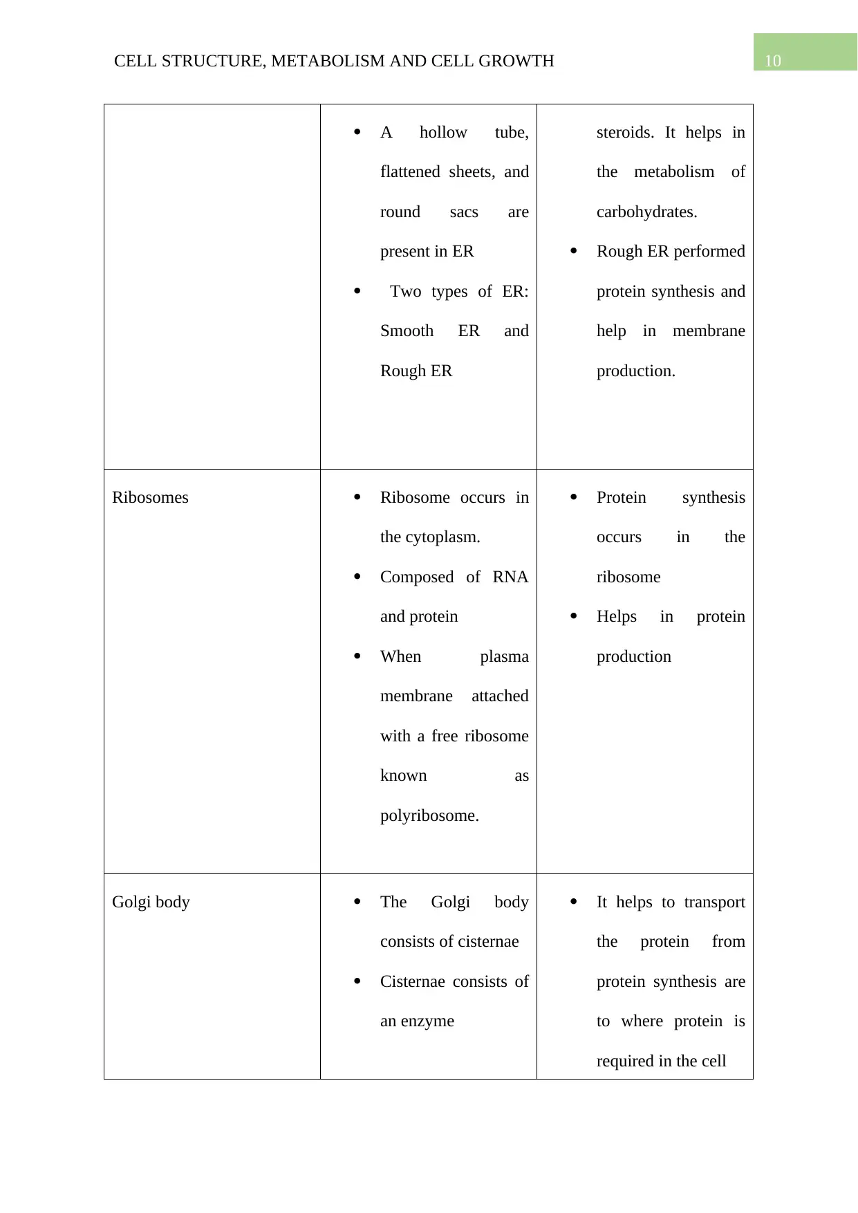
A hollow tube,
flattened sheets, and
round sacs are
present in ER
Two types of ER:
Smooth ER and
Rough ER
steroids. It helps in
the metabolism of
carbohydrates.
Rough ER performed
protein synthesis and
help in membrane
production.
Ribosomes Ribosome occurs in
the cytoplasm.
Composed of RNA
and protein
When plasma
membrane attached
with a free ribosome
known as
polyribosome.
Protein synthesis
occurs in the
ribosome
Helps in protein
production
Golgi body The Golgi body
consists of cisternae
Cisternae consists of
an enzyme
It helps to transport
the protein from
protein synthesis are
to where protein is
required in the cell

11CELL STRUCTURE, METABOLISM AND CELL GROWTH
Vesicles and lysosomes Membrane-bound
spherical sac
Small
Lysosomes are made
by Golgi body
The lysosome
contains digestive
enzymes
Vesicles help in
transportation of
molecules within the
cell
When a cell dies,
then lysosome
secrets the enzyme
which digest the cell
The digestive
enzyme helps to
digest the food, such
as carbohydrates and
protein.
Vacuole Fluid-filled organelle
Membrane-bound
Occur in the
cytoplasm
Absent in plant cell
Contains cell sap
(sugar, amino acid,
minerals, salt)
Helps in the
digestion
Excretion of cellular
waste
Releases water by
osmosis
Maintaining the plant
cell shape.
Centrioles Cylindrical tube-like Centrosome helps in
⊘ This is a preview!⊘
Do you want full access?
Subscribe today to unlock all pages.
Trusted by 1+ million students worldwide
1 out of 35
Related Documents


Your All-in-One AI-Powered Toolkit for Academic Success.
+13062052269
info@desklib.com
Available 24*7 on WhatsApp / Email
![[object Object]](/_next/static/media/star-bottom.7253800d.svg)
Unlock your academic potential
Copyright © 2020–2026 A2Z Services. All Rights Reserved. Developed and managed by ZUCOL.