Cell Biology Report: Microscopes, Eukaryotic Cell and Membranes
VerifiedAdded on 2022/12/28
|10
|1391
|93
Report
AI Summary
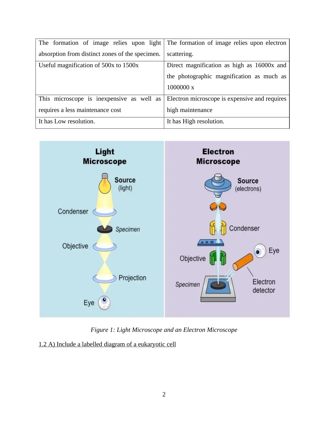
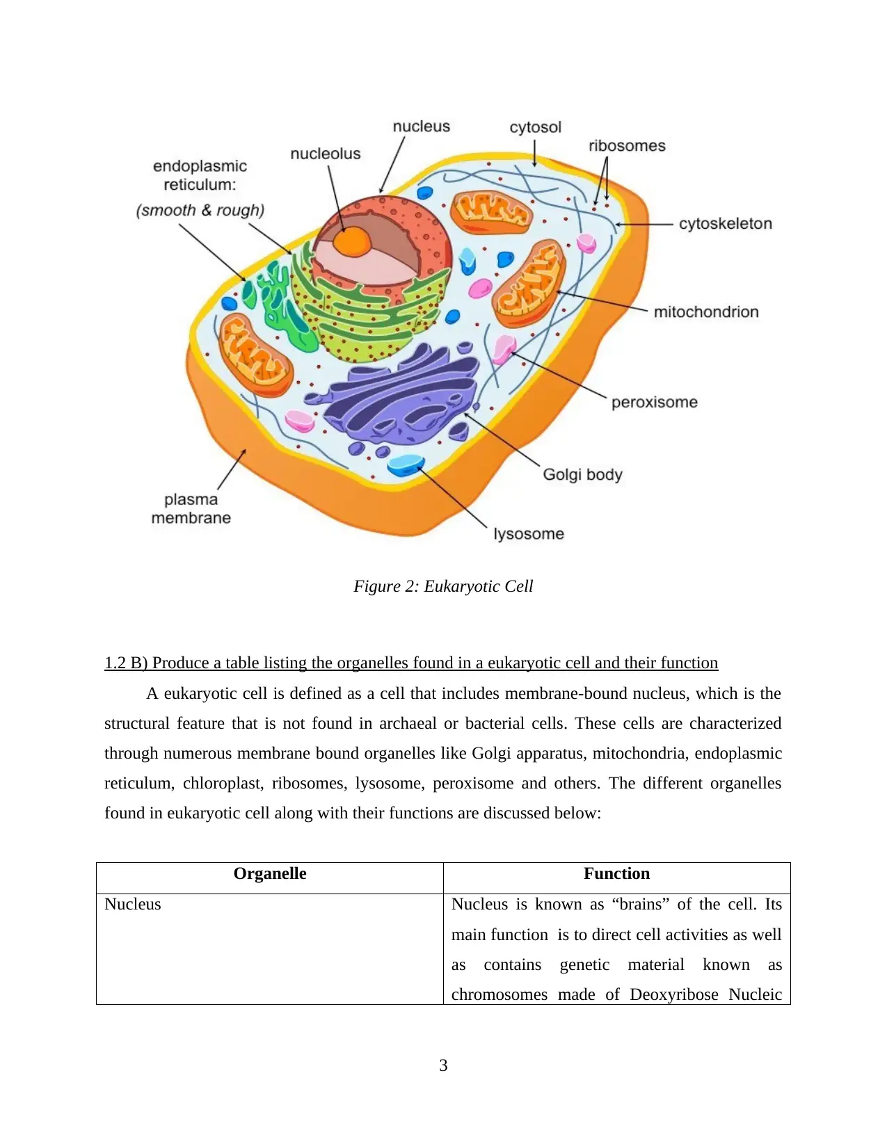
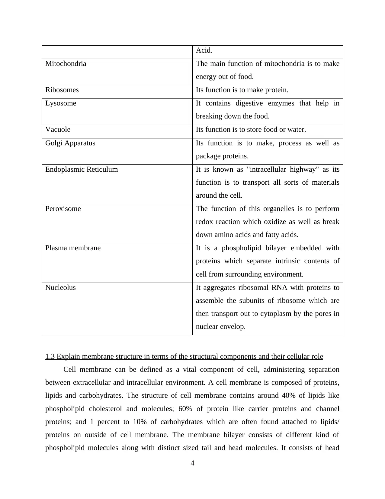
This report delves into the fundamentals of cell biology, beginning with a comparison of light and electron microscopes, highlighting their distinct uses and magnification capabilities. It then provides a detailed examination of the eukaryotic cell, including a labeled diagram and a table outlining the va...

Cell Biology
Secure Best Marks with AI Grader
Need help grading? Try our AI Grader for instant feedback on your assignments.

Contents
INTRODUCTION...........................................................................................................................1
MAIN BODY...................................................................................................................................1
1.1 Explain the differences between a light microscope and an electron microscope and how
these are used for different purposes......................................................................................1
1.2 A) Include a labelled diagram of a eukaryotic cell...........................................................2
1.2 B) Produce a table listing the organelles found in a eukaryotic cell and their function...3
1.3 Explain membrane structure in terms of the structural components and their cellular role
................................................................................................................................................4
CONCLUSION................................................................................................................................6
REFERENCES................................................................................................................................7


INTRODUCTION
Cell biology is defined as the study of structure and function of cell, and it revolves around a
concept that cell is fundamental life unit. This report covers the differences between light and
electron microscopes and its uses for different purposes. It also covers different organelles found
in eukaryotic cell along with their functions and an explanation about cell membrane structure,
its components and their cellular role.
MAIN BODY
1.1 Explain the differences between a light microscope and an electron microscope and how
these are used for different purposes
A light microscope is referred as a tool which is utilised to study micro bodies like fungus,
bacteria etc. to determine their features. It also allows examination of small objects that are not
visible by naked eyes. Whereas, an electron microscope is a tool which utilizes electron beams in
order to capture an image as well as enlarge it. It is a tool which allow an individual to visualise
small entities in high resolution. There are these differences between light microscope and
electron microscope. Some of the differences are provided below -
Light Microscope Electron Microscope
This microscope uses light (around 400-700
nm) as an illuminating source.
It utilizes electron beams (approx. 1 nm) as a
source of illumination.
The magnification of this microscope is lower
than an electron microscope.
The magnification of this microscope is
higher than light microscope.
In this, there is no risk of radiation leakage. In it, there is risk of radiation leakage.
The image is seen by ocular lens in it. No
screen is needed.
The image is received on zinc sulfate
fluorescent screen.
From light microscope, both dead and live
specimen can be seen.
From electron microscope, only the dried and
dead specimen can be seen.
The preparation of specimen takes around a
few minutes or an hour
The preparation of specimen takes several
days.
1
Secure Best Marks with AI Grader
Need help grading? Try our AI Grader for instant feedback on your assignments.

The formation of image relies upon light
absorption from distinct zones of the specimen.
The formation of image relies upon electron
scattering.
Useful magnification of 500x to 1500x Direct magnification as high as 16000x and
the photographic magnification as much as
1000000 x
This microscope is inexpensive as well as
requires a less maintenance cost
Electron microscope is expensive and requires
high maintenance
It has Low resolution. It has High resolution.
Figure 1: Light Microscope and an Electron Microscope
1.2 A) Include a labelled diagram of a eukaryotic cell
2

Figure 2: Eukaryotic Cell
1.2 B) Produce a table listing the organelles found in a eukaryotic cell and their function
A eukaryotic cell is defined as a cell that includes membrane-bound nucleus, which is the
structural feature that is not found in archaeal or bacterial cells. These cells are characterized
through numerous membrane bound organelles like Golgi apparatus, mitochondria, endoplasmic
reticulum, chloroplast, ribosomes, lysosome, peroxisome and others. The different organelles
found in eukaryotic cell along with their functions are discussed below:
Organelle Function
Nucleus Nucleus is known as “brains” of the cell. Its
main function is to direct cell activities as well
as contains genetic material known as
chromosomes made of Deoxyribose Nucleic
3

Acid.
Mitochondria The main function of mitochondria is to make
energy out of food.
Ribosomes Its function is to make protein.
Lysosome It contains digestive enzymes that help in
breaking down the food.
Vacuole Its function is to store food or water.
Golgi Apparatus Its function is to make, process as well as
package proteins.
Endoplasmic Reticulum It is known as "intracellular highway" as its
function is to transport all sorts of materials
around the cell.
Peroxisome The function of this organelles is to perform
redox reaction which oxidize as well as break
down amino acids and fatty acids.
Plasma membrane It is a phospholipid bilayer embedded with
proteins which separate intrinsic contents of
cell from surrounding environment.
Nucleolus It aggregates ribosomal RNA with proteins to
assemble the subunits of ribosome which are
then transport out to cytoplasm by the pores in
nuclear envelop.
1.3 Explain membrane structure in terms of the structural components and their cellular role
Cell membrane can be defined as a vital component of cell, administering separation
between extracellular and intracellular environment. A cell membrane is composed of proteins,
lipids and carbohydrates. The structure of cell membrane contains around 40% of lipids like
phospholipid cholesterol and molecules; 60% of protein like carrier proteins and channel
proteins; and 1 percent to 10% of carbohydrates which are often found attached to lipids/
proteins on outside of cell membrane. The membrane bilayer consists of different kind of
phospholipid molecules along with distinct sized tail and head molecules. It consists of head
4
Paraphrase This Document
Need a fresh take? Get an instant paraphrase of this document with our AI Paraphraser

molecule, a glycerol, a phosphate molecule and two chains of fatty acids. A head group is a polar
group includes choline or sugar, which means that the head end is hydrophilic. The tail group of
2 fatty acid chains consist of 14 to 24 carbons.
The phospholipid molecules are thus amphipathic as it is both hydrophobic and
hydrophilic. This spontaneously develop bilayers in water along with tail groups facing in and
head groups facing out. There is Vander wall forces among fatty acid tails a phospholipid.
Cholesterol is important function in cell and is a major constituent of membrane of cell. It itself
contains a planar steroid ring, a polar head and nonpolar hydrocarbon tail. It is significant in
membrane as it assists in maintaining stability and fluidity in the cell membrane at varying
temperatures. Membrane proteins are present in higher proportion as they are very crucial in
each process within the cell. Some of the functions of membrane proteins includes transporters,
catalysts, energy transducers and receptors for local mediators, neurotransmitters and hormones.
The cell membrane forms a highly selective and continuous permeable barrier around cells as
well as intracellular compartments. It generates signals in response to stimulus developing a
transformation in membrane potential.
Figure 3: Cell Membrane: Components and Structure
5

CONCLUSION
As per the above mentioned report, it has been concluded that cell is fundamental unit of
life. A cell consists of many organelles and each one has its specific function. The uses of light
microscope and electron microscope are different which allow a person to visualize things.
6
As per the above mentioned report, it has been concluded that cell is fundamental unit of
life. A cell consists of many organelles and each one has its specific function. The uses of light
microscope and electron microscope are different which allow a person to visualize things.
6

REFERENCES
Books and Journals
Pollard, T.D., Earnshaw, W.C., Lippincott-Schwartz, J. and Johnson, G., 2016. Cell biology E-
book. Elsevier Health Sciences.
Steinberg, G., Peñalva, M.A., Riquelme, M., Wösten, H.A. and Harris, S.D., 2017. Cell biology
of hyphal growth. The Fungal Kingdom, pp.231-265.
Giacomello, M., Pyakurel, A., Glytsou, C. and Scorrano, L., 2020. The cell biology of
mitochondrial membrane dynamics. Nature reviews Molecular cell biology, 21(4),
pp.204-224.
Goldstein, B. and King, N., 2016. The future of cell biology: emerging model organisms. Trends
in cell biology, 26(11), pp.818-824.
Meents, M.J., Watanabe, Y. and Samuels, A.L., 2018. The cell biology of secondary cell wall
biosynthesis. Annals of Botany, 121(6), pp.1107-1125.
7
1 out of 10
Related Documents

Your All-in-One AI-Powered Toolkit for Academic Success.
+13062052269
info@desklib.com
Available 24*7 on WhatsApp / Email
Unlock your academic potential
© 2024 | Zucol Services PVT LTD | All rights reserved.