Cellular Enzyme Assays and Colony Formation in Toxicity Studies
VerifiedAdded on 2021/06/14
|20
|5159
|95
Homework Assignment
AI Summary
This assignment delves into various aspects of cellular toxicity assessment, including the use of six different cellular enzymes (LDH, ALT, Creatinine kinase, Caspase, Hexokinase, and Beta-hydroxy-butrate) as endpoints for toxicity evaluation. It explores the mechanisms and applications of each enzyme, providing reaction schemes and references to substantiate the statements. The assignment then examines colony formation assays as a method to measure DNA repair in response to ionizing radiation, comparing it with standard colony formation assays used to assess drug toxicity, and discussing advantages, disadvantages, and potential artifacts. Finally, the assignment tasks the development of an assay to measure lactate in cell culture media, including a complete reaction diagram, assay conditions, and a step-by-step protocol, along with a detailed explanation of the cell culture methods.

1
Pharmacy
Name
Date
Pharmacy
Name
Date
Paraphrase This Document
Need a fresh take? Get an instant paraphrase of this document with our AI Paraphraser
2
Question 1
List 6 different cellular enzymes that can be used to assess different endpoints of
toxicity. a reaction scheme and add references to substantiate your statements.
The cells are able to respond very fast to toxic materials through changing of
morphologies, biochemical processes and altered growth behaviours. The biochemical
changes are involved in changing the normal functions of the cells and are thus significant in
endpoint toxicity tests. The enzymes are commonly used in various cellular biochemical
roles, leading to their increased use in cytotoxic tests.
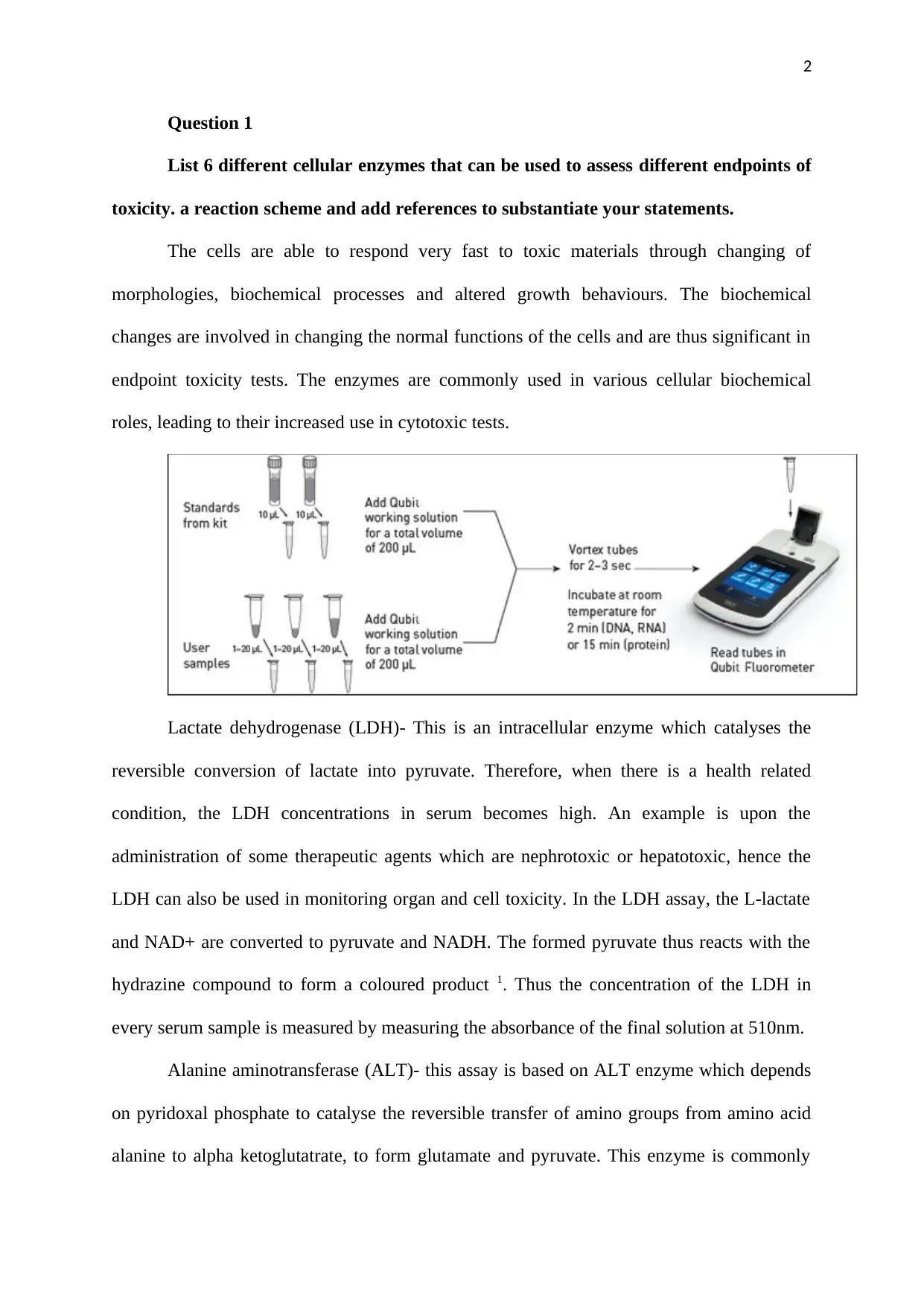
Lactate dehydrogenase (LDH)- This is an intracellular enzyme which catalyses the
reversible conversion of lactate into pyruvate. Therefore, when there is a health related
condition, the LDH concentrations in serum becomes high. An example is upon the
administration of some therapeutic agents which are nephrotoxic or hepatotoxic, hence the
LDH can also be used in monitoring organ and cell toxicity. In the LDH assay, the L-lactate
and NAD+ are converted to pyruvate and NADH. The formed pyruvate thus reacts with the
hydrazine compound to form a coloured product 1. Thus the concentration of the LDH in
every serum sample is measured by measuring the absorbance of the final solution at 510nm.
Alanine aminotransferase (ALT)- this assay is based on ALT enzyme which depends
on pyridoxal phosphate to catalyse the reversible transfer of amino groups from amino acid
alanine to alpha ketoglutatrate, to form glutamate and pyruvate. This enzyme is commonly
Question 1
List 6 different cellular enzymes that can be used to assess different endpoints of
toxicity. a reaction scheme and add references to substantiate your statements.
The cells are able to respond very fast to toxic materials through changing of
morphologies, biochemical processes and altered growth behaviours. The biochemical
changes are involved in changing the normal functions of the cells and are thus significant in
endpoint toxicity tests. The enzymes are commonly used in various cellular biochemical
roles, leading to their increased use in cytotoxic tests.
Lactate dehydrogenase (LDH)- This is an intracellular enzyme which catalyses the
reversible conversion of lactate into pyruvate. Therefore, when there is a health related
condition, the LDH concentrations in serum becomes high. An example is upon the
administration of some therapeutic agents which are nephrotoxic or hepatotoxic, hence the
LDH can also be used in monitoring organ and cell toxicity. In the LDH assay, the L-lactate
and NAD+ are converted to pyruvate and NADH. The formed pyruvate thus reacts with the
hydrazine compound to form a coloured product 1. Thus the concentration of the LDH in
every serum sample is measured by measuring the absorbance of the final solution at 510nm.
Alanine aminotransferase (ALT)- this assay is based on ALT enzyme which depends
on pyridoxal phosphate to catalyse the reversible transfer of amino groups from amino acid
alanine to alpha ketoglutatrate, to form glutamate and pyruvate. This enzyme is commonly
3
found in the liver and to a less extent in other tissues. When injury to hepatocytes occurs, the
concentrations of ALT in the serum becomes high, and hence a biomarker for hepatocytes
toxicity 2. The assay is conducted to detect pyruvate in a reaction that coverts a colourless to a
coloured product whose absorbance is measured at 570nm.
Creatinine kinase- Creatinine kinase is applied in the enzymatic assay of creatine
kinase in patient serum. This enzyme is applied in the diagnosis of diseases that are linked to
heart, central nervous system and the skeletal muscles 3. When this enzyme is present in
blood, it catalyses the transfer of a high energy phosphate from creatine phosphate to ADP to
form ATP. In the muscles, such a reaction is important because the resulting ATP is used in
presence of the enzyme hexokinase to convert glucose to glucose-6-phosphate to make more
energy in glycolysis. At the same time, this reaction involves the reduction of NADP+ to
NADPH. Therefore, the activity of creatine kinase is measured by testing the rate of
formation of NADPH which is monitored at 340nm. These reactions take place in presence of
N-acetyl-L-cysteine which is the enzyme activator.
Caspase: this enzyme assay is used in the detection of cell apoptosis, due to the
activation of the caspase enzymes by the cells 4. These enzymes are activated so that they can
catalyse the cleavage of protein substrates in the cells causing the destruction of the cell wall,
and thus cell death. The caspase assay there facilitates the detection of these enzymes in
living cells on real time basis. The caspase type 3 and 7 cleaves the PARP protein to form
85kDa and 25kDa fragments .in this assay, antibodies against the 85kDa fragment are used as
markers for apoptotic cells.
Hexokinase: this enzyme activity is involved in the first step of glycolysis to convert
glucose to glucose-6-phosphate 5. At the same time, glucose-6-phosphate is oxidized by
glucose-6-phosphate dehydrogenase to produce NADH, a product that can reduce a
colourless molecule to form a coloured product, whose absorbance can be measured at
⊘ This is a preview!⊘
Do you want full access?
Subscribe today to unlock all pages.
Trusted by 1+ million students worldwide
4
450nm. The hexokinase enzyme is very common in many cells and thus critical for glucose
metabolism. Too low levels of hexokinase enzyme are associated with diseases like muscular
dystrophy, while too low levels could be due to tumours. However, with early detection, it is
possible to diagnose, predict and treat the underlying disease.
Beta-hydroxy-butrate: this enzyme assay is used to determine the presence of ketone
bodies in blood. The ketone bodies are formed when the levels of glucose are low especially
during starvation and fasting. Additionally, the ketones like Beta-hydroxy-butyrate can rise in
diabetics and alcoholics 6. Thus, the use of Beta-hydroxy-butrate is used in determination of
the Beta-hydroxy-butyrate levels through a coupled enzymatic reaction, whereby the
absorbance is measured at 450nm, which is proportional to the amount of Beta-hydroxy-
butrate present in a sample. In this assay, Beta-hydroxy-butyrate, which is produced by the
hepatocytes and released into the tissues as a source of energy.
Lactase enzyme is critical for the metabolism of a disaccharide known as lactose. This
is measured using colorimetric and fluorometric methods. The presence or absence of lactose
in blood or urine by hydrolysing lactose by the enzyme lactase 7. Lack of lactase in cells leads
to fructose intolerance.
Question 2
Discuss in detail how a colony formation assay can be used to measure DNA
repair in response to ionizing radiation and how this differs compared to the standard
colony formation assay that assesses toxicity of a drug. Also, list at least 5 advantages, 5
disadvantages and possible artefacts associated with this assay.
The ionizing radiations are associated with negative effects to the human cells such as
cell necrosis, programmed cell death, autophagy and multinucleation among other negative
effects. These processes make the cells lose their colony forming abilities during the
Paraphrase This Document
Need a fresh take? Get an instant paraphrase of this document with our AI Paraphraser
5
clonogenic assays. However, the clonogenic cell death is not advantageous in all cases, for
instance the cancer therapy, it leads to more harm to the cells. An example is the association
between stress induced premature senescence and autophagy which have been found to
influence the nature of cancer cells to have a prolonged growth inhibition leading to regrowth
of a tumour and the disease is likely to occur. Upon exposure to the ionizing radiations, the
double strands of the DNA molecules are the most at risk structures 8. However, when such
damages to the DNA, a repair mechanism is initiated through the involvement of several
steps such as sensors, transducers and effector proteins. The DNA double strand breaks are
initially sensed whereby the sensors recognize a lesion on the chromatins. Then the
transducers are brought at the site of the DNA damage so as to assemble the double strand
repair complex and send signals to the effectors. Once the DNA double strand breaks have
occurred due to exposure to radiations, then the colony formation by such cells can be
assessed in a method known as clonogenic assay 9. Therefore, if the DNA has been repaired
by cellular mechanisms, the colony formation assay will produce viable colonies because the
cellular machinery for growth and cell division is complete. However, in the vent of exposure
to ionizing radiation and the cell machinery was not good enough to repaid the DBA breaks,
then no colonies will grow in the culture plates.
On the other hand, the use of colony assay in testing of drug toxicity is less
cumbersome than the use of colony assay in detecting DNA repair processes. The cell colony
formation is termed as a more sensitive parameter of toxicity studies. This is because this
allows the formation of colonies to be determines when the cells are in the actual state of
division, thus they could be more susceptible to toxic drug effects. The colonies can then be
counted on an agarose bilayer inside the Petri dishes, either manually or by use of the
recently colour detecting automatic colony counters. To be precise, the use of colony
formation techniques for drug sensitivity tests is based on the determination of the number of
6
colonies that go beyond the arbitrary sizes in cultures that have been treated with the drug in
question.
Advantages:
a. It makes it possible for a researcher to determine the number of cells which are
needed to differentiate between a colony and a cluster of cells 10.
b. The colony forming assay can be used in xenobiotic which are known to cause
toxicities in the hematopoietic system, leading to haematological disorders.
c. Evaluates the possible toxic effects of new xenobiotic and thus close the gap
between clinical investigations and pre-clinical investigations 11.
d. Determines the external factors or environment affecting cell growth.
e. It produces accurate results.
Disadvantages
a. The method is time consuming.
b. For tumour cell, this method can only give results for the cells that are
culturable only.
c. It is expensive and requires expertise.
d. Involves the use of clones produced from other cells and hence a possibility of
errors.
e. Counting cells under the microscope is quite tedious 12.
The common artefacts that arise include: an increase in agarose gel pore sizes, due to
wrong gel preparation, an automatic colony counter might not detect the white-coloured
colonies 13.
Question 3
⊘ This is a preview!⊘
Do you want full access?
Subscribe today to unlock all pages.
Trusted by 1+ million students worldwide
7
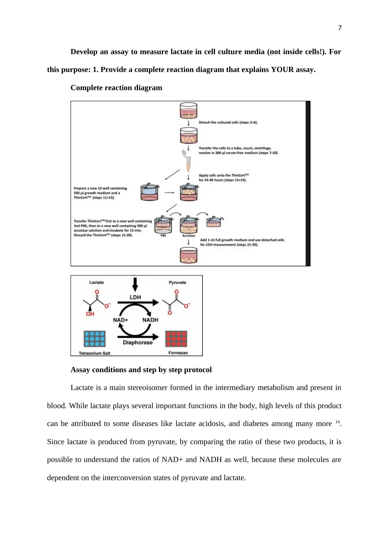
Develop an assay to measure lactate in cell culture media (not inside cells!). For
this purpose: 1. Provide a complete reaction diagram that explains YOUR assay.
Complete reaction diagram
Assay conditions and step by step protocol
Lactate is a main stereoisomer formed in the intermediary metabolism and present in
blood. While lactate plays several important functions in the body, high levels of this product
can be attributed to some diseases like lactate acidosis, and diabetes among many more 14.
Since lactate is produced from pyruvate, by comparing the ratio of these two products, it is
possible to understand the ratios of NAD+ and NADH as well, because these molecules are
dependent on the interconversion states of pyruvate and lactate.
Paraphrase This Document
Need a fresh take? Get an instant paraphrase of this document with our AI Paraphraser
8
In cell cultures, the scientists are faced with the problem of lactic acid accumulation,
to high levels such that this by-product limits the growth and production of the cells 15. Thus
there have been continued efforts to eliminate lactic acids and other toxic metabolites form a
culture medium but no efficiency has ever been achieved. The common method adopted for
reducing the accumulation of lactic acid in the culture medium has been to have controlled
glucose levels. Controlled glucose supply reduces the lactic acid production since this
product is formed from glucose through the process of glycolysis. This means that for
glucose to be properly controlled, there is a need to keep on taking lactic acid measurements
in the cell culture medium from time to time.
a. Cultures
The human MSC cell lines that had been isolated from two healthy adult donors,
following informed consent were bought from Lonza technologies in USA. The hMSC cells
were first cultures in Dulbecco modified Eagles medium (purchased from Lonza
technologies). This medium was supplemented with either 2, 5 or 10% foetal bovine serum
(Hyclone technologies, Belgium). Unless stated otherwise, the growth medium used in this
assay was supplemented with 10% foetal bovine serum 16. The complete culture media was
stored at 4 degrees Celsius inside the fridge for use not later than one month upon
preparation. Using the serum free technique, the PRIME, MSC EXPANSION SFM (USA)
was used based on the manufactures instructions and without supplementing the culture
medium. The aliquots from the serum free experiment were harvested and stored at 4 degrees
Celsius in dark.
Using serum-based media, the hmSC cells were grown in a monolayer culture 17. The
cells were passaged after one week, since this is the normal time for early hMSC cells to
attain confluence as confirmed by the Nikon light microscope (Nikon, USA). After every
passage, the used up media was aspirated and the flasks rinsed twice. Then the cells were
9
enzymatically detached from the surface of the culture area by exposing them to 0.25(v/v of
trypsin EDTA solution, for five minutes and at body temperature. To confirm the detachment
of cells, a light microscope was used to visualize the walls of every culture flask. Then a
fresh culture medium was added and the suspension of every flask was slowly pippted up and
downwards over the flask surfaces to dislodge any remaining cells, down into the fresh
medium.
b. Lactate assay
The measurement of lactate concentration in the cell culture was performed. The
media from the cell cultures was collected following infection if the cells using a HIF-1a
siRNA following exposure for one day under limited oxygen supply 18.
c. Viable cells concentration
Trypan blue, which is the common stain for distinguishing viable and dead cells was
used. In this case, only the dead cells absorb the dye and appear bluish in colour.
Approximately 100microliter of cell suspension was taken from the Petri dishes and dilute in
trypan blue dye, followed by cell counting process. The lactate concentration was then
measured using a commercial kit which measures the activity of lactate dehydrogenase
enzyme which converts pyruvate to lactate. The kit was purchased from Sigma technologies.
Thus the reaction is forced in the direction of lactate formation through the addition of excess
beta-nicotinamide adenine dinucleotide and trapping of pyruvate by use of hydrazine 19. The
absorbance at 340nm as a result of the reduction of dinucleotide is equal to the amount of
lactic acid produced by the cultured cells in the medium. The obtained difference in
absorbance at 340nm was used to calculate the actual concentration of lactate released by the
cultured cells into the medium by the use of beer lambert law 20.
Order number
PRIME, MSC EXPANSION SFM (USA) cells- 91135
⊘ This is a preview!⊘
Do you want full access?
Subscribe today to unlock all pages.
Trusted by 1+ million students worldwide
10
Mesenchymal Stem Cells cryo amp – PT2501
Lactase assay kit (Sigma technologies)- MAK065
Controls
The positive control used in this lactate assay was the lactate supplied with the kit by
the manufacturer, while the negative control was the sterile distilled water.
Question 4
Discuss why the use of Michaelis-Menten diagrams is not able to accurately
predict V0 and Vmax and how Lineweaver-Burk diagrams are biased for a particular
substrate concentration range. Based on your answer, discuss which method of
calculating enzyme activities is better for high substrate concentrations.
In order to determine the function of enzymes, there is a need for getting a kinetic
description of enzyme activity. Majority of the enzymes have the initial velocity Vo, which
varies with changes in enzyme concentrations. The rate of an enzyme catalysed reaction
proceeds in a linear manner as the concentrations of the substrate rises and at some point, it
begins to level off, and approaches a maximum as the substrate concentration increases. This
is the Michaeli’s menten equation which is a plot of initial velocity Vo against the substrate
concentration for enzymes which obey this kinetics 21. The maximal velocity, Amax
approaches in the plot asymptotically. The Amax gives a turnover of the number of enzyme
units, which is the number of substrates and converted into a product, at a point when the
enzymes are fully saturated with the substrate.
In Michaeli’s Menten plot, the Amax can be arrived at only if the substrate
concentration is quite high in order to saturate the enzymes’ active site. Continued increase in
Paraphrase This Document
Need a fresh take? Get an instant paraphrase of this document with our AI Paraphraser
11
the substrate concentration when the enzyme concentration is constant means that all the
active sites of the enzyme will be full. Once all the active sites of the enzyme are occupied,
the enzyme is said to be at its maximum capacity and thus, any increase in the substrate
concentration will not raise the enzyme rate turnover.
The limitation arises when calculating or determining the Vo and Amax from the
Michaeli’s Menden plot since it becomes impossible to estimate the Amax from a plot which
has a hyperbolic shape 22. Therefore, the Michaeli’s Menten plot is converted into its
reciprocal making it possible to determine Amax and Km experimentally using Vo
measurements at various substrate concentrations. Therefore, the lineweaverburk plot is made
by making a plot of 1/vo against 1/substrate concentrations, giving a plot which contains a
straight line. In the lineweaverburk plot, the y-intercept is the 1/Amax, while the x-intercept
is 1/-Km.
The lineweaverburk plot was commonly used to determine various terms in enzyme
kinetics such as the Km and Vmax. However, this plot distorts the error structure of the data
and is hence unreliable for determining the parameters of enzyme kinetics in various enzyme
concentrations 23. This plot can however be used to distinguish between the various types of
enzyme inhibitors i.e. competitive, uncompetitive and non-competitive inhibitors. The bias in
the lineweaverburk plot is that it is quite prone to errors since the y-axis takes the reciprocal
of the reaction rate, thus increasing the small errors that could be common. Majority of the
points on this plot are at a far distance to the right of the y-axis as a result of the limited
solubility properties. This far distance between the points on the plot limits the ability for
plotting larger values of substrate concentration reciprocals, indicating that there are no small
substrate concentrations. In this case, there needs to be a big extrapolation backwards in order
to get the x as well as y intercepts.

12
Michaelis Menten graph of reaction rate against increasing substrate concentration.
Line weaver Burk plot of reaction rate against increasing substrate concentration.
Therefore, the better method of calculating the enzyme activity in high substrate
concentrations is the Line Weaver Burk plot method since it allows the extrapolation of the
straight line graph, for reaction rate with increasing substrate concentrations. This is the
Michaeli’s menten method produces a hyperbolic which limits the determination of enzyme
activity at increasing substrate concentrations.
Question 5
The data below were obtained in the presence and absence of an unknown
inhibitor X.
⊘ This is a preview!⊘
Do you want full access?
Subscribe today to unlock all pages.
Trusted by 1+ million students worldwide
1 out of 20
Your All-in-One AI-Powered Toolkit for Academic Success.
+13062052269
info@desklib.com
Available 24*7 on WhatsApp / Email
![[object Object]](/_next/static/media/star-bottom.7253800d.svg)
Unlock your academic potential
Copyright © 2020–2026 A2Z Services. All Rights Reserved. Developed and managed by ZUCOL.