Ankara Hospital Study: Cerebellar Antibodies in ADHD, ASD, and Control
VerifiedAdded on 2020/04/15
|13
|4681
|85
Report
AI Summary
This report presents a study conducted at Ankara Pediatric Hematology Oncology Training and Research Hospital, investigating the association between cerebellar antibodies and Attention Deficit Hyperactivity Disorder (ADHD) and Autism Spectrum Disorder (ASD) in children aged 4-12 years. The study compared anti-Yo and anti-GAD antibody levels in 39 children with ADHD, 40 children with ASD, and a control group of 40 healthy children. Participants were assessed using DSM-5 criteria, K-SADS-PL, Denver-II or Stanford Binet tests, WISC, Conners’ scales, ABC, AbBC, and CARS. The study found significant differences in anti-Yo levels between children with ADHD and healthy subjects. No significant differences were observed in GAD levels between the groups. Positive correlations were found between GAD levels and children's age, and a negative correlation was observed between age and ABC scores. The study highlights the potential role of cerebellar antibodies in ADHD and ASD, suggesting a need for further research with larger samples to explore etiological factors in neuropsychiatric disorders. The study used ELISA method to determine the anti-Yo and anti-GAD levels from venous samples.
Key findings
Significant differences were observed in anti-Yo levels between children with ADHD and
healthy subjects.
No significant differences were observed in GAD and anti-Yo levels between children with
ADHD and those with ASD.
Probable associations were found between cerebellar antibodies and ADHD.
Cerebellar antibodies in autism spectrum disorder and attention deficit hyperactivity disorder
(ADHD)
Abstract
Compelling evidences suggest that dysfunction in the cerebellar region of the brain plays a
significiant role in the etiology of autism spectrum disorders (ASD) and attention deficit hyperactivity
disorders (ADHD). The study aims to investigate the association between degeneration of the
cerebellum and the etiologies of ASD and ADHD. The study recruited participants comprising of 40
children with ASD and 39 children with ADHD. 40 healthy children were recruited as the control
group. The participants were analysed for an increase in anti-Yo antibodies and anti-glutamic acid
decarboxylase (GAD) levels during cerebellar damage. Each child with ASD was evaluated using the
autism behavior checklist (ABC), aberrant behavior checklist (AbBC) and childhood autism rating scale
(CARS). The Conners’ Parent and Teacher Rating Scales-Revised Long Form (CPRS and CTRS) screening
questionnaires were completed by the parents and teachers of children with ADHD. The anti-Yo and
anti-GAD levels were determined using 10 cc venous samples from the participants. No significant
sociodemographical difference was observed between the groups. Significant differences were found
in the anti-Yo levels between children with ADHD and healthy subjects (p=0.002). Additionally, a
positive correlation was observed between GAD levels and children age. Furthermore, a negative
correlation was found between their age and ABC scores. This was the first study to evaluate levels of
cerebellar antibodies in ADHD and ASD, and compare it to a control group. It found significant
Paraphrase This Document
Need a fresh take? Get an instant paraphrase of this document with our AI Paraphraser
association between cerebellar antibodies and ADHD. Further studies with larger samples and follow-
up periods are necessary in order to investigate possible etiological factors among subjects with
neuropsychiatric disorders.
Key words: Attention deficit-hyperactivity disorder (ADHD); autism (ASD); cerebellar antibodies; anti-
Yo; anti-GAD
1. Introduction
Autism spectrum disorder (ASD) is an umbrella term that encompasses a group of
neurodevelopmental disorders, characterized by developmental delay, abnormalities in langugage
comprehension, social interaction, reciprocity, communication, and repetitive stereotypical
behaviors and interests [1]. Although previoius studies provided evidences for the underlying
genetic, prenatal, early postnatal, and biochemical pathways that are responsible for the disorder,
the etiology and pathogenesis are still unclear. Epidemiological studies suggest that there is not a
single reason that leads to the occurrence of ASD. Mulitfactorial conditions (genetic and
environmental) contribute to the development of autism [2,3]. Recent studies have focused on the
possible role of cerebellar atrophy and loss of Purkinje cells in these neuropsychiatric disorders [4-6].
The most widely known abnormalities associated with ASD are atrophy of the cerebellum and
selective loss of Purkinje cells [7]. ASD appears to decrease the volume of neocerebellar vermis and
results in loss of Purkinje cells in the cerebellar hemispheres. These factors are thought to contribute
to impaired attention, vigilance, and sensorial processes in children with ASD [8-10]. Developmental
abnormalities and damage to the cerebellum result in impaired cognitive functions, poor verbal skills
and increased stereotypical behaviors [11-12]. These findings support the idea that abnormal density
of Purkinje cells could contribute to development of autism phenotype [13].
Attention Deficit Hyperactivity Disorder (ADHD) is another common child neuropsychiatric disorder
that persisits into adulthood. A meta-analysis study showed a frequency of 5-29% among children
[14]. ADHD and ASD have similar biological features and are likely to be found together [15-16].
Although, the etiology of ADHD is unclear, both neurobiological and psychosocial factors are thought
to play a role. Recent studies have mostly evaluated cerebellar atrophy and loss of Purkinje cells [17].
Cerebellar abnormalities are consistently found in ADHD structural neuroimaging studies [18,19].
Additionally, many studies have reported impaired developmental differentiation and decreased
cerebellar volume among ADHD children [20-23]. Studies that investigated the pathophysiology of
both ASD and ADHD pointed out common structural differences in cerebellum [8,9,18,19]. However,
previous studies failed to compare the disorders in terms of cerebellar degeneration. Presence of
Anti-Yo antibodies is the most common and well defined characteristic of cerebellar degeneration
[24]. Additionally, distribution of glutamic acid decarboxylase (GAD) in the neuroendocrine tissues
and antibodies against GAD act on the cerebellar pathways [25]. These findings highlight the need to
(1) compare anti-Yo and anti-GAD serum levels between children with ADHD, ASD, and healthy
control group, and (2) investigate the association between antibody levels, sociodemographical
features and symptom severity among children with ASD.
2. Methods
A total of 119 Caucasian children aged 4 to 12 years of age, who were admitted to the Ankara
Pediatric Hematology Oncology Training and Research Hospital between July 2015 and July 2016,
were included in this study. The children were gender matched and belonged to the same grade
level. They were also matched for their intelligent levels. The sample population consisted of 40
children diagnosed with ASD and 39 children diagnosed with ADHD, according to DSM-5 criteria. A
stratified sampling method was used to select 40 healthy children belonging to the same age group
from three different pre-school institutions and three primary schools. They formed the control
group.
Children with comorbid psychiatric disorders, chronic medical illnesses, mental retardation
(intelligence quotient <70) and developmental delays were excluded from the study. Additionally,
patients with pure ASD or ADHD were included in the appropriate groups. The parents and children
were informed about the study. Verbal and written consent was obtained from the parents. The
study was financed by the Ankara Pediatric Hematology Oncology Training and Research Hospital
⊘ This is a preview!⊘
Do you want full access?
Subscribe today to unlock all pages.
Trusted by 1+ million students worldwide
Scientific Research Support Commission. It followed the principles of the Declaration of Helsinki, and
was approved by the Ethical Committee of Ankara Pediatric Hematology Oncology Training and
Research Hospital.
Researchers determined the socio-demographical features and clinical features of all participants.
The children were assessed by child and adolescent psychiatrists. They were diagnosed with ADHD or
ASD according to the DSM-5 criteria. The Schedule for Affective Disorders and Schizophrenia for
School-Age Children-Present and Lifetime Version (K-SADS PL) was applied to the clinical sample to
evaluate the differential diagnosis of each symptom. The reliability and validity of K-SADS-PL was
assessed by Gökler [26]. An assessment of the children aged between 4-6 years was done using the
Denver-II (Denver Developmental Screening Test) or Stanford Binet test, to exclude developmental
delays from consideration. The revised edition of Wechsler Intelligence Scale for Children was used
to exclude mental retardation among the participants aged between 6-12 years. The Conners’ Parent
Rating Scale-Revised Long Form (CPRS) and Conners’ Teacher Rating Scale-Revised Long Form (CTRS)
were completed by parents and teachers of children diagnosed with ADHD. The Autism Behavior
Checklist (ABC) and Aberrant Behavior Checklist (AbBC) were completed by parents of autistic
children. Researchers applied the Childhood Autism Checklist Scale (CARS) to all participants with
autism. The serum anti-Yo and anti-GAD levels were analysed from all participants via the Enzyme-
Linked ImmunoSorbent Assay (ELISA) method, in a laboratory at the hospital. The method was used
as a quantitative measurement to investigate the antigen-antibody relationship, and the activity of
an enzyme bound to an anti-core antibody.
2.1. Instruments
2.1.1. Conners’ Parent Rating Scale-Revised Long Form (CPRS): It is an assessment tool used by
parents to report behavioral problems and severity of ADHD symptoms in their children aged 3-17
years [27]. This four-point Likert scale consists of 80-items. The translation, validity and reliability of
the Turkish version of the scale were done by Kaner [28].
Paraphrase This Document
Need a fresh take? Get an instant paraphrase of this document with our AI Paraphraser
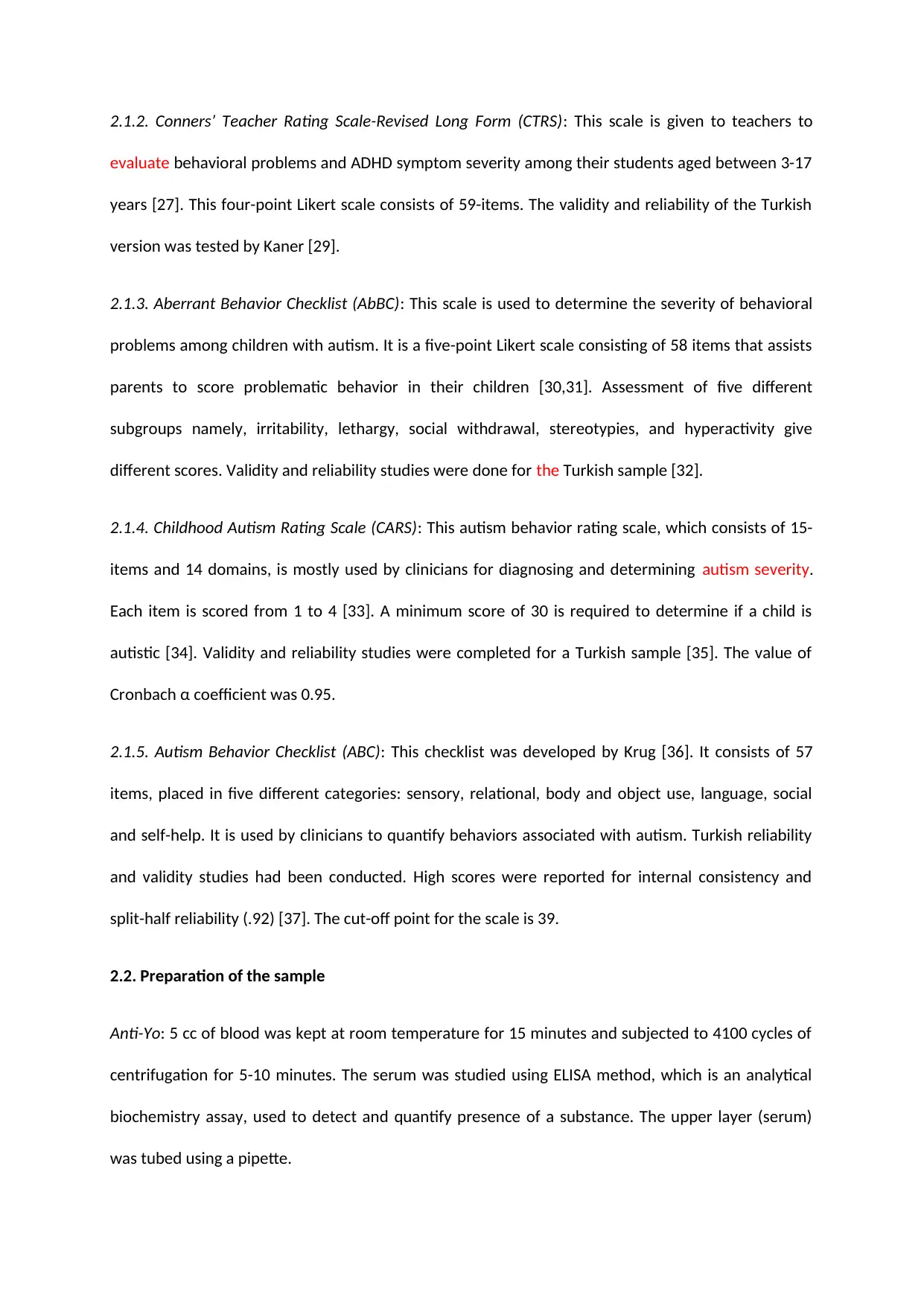
2.1.2. Conners’ Teacher Rating Scale-Revised Long Form (CTRS): This scale is given to teachers to
evaluate behavioral problems and ADHD symptom severity among their students aged between 3-17
years [27]. This four-point Likert scale consists of 59-items. The validity and reliability of the Turkish
version was tested by Kaner [29].
2.1.3. Aberrant Behavior Checklist (AbBC): This scale is used to determine the severity of behavioral
problems among children with autism. It is a five-point Likert scale consisting of 58 items that assists
parents to score problematic behavior in their children [30,31]. Assessment of five different
subgroups namely, irritability, lethargy, social withdrawal, stereotypies, and hyperactivity give
different scores. Validity and reliability studies were done for the Turkish sample [32].
2.1.4. Childhood Autism Rating Scale (CARS): This autism behavior rating scale, which consists of 15-
items and 14 domains, is mostly used by clinicians for diagnosing and determining autism severity.
Each item is scored from 1 to 4 [33]. A minimum score of 30 is required to determine if a child is
autistic [34]. Validity and reliability studies were completed for a Turkish sample [35]. The value of
Cronbach α coefficient was 0.95.
2.1.5. Autism Behavior Checklist (ABC): This checklist was developed by Krug [36]. It consists of 57
items, placed in five different categories: sensory, relational, body and object use, language, social
and self-help. It is used by clinicians to quantify behaviors associated with autism. Turkish reliability
and validity studies had been conducted. High scores were reported for internal consistency and
split-half reliability (.92) [37]. The cut-off point for the scale is 39.
2.2. Preparation of the sample
Anti-Yo: 5 cc of blood was kept at room temperature for 15 minutes and subjected to 4100 cycles of
centrifugation for 5-10 minutes. The serum was studied using ELISA method, which is an analytical
biochemistry assay, used to detect and quantify presence of a substance. The upper layer (serum)
was tubed using a pipette.
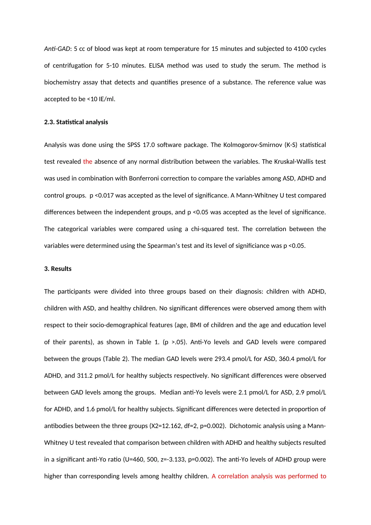
Anti-GAD: 5 cc of blood was kept at room temperature for 15 minutes and subjected to 4100 cycles
of centrifugation for 5-10 minutes. ELISA method was used to study the serum. The method is
biochemistry assay that detects and quantifies presence of a substance. The reference value was
accepted to be <10 IE/ml.
2.3. Statistical analysis
Analysis was done using the SPSS 17.0 software package. The Kolmogorov-Smirnov (K-S) statistical
test revealed the absence of any normal distribution between the variables. The Kruskal-Wallis test
was used in combination with Bonferroni correction to compare the variables among ASD, ADHD and
control groups. p <0.017 was accepted as the level of significance. A Mann-Whitney U test compared
differences between the independent groups, and p <0.05 was accepted as the level of significance.
The categorical variables were compared using a chi-squared test. The correlation between the
variables were determined using the Spearman’s test and its level of significiance was p <0.05.
3. Results
The participants were divided into three groups based on their diagnosis: children with ADHD,
children with ASD, and healthy children. No significant differences were observed among them with
respect to their socio-demographical features (age, BMI of children and the age and education level
of their parents), as shown in Table 1. (p >.05). Anti-Yo levels and GAD levels were compared
between the groups (Table 2). The median GAD levels were 293.4 pmol/L for ASD, 360.4 pmol/L for
ADHD, and 311.2 pmol/L for healthy subjects respectively. No significant differences were observed
between GAD levels among the groups. Median anti-Yo levels were 2.1 pmol/L for ASD, 2.9 pmol/L
for ADHD, and 1.6 pmol/L for healthy subjects. Significant differences were detected in proportion of
antibodies between the three groups (X2=12.162, df=2, p=0.002). Dichotomic analysis using a Mann-
Whitney U test revealed that comparison between children with ADHD and healthy subjects resulted
in a significant anti-Yo ratio (U=460, 500, z=-3.133, p=0.002). The anti-Yo levels of ADHD group were
higher than corresponding levels among healthy children. A correlation analysis was performed to
⊘ This is a preview!⊘
Do you want full access?
Subscribe today to unlock all pages.
Trusted by 1+ million students worldwide
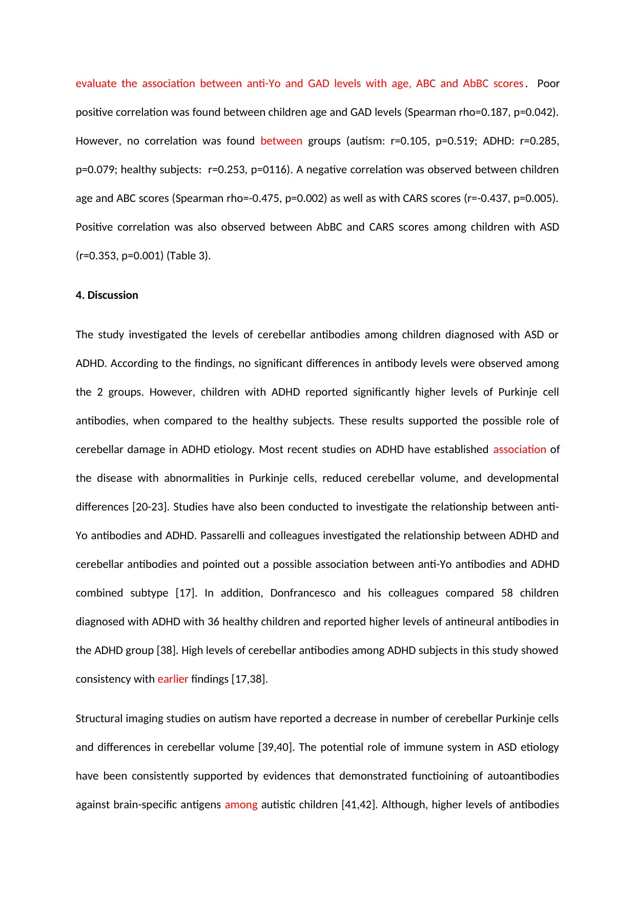
evaluate the association between anti-Yo and GAD levels with age, ABC and AbBC scores . Poor
positive correlation was found between children age and GAD levels (Spearman rho=0.187, p=0.042).
However, no correlation was found between groups (autism: r=0.105, p=0.519; ADHD: r=0.285,
p=0.079; healthy subjects: r=0.253, p=0116). A negative correlation was observed between children
age and ABC scores (Spearman rho=-0.475, p=0.002) as well as with CARS scores (r=-0.437, p=0.005).
Positive correlation was also observed between AbBC and CARS scores among children with ASD
(r=0.353, p=0.001) (Table 3).
4. Discussion
The study investigated the levels of cerebellar antibodies among children diagnosed with ASD or
ADHD. According to the findings, no significant differences in antibody levels were observed among
the 2 groups. However, children with ADHD reported significantly higher levels of Purkinje cell
antibodies, when compared to the healthy subjects. These results supported the possible role of
cerebellar damage in ADHD etiology. Most recent studies on ADHD have established association of
the disease with abnormalities in Purkinje cells, reduced cerebellar volume, and developmental
differences [20-23]. Studies have also been conducted to investigate the relationship between anti-
Yo antibodies and ADHD. Passarelli and colleagues investigated the relationship between ADHD and
cerebellar antibodies and pointed out a possible association between anti-Yo antibodies and ADHD
combined subtype [17]. In addition, Donfrancesco and his colleagues compared 58 children
diagnosed with ADHD with 36 healthy children and reported higher levels of antineural antibodies in
the ADHD group [38]. High levels of cerebellar antibodies among ADHD subjects in this study showed
consistency with earlier findings [17,38].
Structural imaging studies on autism have reported a decrease in number of cerebellar Purkinje cells
and differences in cerebellar volume [39,40]. The potential role of immune system in ASD etiology
have been consistently supported by evidences that demonstrated functioining of autoantibodies
against brain-specific antigens among autistic children [41,42]. Although, higher levels of antibodies
Paraphrase This Document
Need a fresh take? Get an instant paraphrase of this document with our AI Paraphraser
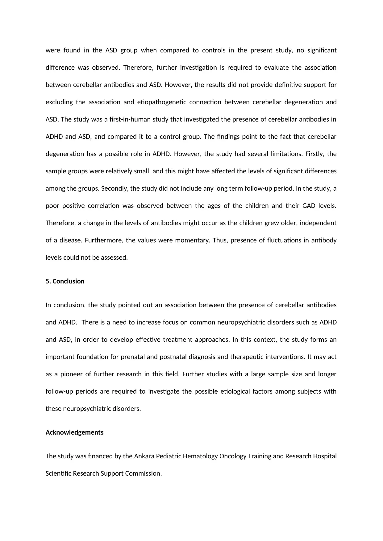
were found in the ASD group when compared to controls in the present study, no significant
difference was observed. Therefore, further investigation is required to evaluate the association
between cerebellar antibodies and ASD. However, the results did not provide definitive support for
excluding the association and etiopathogenetic connection between cerebellar degeneration and
ASD. The study was a first-in-human study that investigated the presence of cerebellar antibodies in
ADHD and ASD, and compared it to a control group. The findings point to the fact that cerebellar
degeneration has a possible role in ADHD. However, the study had several limitations. Firstly, the
sample groups were relatively small, and this might have affected the levels of significant differences
among the groups. Secondly, the study did not include any long term follow-up period. In the study, a
poor positive correlation was observed between the ages of the children and their GAD levels.
Therefore, a change in the levels of antibodies might occur as the children grew older, independent
of a disease. Furthermore, the values were momentary. Thus, presence of fluctuations in antibody
levels could not be assessed.
5. Conclusion
In conclusion, the study pointed out an association between the presence of cerebellar antibodies
and ADHD. There is a need to increase focus on common neuropsychiatric disorders such as ADHD
and ASD, in order to develop effective treatment approaches. In this context, the study forms an
important foundation for prenatal and postnatal diagnosis and therapeutic interventions. It may act
as a pioneer of further research in this field. Further studies with a large sample size and longer
follow-up periods are required to investigate the possible etiological factors among subjects with
these neuropsychiatric disorders.
Acknowledgements
The study was financed by the Ankara Pediatric Hematology Oncology Training and Research Hospital
Scientific Research Support Commission.
Disclosure
The authors did not report any conflict of interest.
References
[1] American Psychiatric Association, Diagnostic and Statistical Manual of Mental Disorders, 4th ed.,
Text-Revision (DSM-IV-TR), American Psychiatric Association, Washington, DC, 2000.
[2] J.J. Cannell, On the aetiology of autism, Acta Paediatrica. 99 (2010) 1128-1130.
[3] M. Coleman, C. Gillberg, The autisms, 4th ed, Oxford University Press, 2011.
[4] P.T. Huerta, C. Kowal, L.A. De Giorgio, B.T. Volpe, B. Diamond, Immunity and behavior: antibodies
alter emotion, Proc Natl Acad Sci USA. 103 (2006) 678-683.
[5] C.B. Yeh, C.H. Wu, H.C. Tsung, C.W. Chen, J.F. Shyu, J.F. Leckman, Antineural antibody in patients
with Tourette’s syndrome and their family members, J Biomed Sci. 13 (2005) 101 112.
[6] L.S. Kiessling, A.C. Marcotte, L. Culpepper, Antineuronal antibodies: tics and obsessive-
compulsive symptoms, J Dev Behav Pediatr. 15 (1994) 421-425.
[7] T.L. Kemper, M.L. Bauman, Neuropathology of infantile autism, Mol Psychiatry. 7 (2002) 12-13.
[Abstract] / [PDF]
[8] D. Riva, C. Giorgi, The contribution of the cerebellum to mental and social functions in
developmental age, Fiziol Cheloveka. 26 (2000) 27-31.
[9] S. Wills, M. Cabanlit, J. Bennett, P. Ashwood, D. Amaral, J. Van de Water, Autoantibodies in
autism spectrum disorders (ASD), Ann NY Acad Sci. 1107 (2007) 79-91.
[10] M. Steinlin, Cerebellar disorders in childhood: cognitive problems, Cerebellum. 7 (2008) 607-610.
⊘ This is a preview!⊘
Do you want full access?
Subscribe today to unlock all pages.
Trusted by 1+ million students worldwide
[11] P.M. Gillig, R.D. Sanders, Psychiatry, neurology, and the role of the cerebellum, Psychiatry
(Edgmont). 7 (2010) 38-43.
[12] L.A. Martin, D. Goldowitz, G. Mittleman, Repetitive behavior and increased activity in mice with
Purkinje cell loss: a model for understanding the role of cerebellar pathology in autism, Eur J
NeuroSci. 31 (2010) 544-555.
[13] J. Skefos, C. Cummings, K. Enzer, J. Holiday, K. Weed, E. Levy, T. Yuce, T. Kemper, M. Bauman,.
Regional alterations in Purkinje cell density in patients with autism, PloS one. 9 (2014) e81255.
[14] G. Polanczyk, M.S. de Lima, B.L. Horta, J. Biederman, L.A. Rohde, The worldwide prevalence of
ADHD: a systematic review and metaregression analysis, Am J Psychiatry. 164 (2007) 942-948.
[15] W.R. McGinnis, Oxidative stress in autism, Altern Ther Health Med. 10 (2004) 22-36.
[16] B.M. Ross, I. McKenzie, I. Glen, C.P.W. Bennett, Increased levels of ethane, a non invasive marker
of n-3 fatty acid oxidation, in breath of children with attention deficit hyperactivity disorder, Nutr
Neurosci. 6 (2003) 277-281.
[17] F. Passarelli, R. Donfrancesco, P. Nativio, E. Pascale, M. Di Trani, A.M. Patti, A. Vulcano, P. Gozzo,
M.P. Villa, Anti-Purkinje cell antibody as a biological marker in attention deficit/hyperactivity
disorder: a pilot study, J Neuroimmunol. 258 (2013) 67-70.
[18] M. Semrud-Clikeman, R.J. Steingard, P. Filipek, J. Biederman, K. Bekken, P.F. Renshaw, Using
MRI to examine brain-behavior relationships inmales with attention deficit disorder with
hyperactivity, J Am Acad Child Adolesc Psychiatry. 39 (2000) 477-484.
[19] M. Ashtari, S. Kumra, S.L. Bhaskar, T. Clarke, E. Thaden, K.L. Cervellione, J. Rhinewine, J.M. Kane,
A. Adesman, R. Milanaik, J. Maytal, A. Diamond, P. Szeszko, B.A. Ardekani,
Attention-deficit/hyperactivity disorder: a preliminary diffusion tensor imaging study, Biol Psychiatry.
57 (2005) 448-455.
Paraphrase This Document
Need a fresh take? Get an instant paraphrase of this document with our AI Paraphraser
[20] P.C. Berquin, J.N. Giedd, L.K. Jacobsen, S.D. Hamburger, A.L. Krain, J.L. Rapoport, F.X.
Castellanos, Cerebellum in attention-deficit hyperactivity disorder: a morphometric MRI study,
Neurology. 50 (1998) 1087-1093.
[21] F.X. Castellanos, M.T. Acosta, Syndrome of attention deficit with hyperactivity as the expression
of an organic functional disorder, Rev Neurol. 35 (2002) 1-11.
[22] F.X. Castellanos, P.P. Lee, W. Sharp, N.O. Jeffries, D.K. Greenstein, L.S. Clasen, J.D. Blumenthal,
R.S. James, C.L. Ebens, J.M. Walter, A. Zijdenbos, A.C. Evans, J.N. Giedd, J.L. Rapoport, Developmental
trajectories of brain volume abnormalities in children andadolescents with attention
deficit/hyperactivity disorder, JAMA. 288 (2002) 1740-1748.
[23] S. Mackie, P. Shaw, R. Lenroot, R. Pierson, D.K. Greenstein, T.F. Nugent, W.S. Sharp, J.N. Giedd,
J.L. Rapoport, Cerebellar development and clinical outcome in attention deficit hyperactivity
disorder, Am J Psychiatry. 164 (2007) 647-655.
[24] S. Ogita, O.H. Llaguna, S.M. Feldman, R. Blum, Paraneoplastic cerebellar degeneration with anti-
Yo antibody in a patient with HER2/neu overexpressing breast cancer: a case report with a current
literature review, Breast J. 14 (2008) 382-384.
[25] M.U. Manto, C.S. Hampe, V. Rogemond, J. Honnorat, Respective implications of glutamate
decarboxylase antibodies in stiff person syndrome and cerebellar ataxia, Orphanet J Rare Dis. 6.1
(2011) 3.
[26] B. Gökler, F. Ünal, B. Pehlivantürk, E.Ç. Kültür, D. Akdemir, Y. Taner, Reliability and validity of
Schedule for Affective Disorders and Schizophrenia for School Age Children Present and Lifetime
Version, Turk J Child and Adolesc Ment Health. 11 (2004) 109-116.
[27] C.K. Conners, Conners’ Rating Scales-Revised technical manual, North Tonawanda, NY, Multi-
Health Systems, 1997.
[28] S. Kaner, S. Buyukozturk, E. Iseri, A. Ak, L. Ozaydın, Conners' Parent Rating Scale Long Form-
Revised: Factor Structure, Reliability and Validity Studies, Turk J Child and Adolesc Ment Health. 18
(2011) 45-58.
[29] S. Kaner, S. Buyukozturk, E. Iseri, A. Ak, L. Ozaydın, The validity and reliability study of the Turkish
version of Conners’ Teacher rating scale-revised (CTRS-R), World Psychiatric Association Congress,
July 12-16, İstanbul. Turk Psikiyatri Derg. 17 (2006) 227.
[30] M.G. Aman, N.N. Singh, A.W. Stewart, C.J. Field, Psychometric characteristics of the Aberrant
Behavior Checklist, Am J Mental Deficiency. 89 (1985) 492-502.
[31] M.G. Aman, N.N. Singh, S.H. Turbott, Reliability of the Aberrant Behavior Checklist and the effect
of variations in instructions, Am J Mental Deficiency. 92 (1987) 237-240.
[32] K. Karabekiroglu, M.G. Aman, Validity of the aberrant behavior checklist in a clinical sample of
toddlers, Child Psychiatry Hum Dev. 40 (2009) 99-110.
[33] E. Schopler, R.J. Reichler, B.R. Renner, The Childhood Autism Rating Scale (CARS), 11th ed,
Western Psychological Services, 2007.
[34] G. Mesibov, E. Schopler, B. Schaffer, N. Michal, Use of childhood autism rating scale with autistic
adolescents and adults, J Am Acad Child Adolesc Psychiatry. 28 (1989) 538-541.
[35] S.İ. Gassologlu, B. Baykara, S. Avcil, Y. Demiral, Validity and Reliability Analysis of Turkish Version
of Childhood Autism Rating Scale, Turk Psikiyatri Derg. 27 (2016) 1-9.
[36] D.A. Krug, J.R. Arick, P.A. Almond, Autism Screening Instrument for Educational Planning, 2th ed,
Pro-ed Inc, Austin, Texas, 1993.
[37] T. Yilmaz-Irmak, S. Tekinsav-Sutcu, A. Aydin, O. Sorias, Validity and Reliability Analysis of Turkish
Version of Autism Behavior Checklist, Turk J Child and Adolesc Ment Health. 1 (2007) 13-23.
⊘ This is a preview!⊘
Do you want full access?
Subscribe today to unlock all pages.
Trusted by 1+ million students worldwide
1 out of 13
Your All-in-One AI-Powered Toolkit for Academic Success.
+13062052269
info@desklib.com
Available 24*7 on WhatsApp / Email
![[object Object]](/_next/static/media/star-bottom.7253800d.svg)
Unlock your academic potential
Copyright © 2020–2026 A2Z Services. All Rights Reserved. Developed and managed by ZUCOL.