Analyzing Wound Management: Case Studies and Treatment Strategies
VerifiedAdded on 2023/06/08
|23
|6159
|132
Case Study
AI Summary
This assignment presents two case studies focusing on comprehensive wound management. The first case involves Mr. Will Jackson, a 77-year-old with multiple health issues including diabetes, ischemic heart disease, and a history of alcohol abuse, who presents with a diabetic foot ulcer, an arterial ulcer, a stage 3 pressure ulcer, and a bruise. The second case focuses on Mrs. Miriam Gold, an 85-year-old with metastatic cervical cancer, a rectovaginal fistula, and a long-standing venous ulcer, admitted for palliative care. The assignment details holistic assessments, including medical history, wound evaluations (wound bed status, characteristics, measurements, surrounding skin condition, and exudates), expected healing processes, and patient health education. Furthermore, it provides specific wound management plans for each type of ulcer and wound, addressing moist wound healing, risk assessment, wound cleansing, pressure support, dressing selection, and pain management strategies, with emphasis on medication, frequency, dose, and justification. The conclusion summarizes the importance of comprehensive and individualized wound care.
Running head: WOUND MANAGEMENT PLANS
WOUND MANAGEMENT PLANS
Name of the Student
Name of the University
Author’s Note
WOUND MANAGEMENT PLANS
Name of the Student
Name of the University
Author’s Note
Paraphrase This Document
Need a fresh take? Get an instant paraphrase of this document with our AI Paraphraser
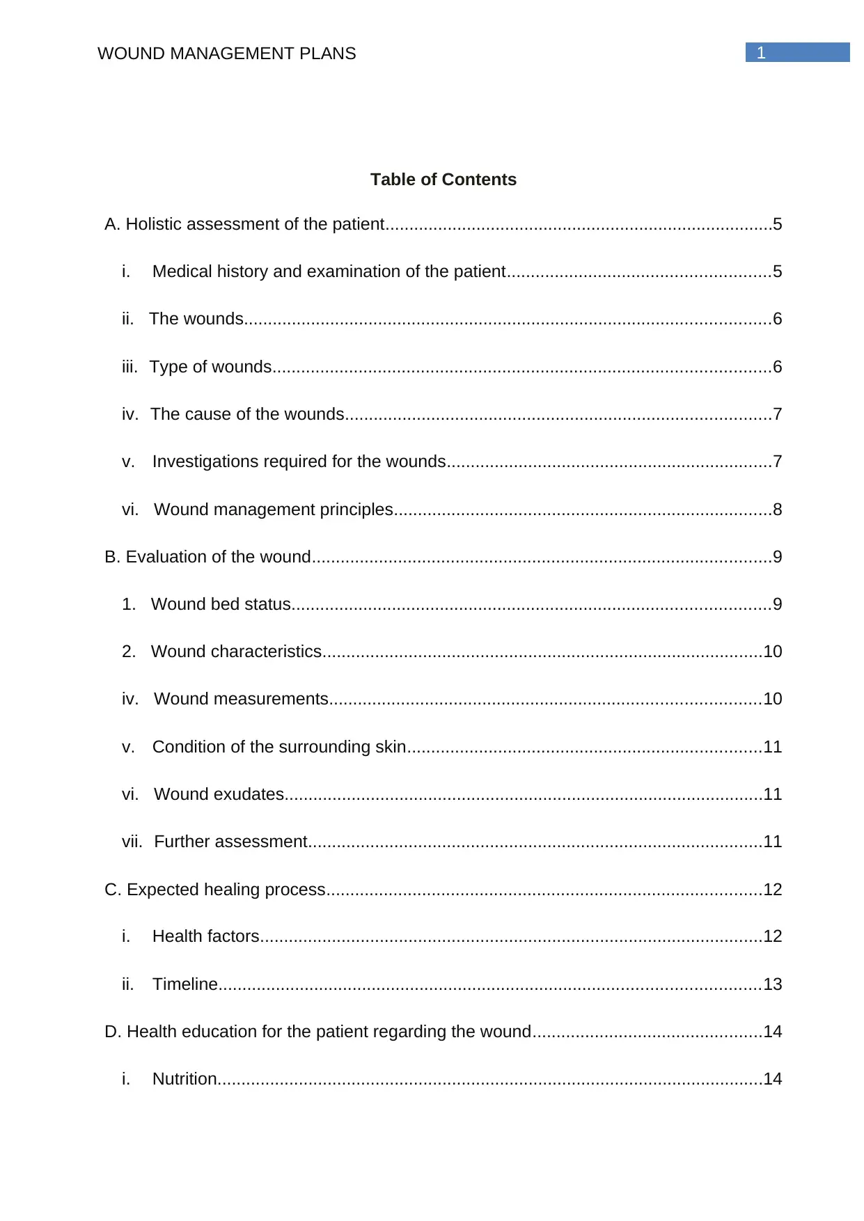
1WOUND MANAGEMENT PLANS
Table of Contents
A. Holistic assessment of the patient.................................................................................5
i. Medical history and examination of the patient.......................................................5
ii. The wounds..............................................................................................................6
iii. Type of wounds........................................................................................................6
iv. The cause of the wounds.........................................................................................7
v. Investigations required for the wounds....................................................................7
vi. Wound management principles...............................................................................8
B. Evaluation of the wound................................................................................................9
1. Wound bed status....................................................................................................9
2. Wound characteristics............................................................................................10
iv. Wound measurements..........................................................................................10
v. Condition of the surrounding skin..........................................................................11
vi. Wound exudates....................................................................................................11
vii. Further assessment...............................................................................................11
C. Expected healing process...........................................................................................12
i. Health factors.........................................................................................................12
ii. Timeline.................................................................................................................13
D. Health education for the patient regarding the wound................................................14
i. Nutrition..................................................................................................................14
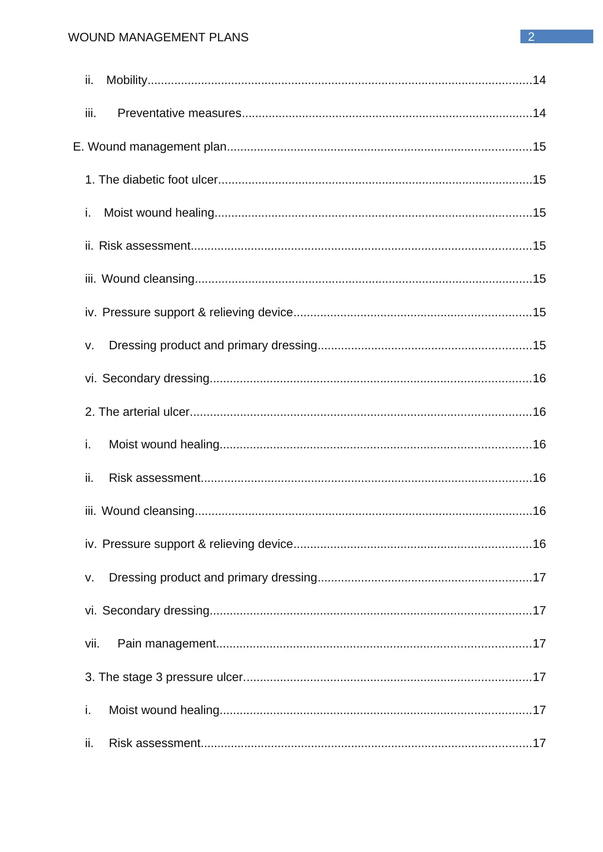
2WOUND MANAGEMENT PLANS
ii. Mobility...................................................................................................................14
iii. Preventative measures.......................................................................................14
E. Wound management plan...........................................................................................15
1. The diabetic foot ulcer..............................................................................................15
i. Moist wound healing...............................................................................................15
ii. Risk assessment......................................................................................................15
iii. Wound cleansing.....................................................................................................15
iv. Pressure support & relieving device.......................................................................15
v. Dressing product and primary dressing................................................................15
vi. Secondary dressing................................................................................................16
2. The arterial ulcer......................................................................................................16
i. Moist wound healing.............................................................................................16
ii. Risk assessment...................................................................................................16
iii. Wound cleansing.....................................................................................................16
iv. Pressure support & relieving device.......................................................................16
v. Dressing product and primary dressing................................................................17
vi. Secondary dressing................................................................................................17
vii. Pain management..............................................................................................17
3. The stage 3 pressure ulcer......................................................................................17
i. Moist wound healing.............................................................................................17
ii. Risk assessment...................................................................................................17
⊘ This is a preview!⊘
Do you want full access?
Subscribe today to unlock all pages.

Trusted by 1+ million students worldwide
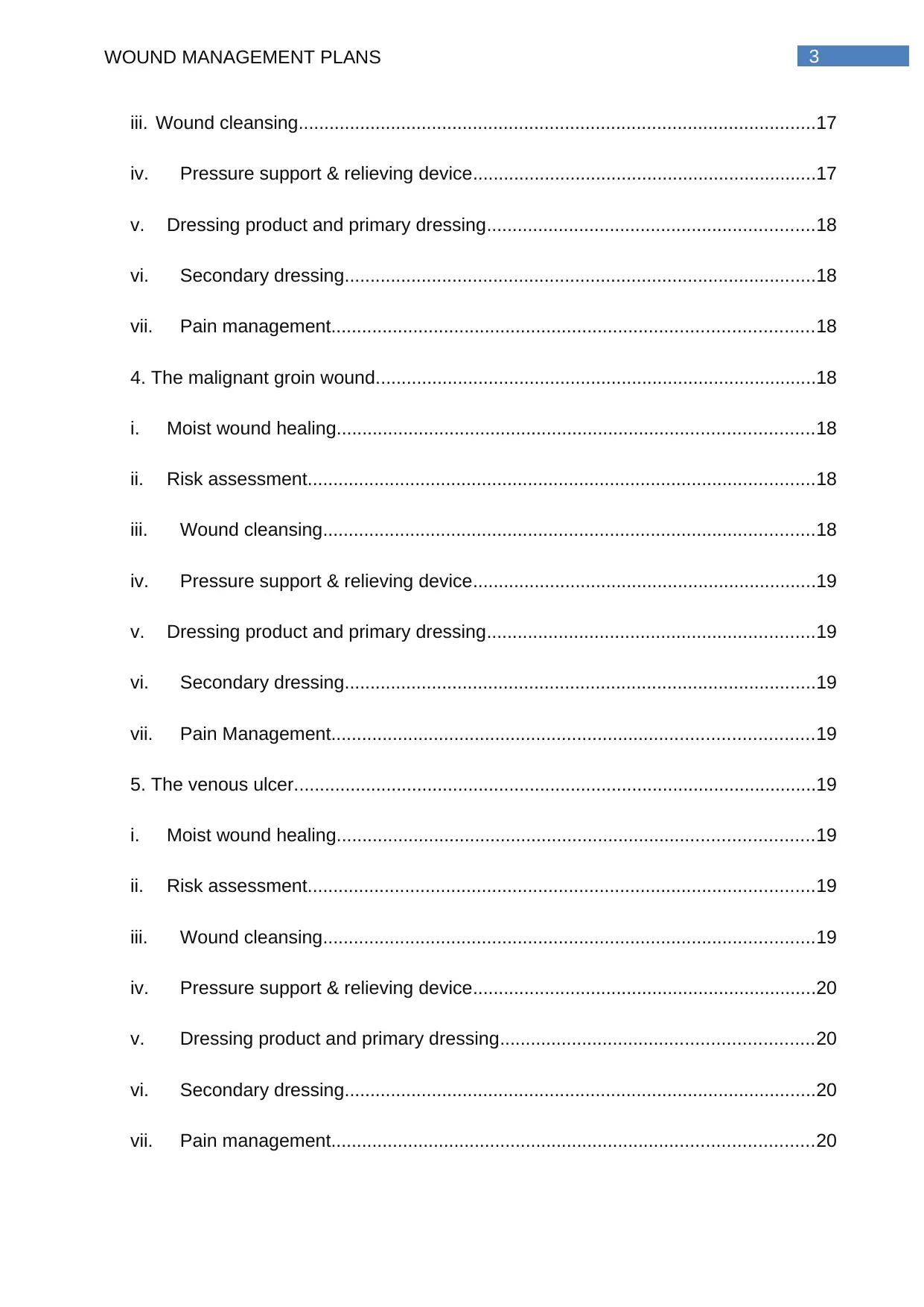
3WOUND MANAGEMENT PLANS
iii. Wound cleansing.....................................................................................................17
iv. Pressure support & relieving device...................................................................17
v. Dressing product and primary dressing................................................................18
vi. Secondary dressing............................................................................................18
vii. Pain management..............................................................................................18
4. The malignant groin wound......................................................................................18
i. Moist wound healing.............................................................................................18
ii. Risk assessment...................................................................................................18
iii. Wound cleansing................................................................................................18
iv. Pressure support & relieving device...................................................................19
v. Dressing product and primary dressing................................................................19
vi. Secondary dressing............................................................................................19
vii. Pain Management..............................................................................................19
5. The venous ulcer......................................................................................................19
i. Moist wound healing.............................................................................................19
ii. Risk assessment...................................................................................................19
iii. Wound cleansing................................................................................................19
iv. Pressure support & relieving device...................................................................20
v. Dressing product and primary dressing.............................................................20
vi. Secondary dressing............................................................................................20
vii. Pain management..............................................................................................20
Paraphrase This Document
Need a fresh take? Get an instant paraphrase of this document with our AI Paraphraser
4WOUND MANAGEMENT PLANS
F. Pain management of the wound..................................................................................20
i. Medication..............................................................................................................20
ii. Frequency and dose..............................................................................................20
iii. Justification for the medication................................................................................21
G. Conclusion..................................................................................................................21
H. References..................................................................................................................22
5WOUND MANAGEMENT PLANS
A. Holistic assessment of the patient
i. Medical history and examination of the patient
Case Study 1:
The first case study is on Mr. Will Jackson, a 77 year old who had been
diagnosed with rectal cancer five years ago. He has a clinical history of ischemic heart
disease (IHD) which attributed to his coronary artery bypass grafting (CABG) two years
ago. He also has Chronic Obstructive Pulmonary Disease (COPD) along with Gastro-
oesophageal reflux disease (GORD) and a history of alcohol abuse. He also suffers
from postural hypotension and takes insulin to control his type 2 diabetes mellitus. He
also suffers from intermittent confusion and mild lack of cognitive integrity at times. He
lives alone in a retirement village since his wife passed away a year ago. His urine
output is incontinent and requires pad in situ. He can walk a few steps for a short
distance with help however requires wheelchair assistance for longer distances.
Mr. Jackson has been admitted with pain, shortness of breath and for
management of his wounds. Prior to his admission, he had struck his head during a fall
at home and had a substantially large bruise over his face. The CT scan of Mr.
Jackson’s head did not reveal any significant damage; however the doctor stopped his
warfarin dose for a week due to this massive bruise as warfarin was being used to
reduce blood clots thereby aiding his CABG.
Case Study 2:
The second case study is on an 85 year old Mrs. Miriam Gold who has been
admitted with fluids overload problem and has pneumonia along with metastatic cervical
cancer. Mrs. Gold has a rectovaginal fistula due to her progressed cancer and
radiotherapy. Additionally, the patient has a long standing venous ulcer that is not
healing. Mrs. Gold has a medical history of Chronic Obstructive Pulmonary Disease
(COPD) and Gastro-oesophageal reflux disease (GORD) along with presence of
coronary artery bypass graft (CABG). Her mobility is restricted to resting in bed and is
cognitively drowsy and vague. She is on a semi liquid diet, comprised of mild thick fluids
and is incontinent, requiring pad in situ.
⊘ This is a preview!⊘
Do you want full access?
Subscribe today to unlock all pages.

Trusted by 1+ million students worldwide
6WOUND MANAGEMENT PLANS
The patient has been admitted for palliative care treatment as the patient is in a
lot of pain due to her malignant wound on her left groin and her family comprised of
husband and children are not coping well with her health issues.
ii. The wounds
Case Study 1:
Mr. Jackson was admitted with three wounds; however he now has four wounds
as follows;
1. One foot ulcer behind his left toe
2. One ulcer on his right lower leg
3. One ulcer on his sacrum bone
4. Bruise on left arm was after admittance to the ward.
Case Study 2:
Mrs Miriam Gold has two wound which require proper management.
1. One wound on the groin
2. One ulcer
iii. Type of wounds
Case Study 1:
The different wounds on Mr. Jackson are categorised below.
1. The foot ulcer behind his left toe is a diabetic ulcer.
2. The ulcer on his right lower leg is an arterial ulcer.
3. The ulcer on his sacrum bone is a stage 3 pressure ulcer.
4. The bruise on the patient’s left arm is a second degree burn bruise.
Case Study 2:
The two wounds on Mrs. Gold are categorised below
1. The wound on the left groin is malignant.
2. The ulcer is a venous ulcer.
Paraphrase This Document
Need a fresh take? Get an instant paraphrase of this document with our AI Paraphraser
7WOUND MANAGEMENT PLANS
iv. The cause of the wounds
Case Study 1:
1. The patient’s diabetic foot ulcer along with the arterial ulcer is both due to
his hyperglycaemic condition as he is a patient diagnosed with type 2 diabetes
mellitus.
2. The arterial ulcer has formed due damage to the arteries caused by
reduced blood flow or lack of blood flow to the tissue.
3. The stage 3 pressure ulcer on his sacrum bone has developed due to
unrelieved pressure on the underlying tissue from the strain from the sacrum bone
because of lying for long intervals in a fixed supine position.
4. Mr. Jackson sustained a second degree burn bruise on his left arm on the
3rd day of hospital stay due to accidental hot coffee spillage.
Case Study 2:
The two wounds of Mrs Gold require attention.
1. The malignant wound on the left groin is a rectovaginal fistula due to her
progressed cervical cancer and radiotherapy.
2. The venous ulcer has developed due to venous valves not functioning
properly.
v. Investigations required for the wounds
Case Study 1:
Mr. Jackson’s blood glucose level needs to be investigated at the earliest to
determine the effect of diabetes on the wounds.
An USG must also be performed on the surrounding tissues of the diabetic foot
ulcer and arterial ulcer along with the stage 3 pressure ulcer to determine the
extent of the ulcers and health of the nearby tissues. pH of the wounds must also
be checked to assess the infections properly.
Wound swabs of the diabetic foot ulcer and arterial ulcer microbial culture
examination are also recommended.
Haemoglobin count, white cell count and platelet count assessment is also vital
to diagnose infections and expected healing time.
8WOUND MANAGEMENT PLANS
The pH of the malignant wound must be assessed to determine the infection and
administer medicines accordingly.
Case Study 2:
Wound swabs of the venous ulcer and malignant groin wound to detect the
specific microbes infecting the wounds or that microbe specific treatment can be
advised.
White cell count, haemoglobin count, and platelet count is required to analyse the
level of infection and assess the healing time.
A biopsy of her groin malignant wound must be done to assess the status of her
cervical cancer.
The pH of the malignant wound must be assessed to determine the infection and
administer medicines accordingly.
vi. Wound management principles
Case Study 1:
The basic wound management principle for diabetic foot ulcer would encompass
debridement of the ulcer, proper therapeutic management of infection, revascularization
measures along with off-loading of the ulcer.
The management of the arterial ulcer on his right lower leg must involve
treatment of the infection while enhancing autolytic debridement along with controlled
exudation of pus, pain reduction and maintenance of a moist sterile healing
environment.
The treatment for pressure ulcer on the patient’s sacrum bone needs
involvement of infection management along with removal of the necrotic tissue, and
proper maintenance of moist environment for rapid wound healing. Repositioning of the
patient is the key to treat pressure ulcers (Moore & Cowman, 2015).
The wound management principle for the second degree burn would include
loose wrapping of a hydrogel based dressing over the bruise which will allow aeration
for fastens the healing process along with application of antibiotic ointment to prevent
infection.
The pH of the malignant wound must be assessed to determine the infection and
administer medicines accordingly.
Case Study 2:
Wound swabs of the venous ulcer and malignant groin wound to detect the
specific microbes infecting the wounds or that microbe specific treatment can be
advised.
White cell count, haemoglobin count, and platelet count is required to analyse the
level of infection and assess the healing time.
A biopsy of her groin malignant wound must be done to assess the status of her
cervical cancer.
The pH of the malignant wound must be assessed to determine the infection and
administer medicines accordingly.
vi. Wound management principles
Case Study 1:
The basic wound management principle for diabetic foot ulcer would encompass
debridement of the ulcer, proper therapeutic management of infection, revascularization
measures along with off-loading of the ulcer.
The management of the arterial ulcer on his right lower leg must involve
treatment of the infection while enhancing autolytic debridement along with controlled
exudation of pus, pain reduction and maintenance of a moist sterile healing
environment.
The treatment for pressure ulcer on the patient’s sacrum bone needs
involvement of infection management along with removal of the necrotic tissue, and
proper maintenance of moist environment for rapid wound healing. Repositioning of the
patient is the key to treat pressure ulcers (Moore & Cowman, 2015).
The wound management principle for the second degree burn would include
loose wrapping of a hydrogel based dressing over the bruise which will allow aeration
for fastens the healing process along with application of antibiotic ointment to prevent
infection.
⊘ This is a preview!⊘
Do you want full access?
Subscribe today to unlock all pages.

Trusted by 1+ million students worldwide
9WOUND MANAGEMENT PLANS
Case Study 2:
Gentle exudation of pus of the malignant wound on the left groin needs to be
performed after warm compression which would also relieve some pain. Therapeutic
treatment of the infection with warm compression with hydrogen borate is required to
treat the malignant wound followed by proper dressing of the wound.
The venous ulcer requires gentle debridement followed by proper management
of infection along with accurate maintenance of moisture to assure that the fresh healthy
tissue does not dry out.
B. Evaluation of the wound
1. Wound bed status
Case Study 1:
i. The wound bed of the diabetic foot ulcer behind the left toe of the patient
contains yellow slough tissue with bone visible at the base and almost no visible
pink healthy tissue and looks like an undermined ulcer.
ii. The wound bed of the arterial ulcer on his right lower leg contains unhealthy
granulation with yellow slough tissue which looks like it will bleed on contact and
looks like sloping edge ulcer and is infected.
iii. The wound bed of the stage 3 pressure ulcer on the sacrum bone on the
patient’s back looks infected with excessive unhealthy granulation and is dark red
in colour and looks like a punched out ulcer. The wound base of the ulcer
consists of 70% slough tissue and 30% granulation tissue.
Case Study 2:
i. The bed of the malignant wound contains pus and the exudate is yellowish white
along with a bit of blackish necrotic tissue. No pink or healthy tissue is visible.
ii. The wound bed of the venous ulcer is of unhealthy granulation with yellow slough
tissue
Case Study 2:
Gentle exudation of pus of the malignant wound on the left groin needs to be
performed after warm compression which would also relieve some pain. Therapeutic
treatment of the infection with warm compression with hydrogen borate is required to
treat the malignant wound followed by proper dressing of the wound.
The venous ulcer requires gentle debridement followed by proper management
of infection along with accurate maintenance of moisture to assure that the fresh healthy
tissue does not dry out.
B. Evaluation of the wound
1. Wound bed status
Case Study 1:
i. The wound bed of the diabetic foot ulcer behind the left toe of the patient
contains yellow slough tissue with bone visible at the base and almost no visible
pink healthy tissue and looks like an undermined ulcer.
ii. The wound bed of the arterial ulcer on his right lower leg contains unhealthy
granulation with yellow slough tissue which looks like it will bleed on contact and
looks like sloping edge ulcer and is infected.
iii. The wound bed of the stage 3 pressure ulcer on the sacrum bone on the
patient’s back looks infected with excessive unhealthy granulation and is dark red
in colour and looks like a punched out ulcer. The wound base of the ulcer
consists of 70% slough tissue and 30% granulation tissue.
Case Study 2:
i. The bed of the malignant wound contains pus and the exudate is yellowish white
along with a bit of blackish necrotic tissue. No pink or healthy tissue is visible.
ii. The wound bed of the venous ulcer is of unhealthy granulation with yellow slough
tissue
Paraphrase This Document
Need a fresh take? Get an instant paraphrase of this document with our AI Paraphraser
10WOUND MANAGEMENT PLANS
2. Wound characteristics
Case Study 1:
i. The wound characteristics of the foot ulcer behind the left toe of the patient is a
diabetic ulcer, undermined, unhealthy granulation with yellow slough tissue, with
the bone visible at the base and almost no visible pink healthy tissue.
ii. The wound characteristics of the ulcer on the right lower leg of the patient is an
arterial sloping edge ulcer with yellow slough tissue and unhealthy granulation
with presence of infection.
iii. The wound characteristics of the ulcer on the sacrum bone is a stage 3 pressure
punched out ulcer on the patient’s back which is very painful and looks infected
with excessive unhealthy granulation and is dark red in colour.
Case Study 2:
i. The malignant wound on the left groin is exudating pus and is giving out
extremely strong odour. It is causing the patient extreme pain and has also
developed a sinus tract which is a narrow passageway under the skin that may
extend in any direction, through the soft tissue, therefore resulting in a dead gap
with high probability for abscess formation. The progress of the wound causes a
burning sensation in the groin and peri-anal area which gets aggravated due to
bowel incontinence & discharge from her cervical cancer.
ii. The venous ulcer is a long standing wound with reduced healing probably due to
radiotherapy and her old age.
iv. Wound measurements
For better assessment of the wounds, the proper area and depth of the wounds
need to be measured. Various techniques and protocols are present for accurate wound
measurement namely the ruler method, water fill technique, the volumetric method,
Acetate Tracing Method, 3D scanner method; however studies have shown that 3D
scanner protocol gives the most accurate result, consistently. The measurements taken
by the 3D scanner are 11% better compared to the water fill technique, 75% better than
the ruler method, 41% better than the acetate tracing method and 52% better in
comparison to other 2D planimetric techniques in average. Consistent accurate
11WOUND MANAGEMENT PLANS
measurements were observed with the 3D scanner and such is required to measure
wounds precisely and treat the wound accordingly.
v. Condition of the surrounding skin
Case Study 1
1. The periwound skin surrounding the diabetic foot ulcer behind the left toe is swollen,
whitish yellow in color with no visible pink healthy tissue and contains pus and slough
tissue.
2. The periwound around the arterial ulcer on his right lower is dry and scaly and pinkish
in colour.
3. The stage 3 pressure ulcer’s periwound is extremely inflamed and pink in colour.
Case Study 2:
1. The periwound region of the patient’s malignant wound on the left groin is pink in
colour and inflamed.
2. The periwound of the venous ulcer is yellowish in color with no visible pink healthy
tissue
vi. Wound exudates
Case Study 1:
1. The diabetic foot ulcer behind the left toe has seropurulent wound exudate.
2. The arterial ulcer on the right lower leg has serous wound drainage.
3. The stage 3 pressure ulcer on the sacrum bone has serosanguineous exudates.
Case Study 2:
1. The malignant wound on the left groin is exudating pus and is giving out strong foul
odour.
2. The venous ulcer has no visible exudate.
vii. Further assessment
Case Study 1:
⊘ This is a preview!⊘
Do you want full access?
Subscribe today to unlock all pages.

Trusted by 1+ million students worldwide
1 out of 23
Related Documents
Your All-in-One AI-Powered Toolkit for Academic Success.
+13062052269
info@desklib.com
Available 24*7 on WhatsApp / Email
![[object Object]](/_next/static/media/star-bottom.7253800d.svg)
Unlock your academic potential
Copyright © 2020–2026 A2Z Services. All Rights Reserved. Developed and managed by ZUCOL.


