MIMIC METHOD: Development and Analysis of Patient-Specific Implants
VerifiedAdded on 2020/06/04
|10
|3405
|175
Report
AI Summary
This report delves into the MIMIC METHOD, a process for developing patient-specific implants. It begins with an introduction to the challenges and advancements in medical imaging, emphasizing the use of Reverse Engineering (RE) and Rapid Prototyping (RP) techniques. The core of the report focuses on converting Computer Tomography (CT) scan data into CAD models. It explores the advantages of using CAD-based solid models, particularly those based on B-Rep methods and NURBS surfaces, over voxel-based approaches. The report details the steps involved in transforming CT images (in DICOM format) into 3D CAD models, including image processing, point cloud data extraction, and segmentation techniques. It also highlights the use of thresholding methods and B-spline curve fitting. The methodology is illustrated with examples, including CT scans of lumbar spines and human skulls. Finally, it discusses the practical aspects of the process, such as data collection from CT images, noise elimination, and the transformation of data to the STL file format for rapid prototyping.
MIMIC METHOD
Paraphrase This Document
Need a fresh take? Get an instant paraphrase of this document with our AI Paraphraser

Table of Contents
Introduction ......................................................................................................................................................... 3
Development of patient specific implants....................................................................................3
CONCLUSION...................................................................................................................................................... 10
REFERENCES...................................................................................................................................................... 11
Introduction
Development of patient specific implants
Reconstruction of three dimensional (3D), Bio-CAD models from
computer tomography (CT) images are some of the problem that has been emerged
because of enhanced development of IT and adverse engineering method. This
model is for medical image. It is required to make attention on such issues that are
being faced. Reverse Engineering (RE) and Rapid Prototyping (RP) are few of the
surgical imagery and free form application. The above techniques are effective to
formulate anatomic models. This model has different features like symptomatic,
therapeutic and rehabilitation medical techniques (Carlesimo, Lombardi and
Caltagirone, 2011).
The above mentioned Bio-CAD models are effective in their own way for
medical practitioners which has number of features like computer-aided surgery,
functional modelling of weave, design of orthopaedic device, organism, tissue
platform and free form fictionalisation or bio-manufacturing [1-4].
This model is also very helpful in non- medical techniques where it is effective in
treatment. Such application consists for also fir the treatment of traveller safety
designing and crash investigation [2, 4-7].
For the reconstruction of 3D Bio-CAD models, mass based or isometric
based approachers are considered at large level. Voxel (Volume image) is used in
better way for the interpretation of CT scan data. This system is used at large in
thought commercialism available bio surgical software (Chang and et. al, 2013).
Voxel model is depicted by cuboidal or optical device elements which is set as per its
tallness, width and extent.
It is transferred to stl (stereolithography) format. When it is transformed
in this format then it is printed on rapid prototyping system. This format is effective
in high resolution surface and it is for visual image of human anatomy. There are
some of its limitation and restriction as it require large memory, loss of geometric
information. It is not preferable in model eating. Triangular patterned theory from
march cube is hard for the production and technology application which is except
pre-processing [8, 10].
The cited limitation of voxel model is replaced by transferring CT image
model into CAD based solid models, that considers isometric based formulation.
Representation of CAD based model can be made by boundaries. It envelops in
'boundary representation' method. B-Spline curves, Non-Uniform Coherent B-Spline
(NURBS) elements are considered to make the boundary surface. This is helpful in
providing some other advantages of taking higher degree continuity. Tangential
(C1 ) and Curvature (C2 ) over Positional (C0 ) continuity is in stl data.
This is effective method that is beneficial as comparison to volume based
approach. It is advantageous because of its editable and limited file size. It ensures
water tight surface model. RE (Reverse Engineering) technique is used in better way
because it comprises of different steps. These stages cover action; pre process for
elimination of noise and out liars; generation of partition and theory from point
cloud data [8,9] (Dreher and Schneider, 2010).
⊘ This is a preview!⊘
Do you want full access?
Subscribe today to unlock all pages.
Trusted by 1+ million students worldwide
Computer Tomography (CT) / Magnetic Resonance Imaging (MRI) in DICOM format
are some of the way that is effectively used in medical diagnostics and surgical
issues. These are the two dimensional non-invasive techniques which are effective in
human anatomical data. Development of 3D model can be made more effectual for
visualization or modelling by considering modern computer graphics and scanning
engineering [10,11].
3D model is utilized in better way by the surgeons so that they can
understand difficult internal anatomy that are faced by the patient. This model is
helpful to provide medical information to doctors. It is considered as powerful
diagnostic tool. This can also be considered to formulate prostheses, to do different
simulation and to perform logical task [1,8,10,11].
It is related to the discovery of Point Cloud Data (PCD) by process Digital
Imagery and Communication theory in Medicines (DICOM). Image processing
techniques are used from non-invasive medical check up picture like CT scans or
MRI. Mental image processing method is projected to pull out point gloom data. It is
from stem of CT scrutiny picture. Point cloud data is the information which is
raggedly separated and it has much noise and the shape. This is presented with full
of intricate information and it is not conferred in formulaic CAD data (Goyal and et.
al, 2011).
It has some additional process that is considered for classification,
smothering and B strip curve adjustment. At pre-processing stage, this data is
classified into two boundaries as internal and external and after that it moves to
smoothening process. When the above steps are covered then this is processed
further so that it can fit to B-spline curve. This method is effective to make CAD
model at high by decreasing its data file sizing and to ensure building of water
binding surface.
The voxel model and multilateral facet triangle based theory are
effectively utilized for bio-modelling and modality. Vast retention space is needed in
this regard. There are many technological advancement that are seen in CAD
engineering help design, model and to formulate object that has free form surface.
CAD based solid modelling is made according to B- rep method (boundary
representation). The data that it covers, converted into 3D CAD model. After that it is
printed by using rapid prototyping.
Paraphrase This Document
Need a fresh take? Get an instant paraphrase of this document with our AI Paraphraser
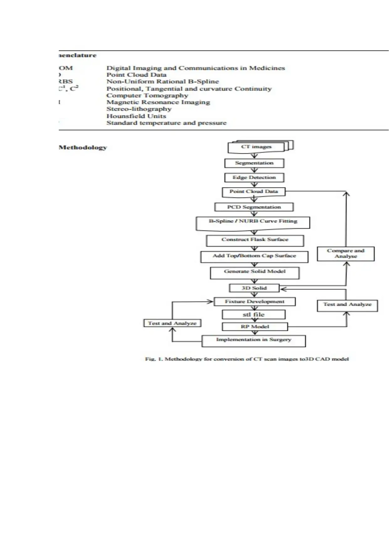
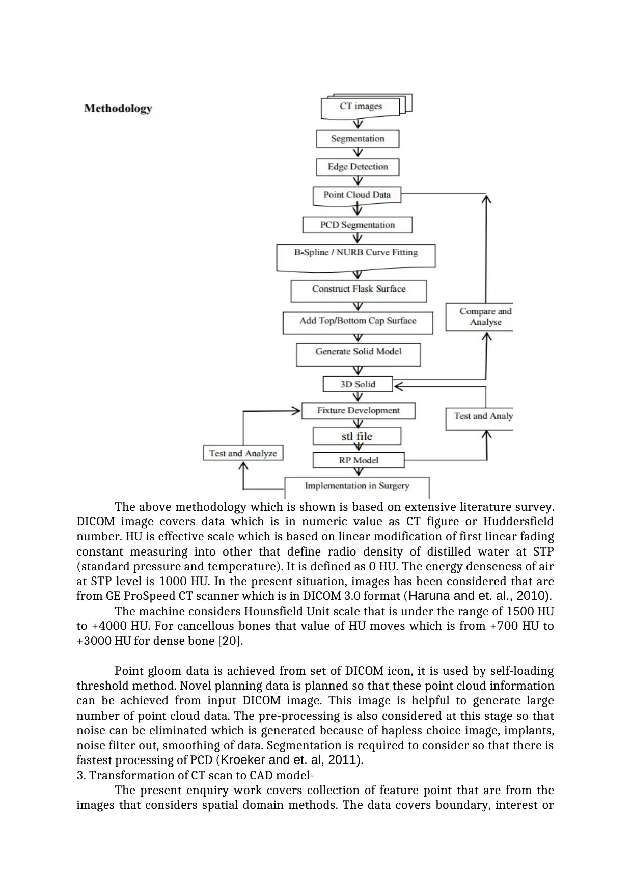
The above methodology which is shown is based on extensive literature survey.
DICOM image covers data which is in numeric value as CT figure or Huddersfield
number. HU is effective scale which is based on linear modification of first linear fading
constant measuring into other that define radio density of distilled water at STP
(standard pressure and temperature). It is defined as 0 HU. The energy denseness of air
at STP level is 1000 HU. In the present situation, images has been considered that are
from GE ProSpeed CT scanner which is in DICOM 3.0 format (Haruna and et. al., 2010).
The machine considers Hounsfield Unit scale that is under the range of 1500 HU
to +4000 HU. For cancellous bones that value of HU moves which is from +700 HU to
+3000 HU for dense bone [20].
Point gloom data is achieved from set of DICOM icon, it is used by self-loading
threshold method. Novel planning data is planned so that these point cloud information
can be achieved from input DICOM image. This image is helpful to generate large
number of point cloud data. The pre-processing is also considered at this stage so that
noise can be eliminated which is generated because of hapless choice image, implants,
noise filter out, smoothing of data. Segmentation is required to consider so that there is
fastest processing of PCD (Kroeker and et. al, 2011).
3. Transformation of CT scan to CAD model-
The present enquiry work covers collection of feature point that are from the
images that considers spatial domain methods. The data covers boundary, interest or
⊘ This is a preview!⊘
Do you want full access?
Subscribe today to unlock all pages.
Trusted by 1+ million students worldwide
corner points, convexity etc. Segmentation method is effective that is most preferable to
acquire part or object of curiosity that are derived from its scene. In current situation,
this is effectively used so that bone can be separated from its surrounding soft muscles
that are in CT image. Te investigator takes threshold based techniques in case of image
based partition [3,4,9,12,16-20]. Such method can be divided into parts like worldwide
and local threshold.
Global threshold is the threshold which value is determined manually or
automatically and its division is based on intensity value. Thresholding is used so that
bones can be separated from its soft tissues because of high level of intensity and HU
belief of bones in CT image. World technique is delicate in volume effect which is
partial; high grey level because of implants and radio beam hardening. localized
thresholding process is based on discovery of various value of apiece pixel. These pixels
are based on local statistics. The methods and theories that are used are of the mean of
higher and lower pixel intensities [3,4] (Lesnik and et. al., 2014).
Mean, normal variation , median of pixel intensities are according to local intensity
gradient magnitude [9].
The above mentioned methods are sensitized that is effective for partial volume
effect and intensity inhomogeneity. There is also effective method which is use of kang
method. It is effective to utilized because it validates bone segmentation. This is made
because of empirically chosen threshold value which is also used along with optional
manual interaction and it is beneficial to polish consequent periosteal surface [13].
According to Changand et. al., they proposed the utility of Separate Curvelet
alteration and FAST (Features from Accelerated Segmentation Test) [3, 4]. these
methods are effective that rely on initial thresholding so that bony area can be cutted
out from each part. It is obligatory for characteristic point ancestry. Curvelet
Transformation is applicable so that it can detect the edges that are in threshold part.
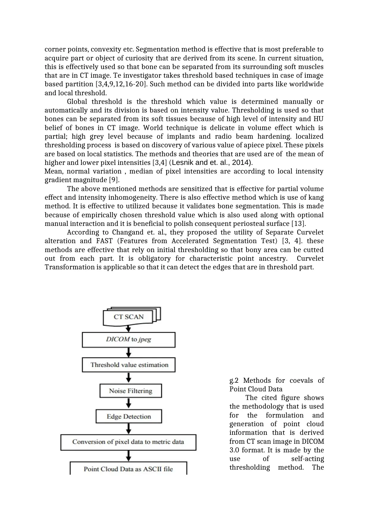
g.2 Methods for coevals of
Point Cloud Data
The cited figure shows
the methodology that is used
for the formulation and
generation of point cloud
information that is derived
from CT scan image in DICOM
3.0 format. It is made by the
use of self-acting
thresholding method. The
acquire part or object of curiosity that are derived from its scene. In current situation,
this is effectively used so that bone can be separated from its surrounding soft muscles
that are in CT image. Te investigator takes threshold based techniques in case of image
based partition [3,4,9,12,16-20]. Such method can be divided into parts like worldwide
and local threshold.
Global threshold is the threshold which value is determined manually or
automatically and its division is based on intensity value. Thresholding is used so that
bones can be separated from its soft tissues because of high level of intensity and HU
belief of bones in CT image. World technique is delicate in volume effect which is
partial; high grey level because of implants and radio beam hardening. localized
thresholding process is based on discovery of various value of apiece pixel. These pixels
are based on local statistics. The methods and theories that are used are of the mean of
higher and lower pixel intensities [3,4] (Lesnik and et. al., 2014).
Mean, normal variation , median of pixel intensities are according to local intensity
gradient magnitude [9].
The above mentioned methods are sensitized that is effective for partial volume
effect and intensity inhomogeneity. There is also effective method which is use of kang
method. It is effective to utilized because it validates bone segmentation. This is made
because of empirically chosen threshold value which is also used along with optional
manual interaction and it is beneficial to polish consequent periosteal surface [13].
According to Changand et. al., they proposed the utility of Separate Curvelet
alteration and FAST (Features from Accelerated Segmentation Test) [3, 4]. these
methods are effective that rely on initial thresholding so that bony area can be cutted
out from each part. It is obligatory for characteristic point ancestry. Curvelet
Transformation is applicable so that it can detect the edges that are in threshold part.
g.2 Methods for coevals of
Point Cloud Data
The cited figure shows
the methodology that is used
for the formulation and
generation of point cloud
information that is derived
from CT scan image in DICOM
3.0 format. It is made by the
use of self-acting
thresholding method. The
Paraphrase This Document
Need a fresh take? Get an instant paraphrase of this document with our AI Paraphraser
scheduling method which is novel programming faculty is improvised in MATLAB. It is
effective to find out or extract point cloud information from input signal DICOM image.
The main way to collect data is CT image which is retrieved by GE (Madison, 2011).
ProSpeed CT scanner is in formulated in way of DICOM 3.0 format. Its grayscale
images is gathered from GE ProSpeed. CT scanner is in format of DICOM 3. 0 where
there is pixel size of format is 512 * 512 and its collection of information diameter is
250 mm and its thickness is around 5 mm.
Fig.3. Shows Stepwise phenomenon of Picture Processing
The cited figure shows the stepwise results that can be gathered from
enhancement of image processing module. 2121 points can be collected from single CT
scan part. The collected points and data is stored in x, y, z format. There is coordination
of amoral record format ‘ASCII’. This file can be utilized in CAD situation (Metter and et.
al, 2011). It shows the very first step of experimental protocol that is effective for the
transformation of CT image to 3D CAD model. This provides assistance to higher order
continuity (C2). It is also effectual in providing some of the benefits like CAD models as
editing. The data of point cloud is extracted from CT scan with the help of new threshold
based algorithm. It is effective way so that such data can be eliminated from CT scan
model. Bones have heterogeneous structure and it also has flexible density. Algorithms
will be beneficial and at this step there is estimation of threshold value. These points are
important to consider so that construction of B-Spline curves and surfaces can be made.
It is effective that unable it to so that it can secure Tangential (C1) and Curvature (C2).
This point out data is transformed to *.stl file because it is good for rapid prototyping. It
is send with only positional continuity (C0). There are some of the major factors that are
faced in this regard like to separate among inner and external bone limit visibility
points. It is also hard to detect nearest point on a surface. The cited figure 4 (a) shows
that the result that are recovered from CT scan of lumbar spine. Where there is spine of
5 year feminine diligent and it covered around 330 slices. Each slice have thickness of at
least 1 mm that outputted into 11,59,752 points. The figure 4(b) shows the point cloud
data which is of 26,564 points and it is discovered from human skull. There was CT
scan which was made on 59 year old male patient where there is use of 11 slices and
each one has thickness of 5 mm (Ou and et. al., 2011).
Different surface are utilized in different reconstruction algorithms that are
fitting to 3D point cloud data as loft aboveground, expanse aboveground, B spline
surface and polynomial surface. In this filling of above mentioned factors are made by
the investigator [11,12,20]. At least 3 degree curve is utilized so that they can achieve
Curvature Continuity (C2). it is considered through point cloud. Three-dimensional
splines, B-spline and Non-Uniform Rational B-Spline (NURBS) are considered for
creating 3 degree curve. In this, B-spline surfaces, particularly NURBs surfaces are
mostly preferable. It is effective because of its power to accurately. Most of the surface
entity is considered in designing and production of new application. DongJin Yoo said
that implicit surface interpolation scheme is used to reconstruct B-spline aboveground.
It is from point physical phenomenon data or line of CT scan data [12]. Closed NURBS
curve is utilized so that there is efficient maintenance of surface accuracy in CAD model.
It is rendered along with shift of each one stratum (Pearce and et. al, 2012).
Olya Grove et al presented that it is better to use medical image data so that
there is construction of surface that is from consecutive antiparallel contours which is
pull out from images [20]. B spline curved shape is most suitable in this way which is
possible through points with circuit of region of interest. These methods are effective to
generate 3D surface theory. Relation based methods are generally considered as B-
splines and NURBs that is effective in providing excellent coverage but it is not effective
sometime because of high level of freedom which outputted in number of control
points. The above mentioned curves shows high level of continuities which is
comparison with ‘stl’ and voxel based method acting.
The figue 5 shows that (a) Point Cloud Data; (b) B-spline curve through PCD
Interpolation and approximation are two effective method that is considered for
line fitting direct sizeable point gloom data. It is not effective due to failure of too many
wiggles in interploting curve. It is nor eliminated because of its high tolerance (Wang
and Shih, 2010).
Dissimilar computation, in reckoning, curve approximates through information points.
Its smooth working calculate on grade of curve. It does not pass because there is failure
of estimating degree of freedom. It also not have complex form. Preprocessing of Point
Cloud Data is used for removal of outliers and it is effective in smoothening due to
enhancement of the modelling process.
The diagram of 5(a) shows sampling Point Cloud Information and figure 5(b)
present closed B-spline bender which is most suitable to improvement through sample
point cloud collection (Postow and et. al, 2012).
Diagram.6. Shows different aspect as
(a) First CT Scan;
(b) Increased CT image;
(c) Point Unreality Data;
(d) NURB’s Worthy;
(e) RP Exemplary;
(f) Abstracted piece of bone from bone after medical science
CONCLUSION
As per above study it can be concluded that CT scan and MRI scan is effective to
use that vary according to the age of the patient. There are different size are used in this
regard. There are different authors that presented their view and this research work is
completely based on some of the content that are effective in different ways. Different
surface are considered in different reconstruction algorithms which are most important
for CAD models. There are different models and theories that are explained in this
regard which is effective in providing solution to the issues that are being faced.
⊘ This is a preview!⊘
Do you want full access?
Subscribe today to unlock all pages.
Trusted by 1+ million students worldwide
REFERENCES
Books and Journals
Carlesimo, G.A., Lombardi, M.G. and Caltagirone, C., 2011. Vascular thalamic amnesia: a
reappraisal. Neuropsychologia. 49(5). pp.777-789.
Chang, B. and et. al, 2013. Natural history of pure ground-glass opacity lung nodules
detected by low-dose CT scan. Chest. 143(1). pp.172-178.
Dreher, A. and Schneider, F., 2010. Corruption and the shadow economy: an empirical
analysis. Public Choice. 144(1). pp.215-238.
Goyal, M. and et. al, 2011. Effect of baseline CT scan appearance and time to
recanalization on clinical outcomes in endovascular thrombectomy of acute
ischemic strokes. Stroke. 42(1). pp.93-97.
Haruna, A. and et. al., 2010. CT scan findings of emphysema predict mortality in COPD.
Chest. 138(3). pp.635-640.
Kroeker, K.I. and et. al, 2011. Patients with IBD are exposed to high levels of ionizing
radiation through CT scan diagnostic imaging: a five-year study. Journal of
clinical gastroenterology. 45(1). pp.34-39.
Lesnik, D. and et. al., 2014. Papillary thyroid carcinoma nodal surgery directed by a
preoperative radiographic map utilizing CT scan and ultrasound in all primary
and reoperative patients. Head & neck. 36(2). pp.191-202.
Madison, D.S., 2011. Critical ethnography: Method, ethics, and performance. Sage.
Metter, R.B. and et. al, 2011. Association between a quantitative CT scan measure of
brain edema and outcome after cardiac arrest. Resuscitation. 82(9). pp.1180-
1185.
Ou, S.H.I., and et. al., 2011. Activity of crizotinib (PF02341066), a dual mesenchymal-
epithelial transition (MET) and anaplastic lymphoma kinase (ALK) inhibitor, in
a non-small cell lung cancer patient with de novo MET amplification. Journal of
thoracic oncology. 6(5). pp.942-946.
Pearce, M.S. and et. al, 2012. Radiation exposure from CT scans in childhood and
subsequent risk of leukaemia and brain tumours: a retrospective cohort study.
The Lancet. 380(9840). pp.499-505.
Postow, M.A. and et. al, 2012. Immunologic correlates of the abscopal effect in a patient
with melanoma. New England Journal of Medicine. 366(10). pp.925-931.
Wang, W.C. and Shih, C.L., 2010. MIMIC methods for assessing differential item
functioning in polytomous items. Applied Psychological Measurement. 34(3).
pp.166-180.
1 out of 10

Your All-in-One AI-Powered Toolkit for Academic Success.
+13062052269
info@desklib.com
Available 24*7 on WhatsApp / Email
![[object Object]](/_next/static/media/star-bottom.7253800d.svg)
Unlock your academic potential
Copyright © 2020–2026 A2Z Services. All Rights Reserved. Developed and managed by ZUCOL.